. 2012 Dec;135(Pt 12):3735-48.
doi: 10.1093/brain/aws289. Epub 2012 Nov 26.

Protection of primary neurons and mouse brain from Alzheimer's pathology by molecular tweezers

Affiliations

Protection of primary neurons and mouse brain from Alzheimer's pathology by molecular tweezers

Aida Attar et al. Brain. 2012 Dec.

Abstract

Alzheimer's disease is a devastating cureless neurodegenerative disorder affecting >35 million people worldwide. The disease is caused by toxic oligomers and aggregates of amyloid β protein and the microtubule-associated protein tau. Recently, the Lys-specific molecular tweezer CLR01 has been shown to inhibit aggregation and toxicity of multiple amyloidogenic proteins, including amyloid β protein and tau, by disrupting key interactions involved in the assembly process. Following up on these encouraging findings, here, we asked whether CLR01 could protect primary neurons from Alzheimer's disease-associated synaptotoxicity and reduce Alzheimer's disease-like pathology in vivo. Using cell culture and brain slices, we found that CLR01 effectively inhibited synaptotoxicity induced by the 42-residue isoform of amyloid β protein, including ∼80% inhibition of changes in dendritic spines density and long-term potentiation and complete inhibition of changes in basal synaptic activity. Using a radiolabelled version of the compound, we found that CLR01 crossed the mouse blood-brain barrier at ∼2% of blood levels. Treatment of 15-month-old triple-transgenic mice for 1 month with CLR01 resulted in a decrease in brain amyloid β protein aggregates, hyperphosphorylated tau and microglia load as observed by immunohistochemistry. Importantly, no signs of toxicity were observed in the treated mice, and CLR01 treatment did not affect the amyloidogenic processing of amyloid β protein precursor. Examining induction or inhibition of the cytochrome P450 metabolism system by CLR01 revealed minimal interaction. Together, these data suggest that CLR01 is safe for use at concentrations well above those showing efficacy in mice. The efficacy and toxicity results support a process-specific mechanism of action of molecular tweezers and suggest that these are promising compounds for developing disease-modifying therapy for Alzheimer's disease and related disorders.


Figures

Figure 1
Figure 1
CLR01 protects neurons from amyloid β protein–induced changes in dendritic spine number and morphology. (A) Rat primary hippocampal neurons were incubated for 72 h with media alone or with amyloid β42 in the absence or presence of molecular tweezers. Yellow arrows point to amyloid β42–induced varicosities. Scale bar = 5 μm. (B) The number of dendritic spines per 100 μm was quantified. ***P < 0.001 compared with control;+++P < 0.001 compared with amyloid β42 + CLR01.
Figure 2
Figure 2
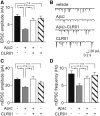
CLR01 rescues amyloid β42–induced inhibition of evoked and spontaneous synaptic neurotransmission in autaptic hippocampal neurons. (A) Mean excitatory postsynaptic current amplitude measured after 24 h of treatment with vehicle, 200 nM amyloid β42, 200 nM amyloid β42 + 2 µM CLR01 or 2 µM CLR01. (B) Representative traces showing miniature excitatory postsynaptic currents recorded in neurons subjected to each treatment. (C and D) Mean miniature excitatory postsynaptic current amplitude (C) and frequency (D) after 24-h treatment with vehicle, 200 nM amyloid β42, 200 nM amyloid β42 + 2 µM CLR01 or 2 µM CLR01. **P < 0.001. ns = not significant.
Figure 3
Figure 3
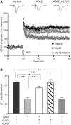
CLR01 attenuates amyloid β42–induced inhibition of long-term potentiation (LTP) at CA3–CA1 synapses. (A) Time course of field EPSP (fEPSP) amplitudes before and after high-frequency stimulation (HFS) (indicated by arrow) in slices treated for 20 min with vehicle (black circles), 200 nM amyloid β42 (grey circles) or 200 nM amyloid β42 + 2 µM CLR01 preincubated for 1 h before application (white circles). Results are expressed as percentages of baseline field EPSP amplitude (=100%). Insets show representative traces of field EPSP at baseline (dotted lines) and during the last 5 min of long-term potentiation recording (solid lines). (B) Bar graph showing mean long-term potentiation changes measured during the last 5 min of recording after slice exposure to vehicle or different combinations of 200 nM amyloid β42, 2 µM CLR01 and 2 µM CLR03. **P < 0.001; *P < 0.05. ns = not significant.
Figure 4
Figure 4
CLR01 decreases amyloid β protein and p-tau deposition and ameliorates microgliosis in transgenic mouse brain. Triple-transgenic mice were treated with 40 μg/kg/d CLR01 or vehicle. (A, D, G and I) Vehicle-treated transgenic mouse hippocampus. (B, E, H and J) CLR01-treated transgenic mouse hippocampus. (A and B) Transgenic mouse brain stained with monoclonal antibody 6E10 showing amyloid plaque deposition. (C) Per cent amyloid β (Aβ) burden was quantified by calculating the total 6E10-stained area divided by the total area measured. (D and E) Transgenic mouse brain showing AT8-positive neurofibrillary tangles in the CA1 region. (F) Per cent aggregated p-tau load was quantified by calculating the total AT8-stained area divided by the total area. (G and H) Transgenic mouse brain stained with monoclonal antibody HT7 for total tau. (I and J) Transgenic mouse brain showing Iba1-positive microglia in the subiculum and CA1 region. (K) Number of stained microglia in a 1.14 mm2 area of hippocampus per treatment condition. Scale bars: bar in B applies to both A and B; bar in J applies to D–J. *P < 0.05, **P < 0.01, ***P < 0.001 compared with vehicle-treated mice. Amyg = amygdala; Ent = entorhinal; Hippo = hippocampus; Peri = perirhinal; Pir = piriform cortices; WT = wild-type.
Figure 5
Figure 5
Weak induction of the cytochrome P450 system by CLR01. African green monkey kidney cells were treated with 10 μM rifampicin (rif; positive control) or CLR01 for 24 h. Cells transfected with luciferase but not PXR (XREM) were used as a negative control. (A) Luciferase activity was determined using a standard luciferase assay system (Promega). (B) β-galactosidase activity was determined using standard methods by the o-nitrophenolgalactoside assay.

References

    1. Thies W, Bleiler L Alzheimer’s Association 2011. Alzheimer's disease facts and figures. Alzheimers Dement. 2011;7:208–44. - PubMed
    1. Abramoff MD, Magelhaes PJ, Ram SJ. Image processing with ImageJ. Biophotonics Intl. 2004;11:36–42.
    1. Akiyama H, Barger S, Barnum S, Bradt B, Bauer J, Cole GM, et al. Inflammation and Alzheimer's disease. Neurobiol Aging. 2000;21:383–421. - PMC - PubMed
    1. Ard MD, Cole GM, Wei J, Mehrle AP, Fratkin JD. Scavenging of Alzheimers amyloid β-protein by microglia in culture. J Neurosci Res. 1996;43:190–202. - PubMed
    1. Arendt T. Synaptic degeneration in Alzheimer's disease. Acta Neuropathol. 2009;118:167–79. - PubMed

Publication types

MeSH terms