

doi: 10.1038/leu.2012.310.
Epub 2012 Nov 6.
Eltrombopag inhibition of acute myeloid leukemia cell survival does not depend on c-Mpl expression
Affiliations
- PMID: 23183425
- PMCID: PMC3987859
- DOI: 10.1038/leu.2012.310
Item in Clipboard
Eltrombopag inhibition of acute myeloid leukemia cell survival does not depend on c-Mpl expression
Leukemia.
2013 Apr.
No abstract available
Conflict of interest statement
This work was supported by the National Cancer Institute grant 1R21CA153018-01, and by support from GlaxoSmithKline.
Figures
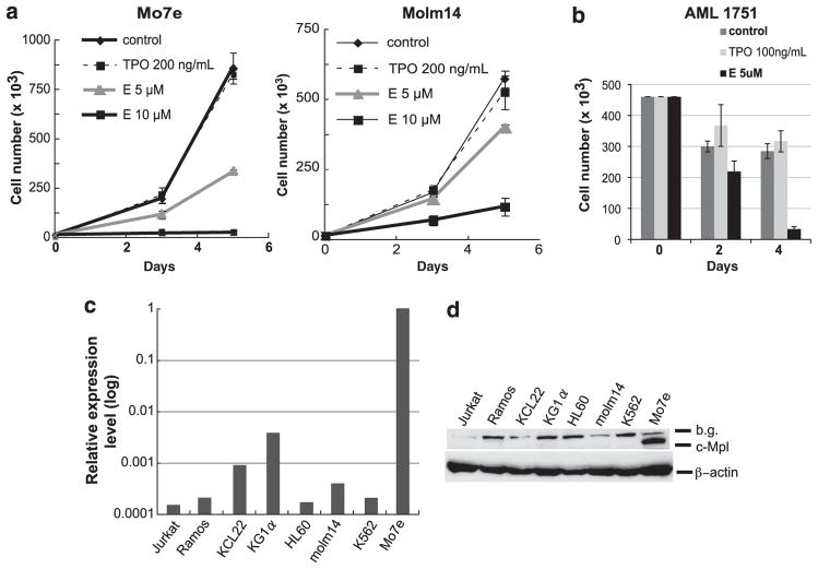
(a) Cell growth analyses of Mo7e and Molm14. A total number of 1000 to 20 000 cells in 250 μl culture medium were seeded in triplicate wells in a 96-well plate and treated with E (5 or 10 μM, supplied by GlaxoSmithKline, Collegeville, PA, USA) or rhTPO (200 ng/ml, R&D Systems, Minneapolis, MN, USA) as a control. The numbers of viable cells were counted using a hemocytometer with trypan blue exclusion on days 3 and 5. Experiments were repeated at least three times with consistent results. Data are shown as the mean±s.d. Mo7e cell line requires cytokines (TPO or granulocyte-macrophage colony-stimulating factor (GM-CSF)) for its proliferation and is maintained with 5 ng/ml of GM-CSF (Immunex Corporation, Seattle, WA, USA). (b) Cell growth analyses of primary AML cells. The proliferation assays from a representative primary AML sample are shown. Primary samples were obtained from the Stem Cell and Xenograft Core facility at the University of Pennsylvania School of Medicine. All samples were collected in accordance to the federal and University guidelines, and provided to us pathologically annotated without patient personal information. Available clinical information about AML’s is provided in Supplementary Table 1. A total number of 100 000 to 450 000 cells were cultured in the presence of E (5 μM), rhTPO (100 ng/ml) or in medium alone. The numbers of viable cells were counted on indicated days and data are shown as the mean±s.d. of three replicates. (c) Relative c-Mpl mRNA expression levels in various leukemia cell lines; Jurkat, Ramos, KCL22, KG1α, HL60, Molm14, K562 and Mo7e. qRT–PCR using a SYBR Green reagent was performed using β-actin and glyceraldehyde 3-phosphate dehydrogenase (GAPDH) as reference genes to normalize mRNA expression in different cell lines. The sequences of primers are as follows. c-Mpl, Forward: 5′-TTTCTCCCGAACATTTGAGG-3′; reverse: 5′-GTGCAGCGGAAAGAAGAGAC-3′. β-Actin, Forward: 5′-AAACTGGAACGGTGAAGGTG-3′; reverse: 5′-AGAGAAGTGGGGTGGCTTTT-3′. GAPDH, Forward: 5′-GACAGTCAGCCGCATCTTCTT-3′; reverse: 5′-CCAATACGACCAAATCCGTTGA-3′. The two-step PCR method was performed according to the following protocol: Enzyme activation at 95 °C, 10 min, 40 cycles of denaturing step at 95 °C for 15 s and annealing/extension step at 60 °C for 1 min. c-Mpl mRNA expression in Mo7e is denoted as 1, and the data are presented on a log scale. (d) c-Mpl protein expression in leukemia cell lines. Cell pellets of each cell line were lysed in ice-cold lysis buffer (50 mM Tris–HCl (pH 7.6), 150 mM NaCl, 0.1% SDS, 0.5% sodium deoxycholate, 1% NP-40) containing a cocktail of phosphatase inhibitors and protease inhibitors (Pierce, Rockford, IL, USA). Protein lysates were separated using SDS-polyacrylamide gel electrophoresis and were transferred onto polyvinylidene fluoride membrane. Transferred proteins were probed with the following primary antibodies and horseradish peroxidase (HRP)-conjugated secondary antibodies: anti-c-Mpl antibody (Millipore, Billerica, MA, USA, no.06-944), anti-β-actin antibody (Sigma-Aldrich, St. Louis, MO, USA), HRP -conjugated anti-rabbit immunoglobulinG (IgG; Cell Signaling, Danvers, MA, USA, no.7074) and HRP-conjugated anti-mouse IgG (Cell Signaling, no.7076). Protein–antibody signals were detected using enhanced chemiluminescence detection reagent (GE Healthcare, Piscataway, NJ, USA).
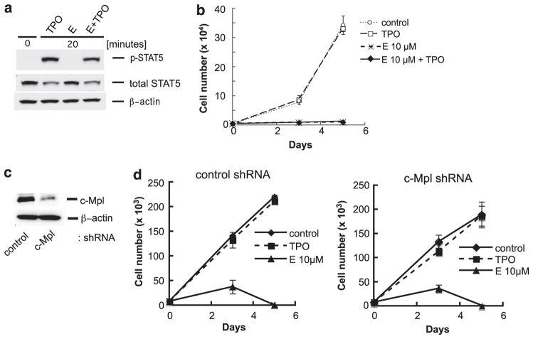
(a) Western blot analyses to detect p-STAT5 in Mo7e cells. After 24 h starvation of serum and cytokines, Mo7e cells were stimulated with E (10 μM) alone, rhTPO (100 ng/ml) alone or with combination of E and rhTPO for 20 min. Western blot was performed as described in Figure 1 using the following antibodies; anti-phosphorylated STAT5 (Tyr694) antibody and anti-STAT5 antibody (nos 9356 and 9363, respectively, Cell Signaling). (b) Cell growth analyses (drug competition assay) of Mo7e cells after treatment of E (5 μM or 10 μM) alone, rhTPO (200 ng/ml) alone, or combination of E and rhTPO. Cell culture conditions, methods, and analyses were performed as described in Figure 1. Data are shown as the mean±s.d. Experiment was repeated at least three times with consistent results. (c) Western blot of c-Mpl protein expression in Mo7e cells without and with the c-Mpl knockdown. The targeted sequence was selected from the previous report and shRNA constructs were designed with two base modifications in the sense strand. The c-Mpl small interfering RNA (siRNA) targeting sequence and control siRNA sequence were as follows: c-Mpl: 5′-gcacctctgggtgaagaatgt-3′, control: 5′-ctttatacgtagtcataag-3′. The shRNA constructs were cloned into the HIUG lentiviral vector, which was kindly provided by Dr EJ. Brown in the University of Pennsylvania, and the lentivirus clones were stably transduced into Mo7e cells. The western blot was performed as described in Figure 1, and protein–antibody signals detected were quantified using ImageQuant TL software (GE Healthcare, Piscataway, NJ, USA). (d) Cell growth analyses in Mo7e cells without and with the c-Mpl knockdown. Knockingdown of c-Mpl did not change E’s inhibitory effect. The manipulated control- and c-Mpl knocked-down Mo7e were cultured in the same conditions as described in Figure 1. After treatment with rhTPO (200 ng/ml) or E (10 μM), the number of viable cells in triplicate wells were counted on days 3 and 5. Cell growth assay data are shown as the mean±s.d.
References
-
- Kalota A, Gewirtz AM. A prototype nonpeptidyl, hydrazone class, thrombopoietin receptor agonist, SB-559457, is toxic to primary human myeloid leukemia cells. Blood. 2010;115:89–93. - PubMed
-
- Takeshita A, Shinjo K, Izumi M, Ling P, Nakamura S, Naito K, et al. Quantitative expression of thrombopoietin receptor on leukaemia cells from patients with acute myeloid leukaemia and acute lymphoblastic leukaemia. Br J Haematol. 1998;100:283–290. - PubMed
-
- Corazza F, Hermans C, D’Hondt S, Ferster A, Kentos A, Benoit Y, et al. Circulating thrombopoietin as an in vivo growth factor for blast cells in acute myeloid leukemia. Blood. 2006;107:2525–2530. - PubMed
Publication types
MeSH terms
Substances
Grants and funding
LinkOut - more resources
Full Text Sources
Other Literature Sources
Medical
