Review
. 2013 Mar;394(3):335-45.
doi: 10.1515/hsz-2012-0290.

Carboxypeptidase M augments kinin B1 receptor signaling by conformational crosstalk and enhances endothelial nitric oxide output

Affiliations
Review

Carboxypeptidase M augments kinin B1 receptor signaling by conformational crosstalk and enhances endothelial nitric oxide output

Xianming Zhang et al. Biol Chem. 2013 Mar.

Abstract

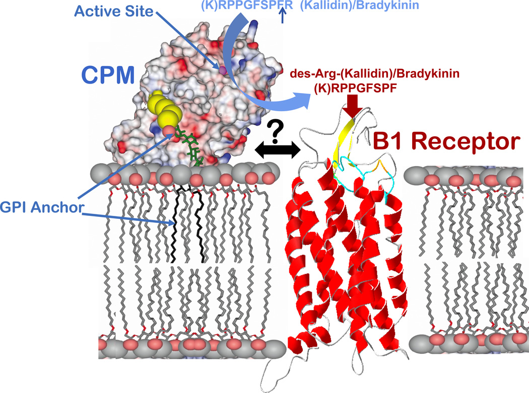
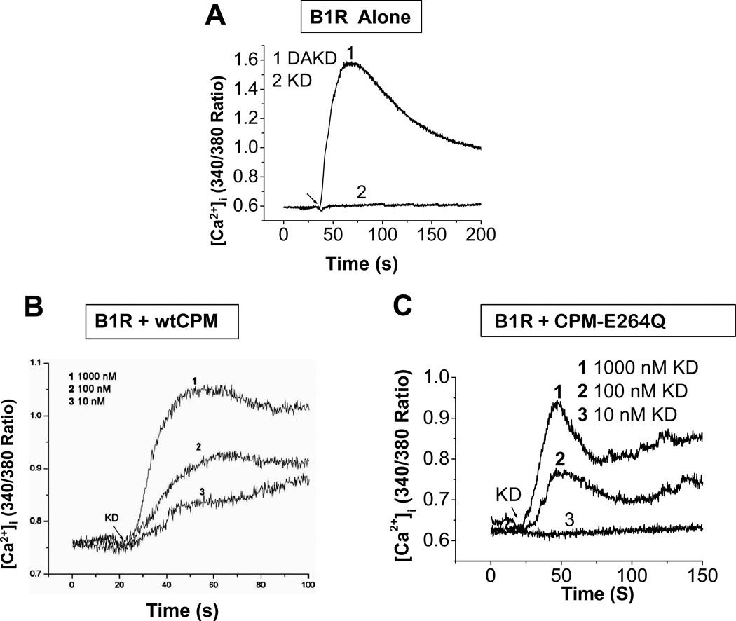
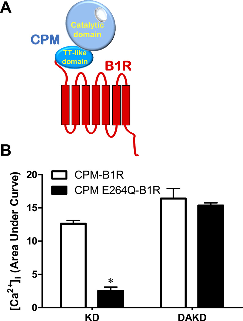
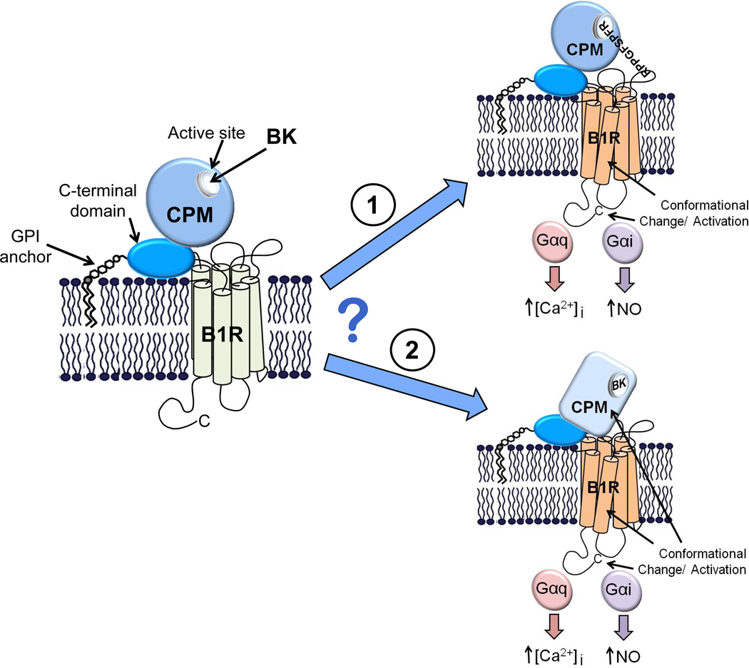
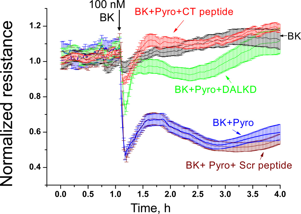
The G protein-coupled receptors (GPCRs) are the largest class of membrane proteins that play key roles in transducing extracellular signals to intracellular proteins to generate cellular responses. The kinin GPCRs, named B1 (B1R) and B2 (B2R), are responsible for mediating the biological responses to kinin peptides released from the precursor kininogens. Bradykinin (BK) or kallidin (KD) are agonists for B2Rs, whereas their carboxypeptidase (CP)-generated metabolites, des-Arg(9)-BK or des-Arg(10)-KD, are specific agonists for B1Rs. Here, we review the evidence for a critical role of membrane-bound CPM in facilitating B1R signaling by its ability to directly activate the receptor via conformational crosstalk as well as generate its specific agonist. In endothelial cells, the CPM/B1R interaction facilitates B1R-dependent high-output nitric oxide under inflammatory conditions.


Figures

Figure 1
Figure 1
Schematic diagram of the possible orientation and interaction of CPM and B1R on the membrane. The CPM model shows its electrostatic surface potential based on the crystal structure and probable membrane association via the GPI anchor and electrostatic interactions (Reverter et al., 2004). The active-site groove is indicated by the zinc ion (magenta sphere). A ribbon model of the B1R is shown at right. CPM cleaves the C-terminal Arg from kinins and its close association with the B1R facilitates delivery of des-Arg-kinin agonists to the receptor.
Figure 2
Figure 2
Co-expression of the B1R with either CPM or CPM-E264Q generates a B1R-dependent calcium response to B2R agonist kallidin. Tracings showing the increase in [Ca2+]i induced by KD or DAKD in HEK cells stably expressing B1R (A), B1R and wtCPM (B), or B1R and CPM-E264Q (C). The concentrations of agonists in (A) were 1 µM. The results are representative of three independent experiments. This research was originally published in the Journal of Biological Chemistry. Zhang, X., Tan, F., Zhang, Y., and Skidgel, R. A. Carboxypeptidase M and kinin B1 receptors interact to facilitate efficient B1 signaling from B2 agonists. J Biol Chem 2008; 283: 7994–8004 (B). Zhang X, Tan F, Brovkovych V, Zhang Y, Skidgel RA. Cross-talk between carboxypeptidase M and the kinin B1 receptor mediates a new mode of G protein-coupled receptor signaling. J Biol Chem 2011; 286:18547 – 18561 (A and C). © the American Society for Biochemistry and Molecular Biology.
Figure 3
Figure 3
The calcium response to kinin peptides in HEK cells stably expressing CPM-B1R or CPM-E264Q-B1R fusion proteins. (A) Schematic diagram of the chimera generated by fusing the C-terminus of CPM to the N-terminus of the B1R. (B) HEK cells stably expressing the B1R chimera with either wtCPM (CPM-B1R) or CPM-E264Q (CPM E264Q-B1R) were stimulated with 1 µM KD or DAKD and the increase in [Ca2+]i was quantified by integrating the area under the curve (expressed as mean ± SE (n=3). * = p<0.05 vs. CPM-B1R). This research was originally published in the Journal of Biological Chemistry. Zhang X, Tan F, Brovkovych V, Zhang Y, Skidgel RA. Cross-talk between carboxypeptidase M and the kinin B1 receptor mediates a new mode of G protein-coupled receptor signaling. J Biol Chem 2011; 286:18547 – 18561. © the American Society for Biochemistry and Molecular Biology.
Figure 4
Figure 4
Potential mechanisms by which BK binding to CPM could activate the B1R. A model of CPM and its potential membrane orientation and basal interaction with the B1R is shown in the left panel. BK (or KD) released from the kininogen precursor could stimulate B1R signaling in two ways via CPM. 1. CPM binding of the C-terminal Arg might shield the positive charge, which is typically repelled by Lys118, allowing the kinin N-terminus to bind and activate the B1R. 2. Binding as a substrate could cause a conformational change in CPM that is transmitted via protein-protein interaction to the B1R, resulting in activation. Our data are consistent with the second (bottom) mechanism, but not the first (top). For further details, see text.
Figure 5
Figure 5
Effect of kinin peptides on B1R conformational change. (A) Schematic representation of the intramolecular FRET reporter, B1R-TC-CFP. An agonist -induced conformational change that brings FlAsH and CFP closer together would result in an increase in the efficiency of FRET as detected by an increase in emission at 530 nm. Lower box: Sequence of the 3rd intracellular loop of the B1R showing the insertion of the tetracysteine biarsenical binding tag in between Gly242 and Arg243. (B) An increase in FRET of B1R-TC-CFP is induced by B1R agonist and is inhibited by B1R antagonist DALKD. The data are expressed as mean ± SE (n=3). * = p<0.05 vs. DAKD. (C) The change in intramolecular FRET of B1R-TC-CFP in response to BK. HEK cells stably expressing B1R-TC-CFP alone or with wtCPM, CPM-E264Q or CPD DIII were stimulated with 1 µM BK and the FlAsH emission at 530 nm was recorded in real-time while exciting CFP at 430 nm. The data are expressed as mean ± SE (n=4). * = p<0.05 vs. B1R-TC-CFP. This research was originally published in the Journal of Biological Chemistry. Zhang X, Tan F, Brovkovych V, Zhang Y, Skidgel RA. Cross-talk between carboxypeptidase M and the kinin B1 receptor mediates a new mode of G protein-coupled receptor signaling. J Biol Chem 2011; 286:18547 – 18561. © the American Society for Biochemistry and Molecular Biology.
Figure 6
Figure 6
Model of how CPM/B1R interactions mediate signaling in response to BK. CPM and its potential membrane orientation and basal interaction with the B1R is shown in the left panel. Based on our results, BK (or KD) released from the kininogen precursor can stimulate B1R signaling in two ways via CPM. 1. Binding as a substrate causes a conformational change in CPM that is transmitted via protein-protein interaction to the B1R, resulting in G protein coupling and activation of calcium signaling. 2. Catalytic conversion of BK (or KD) to B1R agonist that can further activate the associated receptor or additional B1Rs. For the catalytically inactive CPM-E264Q mutant, only the first mechanism of activation is possible. This research was originally published in the Journal of Biological Chemistry. Zhang X, Tan F, Brovkovych V, Zhang Y, Skidgel RA. Cross-talk between carboxypeptidase M and the kinin B1 receptor mediates a new mode of G protein-coupled receptor signaling. J Biol Chem 2011; 286:18547 – 18561. © the American Society for Biochemistry and Molecular Biology.
Figure 7
Figure 7
The relative efficiency of the BK-stimulated increase in [Ca2+]i in three experimental models. In the first two groups (the first 6 bars) the increase in [Ca2+]i was recorded and quantified in HEK cells stably co-expressing B1Rs and either wtCPM or CPM-E264Q and B1R in response to treatment with various concentrations of B2R agonist BK. In the last group (last 3 bars), the calcium response was quantified in mixed co-cultures of cells singly expressing either wtCPM or B1R after stimulation with the indicated concentration of BK. The results were normalized by defining the calcium response of cells stimulated by 1000 nM B1R agonist DABK as 100% in the corresponding experimental system. The data are expressed as mean ± SE (n=3). This research was originally published in the Journal of Biological Chemistry. Zhang X, Tan F, Brovkovych V, Zhang Y, Skidgel RA. Cross-talk between carboxypeptidase M and the kinin B1 receptor mediates a new mode of G protein-coupled receptor signaling. J Biol Chem 2011; 286:18547 – 18561. © the American Society for Biochemistry and Molecular Biology.
Figure 8
Figure 8
Disruption of the CPM/B1R interaction inhibits B1R-mediated NO production in response to BK in human endothelial cells. (A) Cytokine-treated HLMVEC were pre-incubated for 30 min with 1 µM HOE140 (B2R antagonist) without (solid line) or with (dotted line) 1 µM DALKD (B1R antagonist). At time zero, 100 nM BK was added and NO production was measured in real time for 20 min with a porphyrinic microsensor. B, Cells were pretreated with 1 µM HOE140 without or with 500 ng/ml CPM monoclonal antibody, 50 µM CT peptide or scrambled CT peptide (Scr. peptide) for 30 min. Cells were stimulated with 100 nM or 1 µM BK and NO production measured for 20 min. Shown are mean values as % control (100 nM BK alone = 100%) ± SE (n = 3). This research was originally published in the Journal of Biological Chemistry. Zhang X, Tan F, Brovkovych V, Zhang Y, Skidgel RA. Cross-talk between carboxypeptidase M and the kinin B1 receptor mediates a new mode of G protein-coupled receptor signaling. J Biol Chem 2011; 286:18547 – 18561. © the American Society for Biochemistry and Molecular Biology.
Figure 9
Figure 9
B1R-dependent increase in endothelial permeability caused by BK combined with superoxide depends on CPM/B1R interaction. HLMVEC, grown to confluence on gold electrodes coated with 10 µg/ml fibronectin, were cytokine pretreated (10ng/ml IL-1β + 100u/ml IFN-γ, 16 h). Medium was changed, cells were allowed to stabilize, and all samples were pretreated with 10 µM HOE140 to block B2R responses. Cells were then treated with 1 µM BK (added at the black arrow) alone (black line) or combined with 200 µM pyrogallol (Pyro; superoxide generator) without (blue line) or with 1 µM B1R antagonist DALKD (green line), 50 µM CT peptide (red line) or 50 µM scrambled (Scr) peptide (brown line) and transendothelial electrical resistance was measured. Results show mean values ± S.E. for n = 4. This research was originally published in the Journal of Biological Chemistry. Zhang X, Tan F, Brovkovych V, Zhang Y, Skidgel RA. Cross-talk between carboxypeptidase M and the kinin B1 receptor mediates a new mode of G protein-coupled receptor signaling. J Biol Chem 2011; 286:18547 – 18561. © the American Society for Biochemistry and Molecular Biology.

Similar articles

Cited by

References

    1. Brovkovych V, Zhang Y, Brovkovych S, Minshall RD, Skidgel RA. A novel pathway for receptor-mediated post-translational activation of inducible nitric oxide synthase. J. Cell. Mol. Med. 2011;15:258–269. - PMC - PubMed
    1. Carretero OA, Scicli AG. Local hormonal factors (intracrine, autocrine, and paracrine) in hypertension. Hypertension. 1991;18:I58–I69. - PubMed
    1. Chen Z, Deddish PA, Minshall RD, Becker RP, Erdös EG, Tan F. Human ACE and bradykinin B2 receptors form a complex at the plasma membrane. FASEB J. 2006;20:2261–2270. - PMC - PubMed
    1. Deddish PA, Skidgel RA, Kriho VB, Li XY, Becker RP, Erdös EG. Carboxypeptidase M in Madin-Darby canine kidney cells. Evidence that carboxypeptidase M has a phosphatidylinositol glycan anchor. J. Biol. Chem. 1990;265:15083–15089. - PubMed
    1. Erdös EG. Kinins, the long march--a personal view. Cardiovasc. Res. 2002;54:485–491. - PubMed

Publication types

MeSH terms