. 2012 Nov;3(11):1439-54.
doi: 10.18632/oncotarget.683.

Epigenetic regulation of miRNA-211 by MMP-9 governs glioma cell apoptosis, chemosensitivity and radiosensitivity

Affiliations

Epigenetic regulation of miRNA-211 by MMP-9 governs glioma cell apoptosis, chemosensitivity and radiosensitivity

Swapna Asuthkar et al. Oncotarget. 2012 Nov.

Abstract

Glioblastoma multiforme (GBM) is the most aggressive brain cancer, and to date, no curative treatment has been developed. In this study, we report that miR-211, a microRNA predicted to target MMP-9, is suppressed in grade IV GBM specimens. Furthermore, we found that miR-211 suppression in GBM involves aberrant methylation-mediated epigenetic silencing of the miR-211 promoter. Indeed, we observed a highly significant inverse correlation between miR-211 expression and MMP-9 protein levels, which is indicative of post-transcriptional control of gene expression. Additionally, shRNA specific for MMP-9 (pM) promoted miR-211 expression via demethylation of miR-211 promoter-associated CpG islands (-140 to +56). In independent experiments, we confirmed that miR-211 overexpression and pM treatments led to the activation of the intrinsic mitochondrial/Caspase-9/3-mediated apoptotic pathway in both glioma cells and cancer stem cells (CSC). We also investigated whether miR-211 is involved in the regulation of MMP-9 and thus plays a functional role in GBM. We found an acute inhibitory effect of miR-211 on glioma cell invasion and migration via suppression of MMP-9. Given the insensitivity of some GBMs to radiation and chemotherapy (temozolomide) along with the hypothesis that glioma CSC cause resistance to therapy, our study indicates that miR-211 or pM in combination with ionizing radiation (IR) and temozolomide significantly induces apoptosis and DNA fragmentation. Of note, miR-211- and pM-treated CSC demonstrated increased drug retention capacity, as observed by MDR1/P-gp mediated-Rhodamine 123 drug efflux activity assay. These results suggest that either rescuing miR-211 expression or downregulation of MMP-9 may have a new therapeutic application for GBM patients in the future.


Conflict of interest statement

Authors declare no conflict of interest exists with this manuscript.

Figures
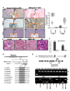
Figure 1
Figure 1. miR-211 negatively regulates MMP-9 in GBM and methylation of the CpG island (-140 to +56) promoter region of miR-211 is associated with MMP-9
(A) Hematoxylin and eosin (H&E) staining of brain sections displaying the neoplastic growth of tumors were further analyzed through in situ and DAB staining to detect the basal expression levels of miR-211 and MMP-9 respectively in normal brain (H6 & H7) and GBM grade IV (F3 & F4) specimens. The relative expression of miR-211 and MMP-9 protein in GBM tissue microarray (containing 33 cases of brain glioblastoma and 5 normal brain tissues, duplicate cores per case) was quantified and is represented graphically (p<0.001). (B) In situ hybridization studies showing the expression of miR-211 in intracranial tumors induced by control, pSV-, MMP-9 OE-, and pM-treated 4910 cells. (C) Relative expression levels of miR-211 using stem and loop primers by miR-quantitative RT-PCR analysis in control, pM-, and MMP-9 OE-treated 4910 and U87 cells (mean + SD, n=3; p<0.001). (D) Sequence alignment of miR-211 and the predicted sequence pairing with a region of MMP-9 mRNA 3'UTR that may interact with miR-211. Alignment of nucleotide sequences of MMP-9 3′-UTR corresponding to the targets for miR-211 across nine different species. A high level of conservation suggests a functional role for these sequences. (E) Schematic map of the miR-211 promoter-associated CpG island region indicating the location of PCR primers used for MSP analysis. Each of the CpG dinucleotide residues with respect to the transcriptional start site ATG is shown as numbers in circles from 1 to 10, which can serve as potential DNA methylation sites in miR-211 promoter. Four potential CpG sites (5/-111 to 2/+33, genomic positions 49731, 49844, 49860 and 49874; Accession No. AEKP01201896) were investigated by MSP as mentioned in Materials and Methods. Bisulfite-modified DNA derived from human glioblastoma grade IV specimens (GB1-GB10) was amplified using MSP primers and was compared with normal human brain specimen (NB). MSP assay was also performed on DNA isolated from control, pM, 5-Aza-CdR and MMP-9 OE-treated 4910 and U87 cells. The bisulfite-modified DNA was amplified with miR-211 promoter primers specific for methylated (top) and unmethylated DNA (bottom). Methylation-specific primers generated the PCR products labeled with “M.” Those labeled with “U” were generated by primers specific for unmethylated DNA.
Figure 2
Figure 2. miR-211 induces apoptosis in human xenograft cell lines
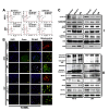
(A) Fluorescence-activated cell sorting (FACS) analysis of cell cycle progression in 4910 and U87 xenograft cells 48 h after miR-211 transfection. The y axis denotes cell count and the x axis represents DNA content. The percentages of cells in the G1 (M2), S (M3) and G2/M (M4) phases of the cell cycle were calculated using CellQuest Pro software. EV represents the empty vector control. (B) Whole cell lysates were prepared from control, miR-211 and pM-treated cells and subjected to western blot analysis to determine the expression of various cell cycle (Panel I), pro-apoptotic and anti-apoptotic (Pane II) proteins. GAPDH was used as a loading control. (C) The 4910 and U87 xenograft cells were seeded in 8-well chamber slides and transfected with miR-211 or pM and incubated further for 48 h. The cells were fixed in 4% paraformaldehyde and subjected to TUNEL assay as described in Materials and Methods. Green fluorescence represents apoptotic cells and blue (DAPI) fluorescence represents the nucleus. Mitochondrial apoptosis in control, miR-211 and pM-treated 4910 and U87 cells was examined by MitoLight® Mitochondrial Apoptosis Detection Kit as described in Materials and Methods. Only the merged figures are represented for mitochondrial apoptosis. In healthy cells, the dye accumulates and aggregates in the mitochondria, giving off a bright red fluorescence. In apoptotic cells with altered mitochondrial membrane potential, the dye in its monomeric form stays in the cytoplasm, fluorescing green, providing a ready discrimination between apoptotic and nonapoptotic cells.
Figure 3
Figure 3. miR-211 inhibits radiation-induced MMP-9 and CSC-like property in glioma cells
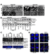
(A) Effect of miR-211 overexpression on the ionizing radiation (IR)-treated glioma 4302 and 4910 CSC spheres. (B) Immunoblots using cell lysates (40 μg) from control, empty vector (EV), miR-211 alone and in combination with IR-treated 4302 and 4910 CSC were developed using antibodies specific for cleaved Caspase-3 and cleaved PARP. GAPDH was used as a loading control. (C) MMP-9 mRNA and protein levels were examined by RT-PCR and immunoblot analysis respectively. Conditioned medium, collected from the above treated glioma CSC was subjected to gelatin zymography to determine MMP-9 activity. (D) TUNEL nuclear staining in 4302 and 4910 CSC spheres. The control CSC with overexpression of miR-211 alone and in combination with IR treatment in 8-well chamber slides were subjected to TUNEL nuclear staining and viewed by fluorescence microscopy. Green fluorescence represents apoptotic cells and blue (DAPI) fluorescence represents the nucleus.
Figure 4
Figure 4. miR-211 suppresses the migratory and invasive potential of glioma CSC
(A) 4302 and 4910 CSC spheres were seeded in 24-well plates. The CSC spheroids were either treated with empty vector (EV) or miR-211 alone or in combination with IR treatment. After 16 h, the media was replaced with fresh complete media. The spheroid migration distance was measured after 48 h and is represented graphically in terms of relative migration (*, p<0.01; **, p<0.001). (B) The treated 4302 and 4910 CSC were disintegrated into single cell suspension by trituration and an equal number (5×10⁴) of spheres were suspended in serum-free media and plated onto Matrigel-coated Transwell inserts as described in Materials and Methods. After a 24-hr incubation period, lower invaded spheres were stained with Hema-3. Images of invaded spheres were taken under a light microscope (Olympus IX-71). The invasive potential of treated spheres was quantified and the percentage of cells invading from 3 independent experiments is graphically represented as bar diagrams. Error bar represents mean + standard deviation (SD) (*, p<0.01; **, p<0.001). (C) Immunoblot analysis of cell lysates (40 μg) from control, EV and miR-211-treated 4302 and 4910 CSC to assess the expression of various EMT-associated proteins. GAPDH was used as a loading control. (D) Schematic representation shows that miR-211 mediates negative regulation of MMP-9 and plays a functional role in inhibition of glioma cell proliferation, migration and invasion. The miR-211, which is predicted to target MMP-9, is suppressed in GBM grade IV specimens. Furthermore, we observed that downregulation of MMP-9 using shRNA specific for MMP-9 (pM) promoted miR-211 expression via demethylation of miR-211 promoter-associated CpG island (-140 to +56). In light of these observations, we interpret a negative feedback loop between miR-211 and MMP-9 and suggest that either rescuing miR-211 expression or downregulation of MMP-9 may have a new therapeutic application for the treatment of GBM patients in the future.
Figure 5
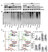
Figure 5. (A) 4302 and 4910 cancer stem cells (CSC) were treated with a chemotherapy compound, temozolomide (500 uM), or transfected with miR-211 and pM plasmids
Immunoblot analysis was performed to determine CD133 levels in the treated CSC, and the results were compared with untreated CSC and normal cancer cells (non-CSC). GAPDH was used as a loading control. (B) Apoptotic DNA laddering profile of temozolomide, miR-211 and pM-treated glioma CSC and non-CSC. (D) Rhodamine 123 efflux assay to determine the role of miR-211 and pM in chemosensitivity. The assay measures P-gp-mediated efflux by the decrease in the intracellular fluorescence of rhodamine 123 (R123, a MDR1/P-pg substrate) using flow cytometry. Intracellular rhodamine 123 was measured at time 0 (load) and after 2 h of efflux in control, miR-211- and pM-treated glioma CSC (panel a). The efflux was measured by the number of cells in the M1 region of the plot. To confirm the specificity of the R123 efflux from the control and treated CSC, we employed a P-gp blocker, vinblastin. The R123 efflux assays were performed in the presence of this inhibitor (panel b). The percentage of cells with R123 retention was quantified both in the absence (panel a) and in the presence of P-gp blocker, vinblastine (panel b) from 3 independent experiments and is graphically represented as bar diagrams. Error bar represents mean + standard deviation (SD) (*, p<0.01).

Similar articles

Cited by

References

    1. Calin GA, Liu CG, Sevignani C, Ferracin M, Felli N, Dumitru CD, Shimizu M, Cimmino A, Zupo S, Dono M, Dell'Aquila ML, Alder H, Rassenti L, Kipps TJ, Bullrich F, Negrini M, et al. MicroRNA profiling reveals distinct signatures in B cell chronic lymphocytic leukemias. Proc Natl Acad Sci U S A. 2004;101:11755–60. - PMC - PubMed
    1. Jovanovic M, Hengartner MO. miRNAs and apoptosis: RNAs to die for. Oncogene. 2006;25:6176–87. - PubMed
    1. Kent OA, Mendell JT. A small piece in the cancer puzzle: microRNAs as tumor suppressors and oncogenes. Oncogene. 2006;25:6188–96. - PubMed
    1. Volinia S, Calin GA, Liu CG, Ambs S, Cimmino A, Petrocca F, Visone R, Iorio M, Roldo C, Ferracin M, Prueitt RL, Yanaihara N, Lanza G, Scarpa A, Vecchione A, Negrini M, et al. A microRNA expression signature of human solid tumors defines cancer gene targets. Proc Natl Acad Sci U S A. 2006;103:2257–61. - PMC - PubMed
    1. Esquela-Kerscher A, Slack FJ. Oncomirs - microRNAs with a role in cancer. Nat Rev Cancer. 2006;6:259–69. - PubMed

Publication types

MeSH terms