

Epstein-Barr virus, human papillomavirus and mouse mammary tumour virus as multiple viruses in breast cancer
- PMID: 23183846
- PMCID: PMC3501510
- DOI: 10.1371/journal.pone.0048788
Epstein-Barr virus, human papillomavirus and mouse mammary tumour virus as multiple viruses in breast cancer
Abstract
Background: The purpose of this investigation is to determine if Epstein Barr virus (EBV), high risk human papillomavirus (HPV), and mouse mammary tumour viruses (MMTV) co-exist in some breast cancers.
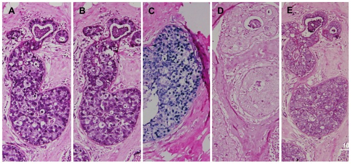
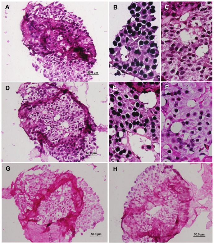
Materials and methods: All the specimens were from women residing in Australia. For investigations based on standard PCR, we used fresh frozen DNA extracts from 50 unselected invasive breast cancers. For normal breast specimens, we used DNA extracts from epithelial cells from milk donated by 40 lactating women. For investigations based on in situ PCR we used 27 unselected archival formalin fixed breast cancer specimens and 18 unselected archival formalin fixed normal breast specimens from women who had breast reduction surgery. Thirteen of these fixed breast cancer specimens were ductal carcinoma in situ (dcis) and 14 were predominantly invasive ductal carcinomas (idc).
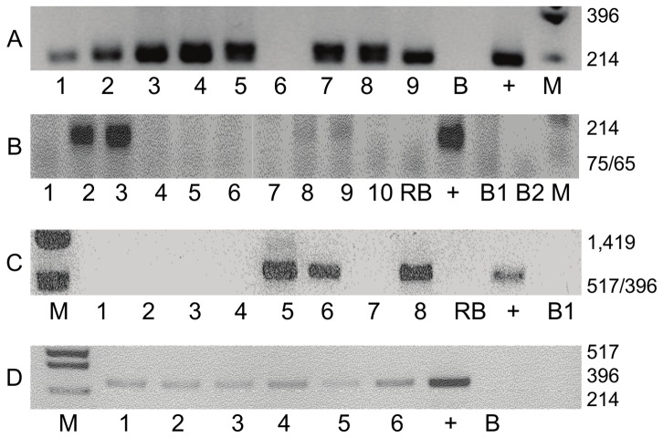
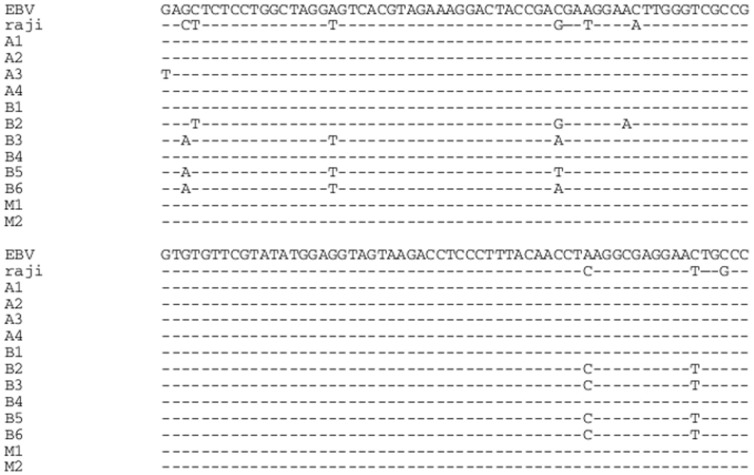
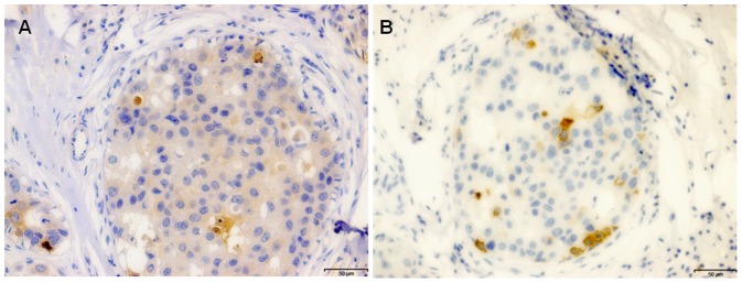
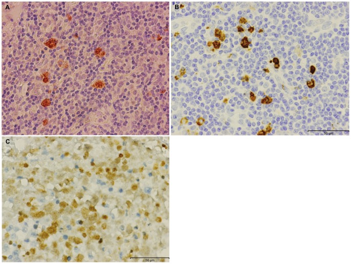
Results: EBV sequences were identified in 68%, high risk HPV sequences in 50%, and MMTV sequences in 78% of DNA extracted from 50 invasive breast cancer specimens. These same viruses were identified in selected normal and breast cancer specimens by in situ PCR. Sequences from more than one viral type were identified in 72% of the same breast cancer specimens. Normal controls showed these viruses were also present in epithelial cells in human milk - EBV (35%), HPV, 20%) and MMTV (32%) of 40 milk samples from normal lactating women, with multiple viruses being identified in 13% of the same milk samples.
Conclusions: We conclude that (i) EBV, HPV and MMTV gene sequences are present and co-exist in many human breast cancers, (ii) the presence of these viruses in breast cancer is associated with young age of diagnosis and possibly an increased grade of breast cancer.
Conflict of interest statement
Figures
Similar articles
-
Elevated expression of the tumor suppressing protein p53 is associated with the presence of mouse mammary tumor-like env gene sequences (MMTV-like) in human breast cancer.Breast Cancer Res Treat. 2004 Sep;87(1):13-7. doi: 10.1023/B:BREA.0000041573.09142.00. Breast Cancer Res Treat. 2004. PMID: 15377846
-
Presence of mouse mammary tumour-like virus gene sequences may be associated with morphology of specific human breast cancer.J Clin Pathol. 2006 Dec;59(12):1287-92. doi: 10.1136/jcp.2005.035907. Epub 2006 May 12. J Clin Pathol. 2006. PMID: 16698952 Free PMC article.
-
Prevalence of EBV, HPV and MMTV in Pakistani breast cancer patients: A possible etiological role of viruses in breast cancer.Infect Genet Evol. 2017 Oct;54:230-237. doi: 10.1016/j.meegid.2017.07.010. Epub 2017 Jul 10. Infect Genet Evol. 2017. PMID: 28705719
-
Are viruses associated with human breast cancer? Scrutinizing the molecular evidence.Breast Cancer Res Treat. 2012 Aug;135(1):1-15. doi: 10.1007/s10549-011-1921-4. Epub 2012 Jan 25. Breast Cancer Res Treat. 2012. PMID: 22274134 Review.
-
Mouse Mammary Tumour Virus (MMTV) in Human Breast Cancer-The Value of Bradford Hill Criteria.Viruses. 2022 Mar 30;14(4):721. doi: 10.3390/v14040721. Viruses. 2022. PMID: 35458452 Free PMC article. Review.
Cited by
-
Non-Response of Epstein-Barr Virus-Associated Breast Cancer after Primary Chemotherapy: Report of Two Cases.Pathogens. 2023 Nov 24;12(12):1387. doi: 10.3390/pathogens12121387. Pathogens. 2023. PMID: 38133273 Free PMC article.
-
Molecular Identification of Human Papillomavirus DNA in Thyroid Neoplasms: Association or Serendipity?Cureus. 2021 Apr 20;13(4):e14578. doi: 10.7759/cureus.14578. Cureus. 2021. PMID: 33898151 Free PMC article.
-
Human Papillomaviruses and Epstein-Barr Virus Interactions in Colorectal Cancer: A Brief Review.Pathogens. 2020 Apr 20;9(4):300. doi: 10.3390/pathogens9040300. Pathogens. 2020. PMID: 32325943 Free PMC article. Review.
-
Bovine leukemia virus DNA associated with breast cancer in women from South Brazil.Sci Rep. 2019 Feb 27;9(1):2949. doi: 10.1038/s41598-019-39834-7. Sci Rep. 2019. PMID: 30814631 Free PMC article.
-
Human papillomaviruses-related cancers. Presence and prevention strategies in the Middle east and north African regions.Hum Vaccin Immunother. 2014;10(7):1812-21. doi: 10.4161/hv.28742. Hum Vaccin Immunother. 2014. PMID: 25424787 Free PMC article. Review.
References
-
- Buehring GC, Shen HM, Jensen HM, Block G (2007) Bovine leukemia virus infection is significantly associated with risk of breast cancer. Proc American Assoc Cancer Res 48: 1747.
-
- Park DJ, Southey MC, Giles GG, Hopper JL (2012) No evidence of MMTV-like env sequences in specimens from the Australian Breast Cancer Family Study. Breast Cancer Res Treat doi:10.1007/s10549-010-0946-4. - DOI - PubMed
-
- Murray PG (2006) Epstein–Barr virus in breast cancer: artefact or aetiological agent? J Pathol 209: 427–429. - PubMed
MeSH terms
Substances
LinkOut - more resources
Full Text Sources
Other Literature Sources
Medical
Research Materials
Miscellaneous
