

doi: 10.3791/50053.
Thinned-skull cortical window technique for in vivo optical coherence tomography imaging
Affiliations
- PMID: 23183913
- PMCID: PMC3529515
- DOI: 10.3791/50053
Item in Clipboard
Thinned-skull cortical window technique for in vivo optical coherence tomography imaging
J Vis Exp.
.
Abstract
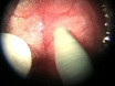
Optical coherence tomography (OCT) is a biomedical imaging technique with high spatial-temporal resolution. With its minimally invasive approach OCT has been used extensively in ophthalmology, dermatology, and gastroenterology. Using a thinned-skull cortical window (TSCW), we employ spectral-domain OCT (SD-OCT) modality as a tool to image the cortex in vivo. Commonly, an opened-skull has been used for neuro-imaging as it provides more versatility, however, a TSCW approach is less invasive and is an effective mean for long term imaging in neuropathology studies. Here, we present a method of creating a TSCW in a mouse model for in vivo OCT imaging of the cerebral cortex.
References
-
- Bizheva K, Unterhuber A, Hermann B, Povazay B, Sattmann H, Drexler W. Imaging ex vivo and in vitro brain morphology in animal models with ultrahigh resolution optical coherence tomography. Journal of Biomedical Optics. 2004;9:719–724. - PubMed
-
- Fujimoto JG. Optical coherence tomography for ultrahigh resolution in vivo imaging. Nature Biotechnology. 2003;21:1361–1367. - PubMed
-
- Wantanabe H, Rajagopalan UM, Nakamichi Y, Igarashi KM, Kadono H, Tanifuji M. Swept source optical coherence tomography as a tool for real time visualization and localization of electrodes used in electrophysiological studies of brain in vivo. Biomedical Optics Express. 2011;2:3129–3134. - PMC - PubMed
-
- Mitsui T. Dynamic range of optical reflectometry with spectral interferometry. Japanese Journal of Applied Physics. 1999;38:6133–6137.
Publication types
MeSH terms
Grants and funding
LinkOut - more resources
Full Text Sources
