

Substantia nigra volume loss before basal forebrain degeneration in early Parkinson disease
- PMID: 23183921
- PMCID: PMC5751707
- DOI: 10.1001/jamaneurol.2013.597
Substantia nigra volume loss before basal forebrain degeneration in early Parkinson disease
Abstract
Objective: To test the hypothesis that degeneration of the substantia nigra pars compacta (SNc) precedes that of the cholinergic basal forebrain (BF) in Parkinson disease (PD) using new multispectral structural magnetic resonance (MR) imaging tools to measure the volumes of the SNc and BF.
Design: Matched case-control study.
Setting: The Athinoula A. Martinos Imaging Center at the McGovern Institute for Brain Research, Massachusetts Institute of Technology (MIT), and the Massachusetts General Hospital/MIT Morris Udall Center of Excellence in Parkinson Disease Research.
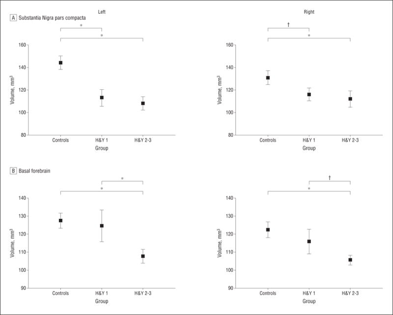
Patients: Participants included 29 patients with PD (Hoehn and Yahr [H&Y] stages 1-3) and 27 matched healthy control subjects.
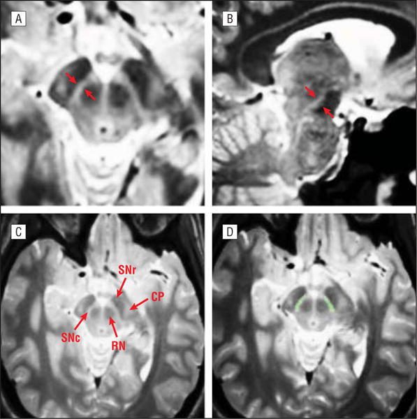
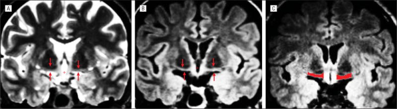
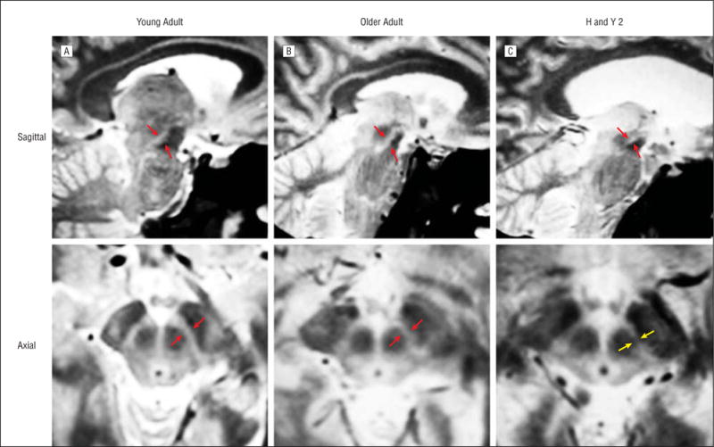
Main outcome measures: We acquired multiecho T1-weighted, multiecho proton density, T2-weighted, and T2-weighted fluid-attenuated inversion recovery (FLAIR) sequences from each participant. For the SNc, we created a weighted mean of the multiple echoes, yielding a single volume with a high ratio of contrast to noise. We visualized the BF using T2-weighted FLAIR images. For each participant, we manually labeled the 2 structures and calculated their volumes.
Results: Relative to the controls, 13 patients with H&Y stage 1 PD had significantly decreased SNc volumes. Sixteen patients with H&Y stage 2 or 3 PD showed little additional volume loss. In contrast, the BF volume loss occurred later in the disease, with a significant decrease apparent in patients having H&Y stage 2 or 3 PD compared with the controls and the patients having H&Y stage 1 PD. The latter group did not differ significantly from the controls.
Conclusion: Our results support the proposed neuropathological trajectory in PD and establish novel multispectral methods as MR imaging biomarkers for tracking the degeneration of the SNc and BF.
Conflict of interest statement
Figures
Similar articles
-
Quantitative assessment of iron deposition in the midbrain using 3D-enhanced T2 star weighted angiography (ESWAN): a preliminary cross-sectional study of 20 Parkinson's disease patients.Magn Reson Imaging. 2013 Sep;31(7):1068-73. doi: 10.1016/j.mri.2013.04.015. Epub 2013 Jun 6. Magn Reson Imaging. 2013. PMID: 23746648
-
Evaluation of striatonigral connectivity using probabilistic tractography in Parkinson's disease.Neuroimage Clin. 2017 Sep 9;16:557-563. doi: 10.1016/j.nicl.2017.09.009. eCollection 2017. Neuroimage Clin. 2017. PMID: 28971007 Free PMC article.
-
Neuromelanin magnetic resonance imaging of nigral volume loss in patients with Parkinson's disease.J Clin Neurosci. 2011 Aug;18(8):1093-6. doi: 10.1016/j.jocn.2010.08.043. Epub 2011 Jun 29. J Clin Neurosci. 2011. PMID: 21719292
-
Reduction of neuromelanin-positive nigral volume in patients with MSA, PSP and CBD.Intern Med. 2011;50(16):1683-7. doi: 10.2169/internalmedicine.50.5101. Epub 2011 Aug 15. Intern Med. 2011. PMID: 21841326
-
7 Tesla magnetic resonance imaging: a closer look at substantia nigra anatomy in Parkinson's disease.Mov Disord. 2014 Nov;29(13):1574-81. doi: 10.1002/mds.26043. Epub 2014 Oct 12. Mov Disord. 2014. PMID: 25308960 Review.
Cited by
-
Magnetic resonance imaging for the diagnosis of Parkinson's disease.J Neural Transm (Vienna). 2017 Aug;124(8):915-964. doi: 10.1007/s00702-017-1717-8. Epub 2017 Apr 4. J Neural Transm (Vienna). 2017. PMID: 28378231 Free PMC article. Review.
-
Radiomic Features of the Nigrosome-1 Region of the Substantia Nigra: Using Quantitative Susceptibility Mapping to Assist the Diagnosis of Idiopathic Parkinson's Disease.Front Aging Neurosci. 2019 Jul 16;11:167. doi: 10.3389/fnagi.2019.00167. eCollection 2019. Front Aging Neurosci. 2019. PMID: 31379555 Free PMC article.
-
Brain MRI Reveals Ascending Atrophy in Parkinson's Disease Across Severity.Front Neurol. 2019 Dec 18;10:1329. doi: 10.3389/fneur.2019.01329. eCollection 2019. Front Neurol. 2019. PMID: 31920949 Free PMC article.
-
Technology: Multiple exposure.Nature. 2013 Oct 31;502(7473):S90-1. doi: 10.1038/502s90a. Nature. 2013. PMID: 24187703 No abstract available.
-
Differential effects of intrastriatal 6-hydroxydopamine on cell number and morphology in midbrain dopaminergic subregions of the rat.Brain Res. 2014 Jul 29;1574:113-9. doi: 10.1016/j.brainres.2014.05.045. Epub 2014 Jun 9. Brain Res. 2014. PMID: 24924804 Free PMC article.
References
-
- Jellinger K. The pathology of Parkinson’s disease: recent advances. In: Galvez-Jimenez N, editor. Scientific Basis for the Treatment of Parkinson’s Disease. 2nd. New York, NY: Taylor & Francis; 2005. pp. 53–86.
-
- Fearnley JM, Lees AJ. Ageing and Parkinson’s disease: substantia nigra regional selectivity. Brain. 1991;114(pt 5):2283–2301. - PubMed
-
- Poewe W. Treatments for Parkinson disease—past achievements and current clinical needs. Neurology. 2009;72(7 suppl):S65–S73. - PubMed
-
- Calabresi P, Picconi B, Parnetti L, Di Filippo M. A convergent model for cognitive dysfunctions in Parkinson’s disease: the critical dopamine-acetylcholine synaptic balance. Lancet Neurol. 2006;5(11):974–983. - PubMed
Publication types
MeSH terms
Grants and funding
LinkOut - more resources
Full Text Sources
Other Literature Sources
Medical
Miscellaneous
