. 2013 Feb;79(3):877-85.
doi: 10.1128/AEM.03323-12. Epub 2012 Nov 26.

Inhibition of Staphylococcus aureus invasion into bovine mammary epithelial cells by contact with live Lactobacillus casei

Affiliations

Inhibition of Staphylococcus aureus invasion into bovine mammary epithelial cells by contact with live Lactobacillus casei

Damien S Bouchard et al. Appl Environ Microbiol. 2013 Feb.

Abstract

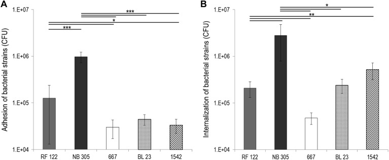
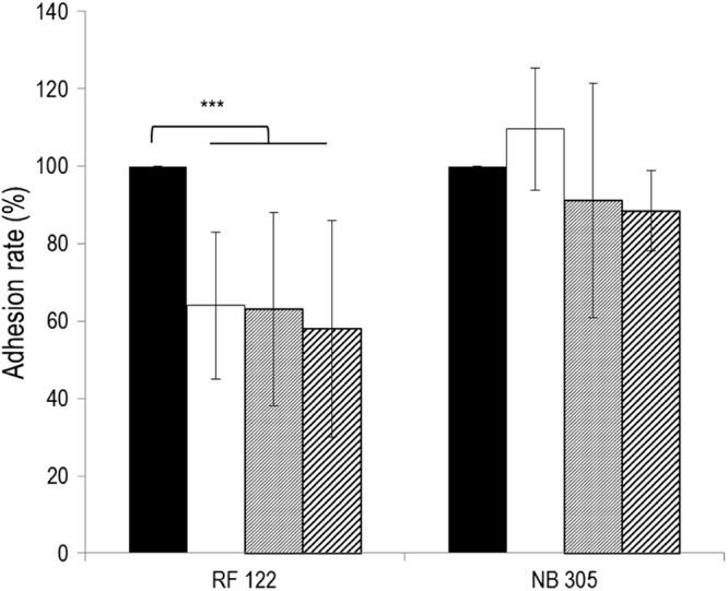
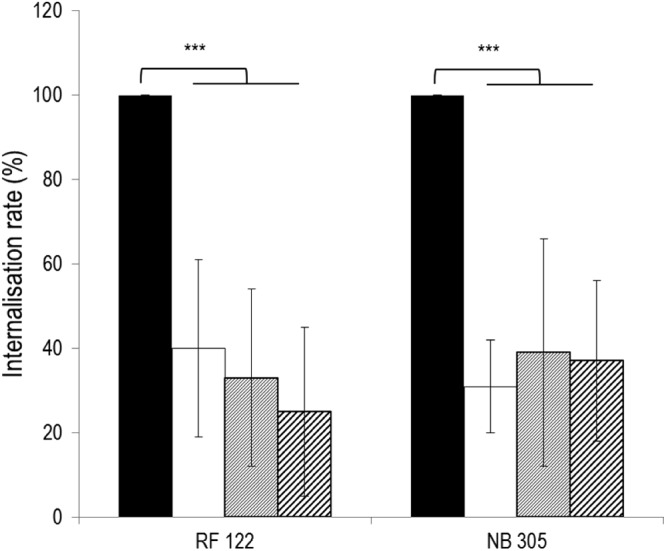
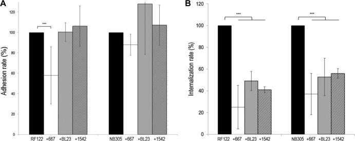
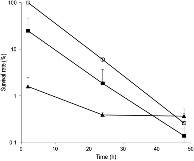
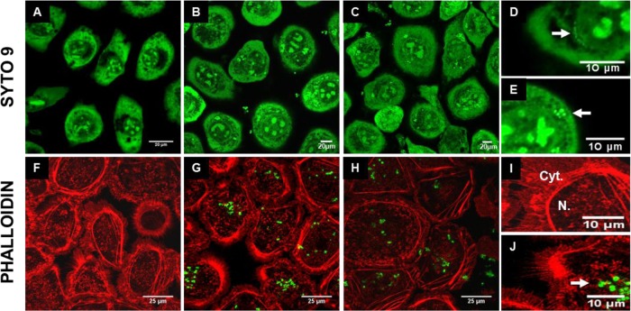
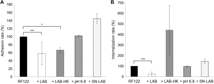
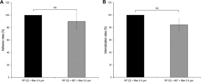
Staphylococcus aureus is a major pathogen that is responsible for mastitis in dairy herds. S. aureus mastitis is difficult to treat and prone to recurrence despite antibiotic treatment. The ability of S. aureus to invade bovine mammary epithelial cells (bMEC) is evoked to explain this chronicity. One sustainable alternative to treat or prevent mastitis is the use of lactic acid bacteria (LAB) as mammary probiotics. In this study, we tested the ability of Lactobacillus casei strains to prevent invasion of bMEC by two S. aureus bovine strains, RF122 and Newbould305, which reproducibly induce acute and moderate mastitis, respectively. L. casei strains affected adhesion and/or internalization of S. aureus in a strain-dependent manner. Interestingly, L. casei CIRM-BIA 667 reduced S. aureus Newbould305 and RF122 internalization by 60 to 80%, and this inhibition was confirmed for two other L. casei strains, including one isolated from bovine teat canal. The protective effect occurred without affecting bMEC morphology and viability. Once internalized, the fate of S. aureus was not affected by L. casei. It should be noted that L. casei was internalized at a low rate but survived in bMEC cells with a better efficiency than that of S. aureus RF122. Inhibition of S. aureus adhesion was maintained with heat-killed L. casei, whereas contact between live L. casei and S. aureus or bMEC was required to prevent S. aureus internalization. This first study of the antagonism of LAB toward S. aureus in a mammary context opens avenues for the development of novel control strategies against this major pathogen.


Figures

Fig 1
Fig 1
Adhesion to and internalization into bMEC of S. aureus strains RF122 and NB305 and L. casei strains CIRM-BIA 667, BL23, and CIRM-BIA 1542. S. aureus (MOI of 100:1) and L. casei (ROI of 2,000:1) populations adhered to (A) and internalized into (B) bMEC were determined after 1 and 2 h of interaction, respectively. Data are presented as mean populations per well (i.e., corresponding to 2.5 × 10⁵ bMEC) ± standard deviations. Each experiment was done in triplicate, and differences between groups were compared using Student's t test. *, P < 0.05; ***, P < 0.0005.
Fig 2
Fig 2
Inhibition of S. aureus RF122 and NB305 adhesion to bMEC by L. casei CIRM-BIA 667. Shown are adhesion rates of S. aureus strains after 1 h of interaction with bMEC and following 2 h of preincubation of cells with L. casei at an ROI of 200:1 (white bars), 400:1 (gray bars), and 2,000:1 (hatched bars). S. aureus was used at an MOI of 100:1. An adhesion assay of S. aureus alone was used as a reference (black bars). Adhesion rates were then defined as the adhered S. aureus population in the presence of L. casei relative to the adhered S. aureus population in the reference experiment. Data are presented as means ± standard deviations. Each experiment was done in triplicate, and differences between groups were compared using Student's t test with Bonferroni's correction. ***, P < 0.0005.
Fig 3
Fig 3
Inhibition of S. aureus RF122 and NB305 internalization into bMEC by L. casei CIRM-BIA 667. Shown are internalization rates of S. aureus strains after 2 h of interaction with bMEC with coincubation with L. casei at an ROI of 2,000:1. S. aureus strains were used at an MOI of 10:1 (white bars), 40:1 (gray bars), or 100:1 (hatched bars). The internalization assay of S. aureus alone was used as a reference (black bars). Internalization rates were then defined as the internalized S. aureus population in the presence of L. casei relative to the internalized S. aureus population in the reference experiment. Data are presented as means ± standard deviations. Each experiment was done in triplicate, and differences between groups were compared using Student's t test with Bonferroni's correction. ***, P < 0.0005.
Fig 4
Fig 4
Inhibition of adhesion and internalization of S. aureus RF122 and NB305 by L. casei strains. (A) Rates of adhesion of S. aureus RF122 and NB305 strains to bMEC following preincubation of cells with L. casei CIRM-BIA 667 (white bars), BL23 (gray bars), and CIRM-BIA 1542 (hatched bars) at an ROI of 2,000:1. (B) Rates of internalization of S. aureus RF122 and NB305 into bMEC in the presence of L. casei CIRM-BIA 667 (white bars) and BL23 (gray bars) at an ROI of 2,000:1 and CIRM-BIA 1542 (hatched bars) at an ROI of 400:1. Adhesion and internalization assays were performed with an S. aureus MOI of 100:1. Adhesion/internalization assays of S. aureus alone were used as a reference (black bars). Adhesion/internalization rates were then defined as the adhered/internalized S. aureus population in the presence of L. casei relative to the adhered/internalized S. aureus population in the reference experiment. Data are presented as means ± standard deviations. Each experiment was done in triplicate, and differences between groups were compared using Student's t test with Bonferroni's correction. ***, P < 0.0005.
Fig 5
Fig 5
Survival rate of S. aureus RF122 and L. casei CIRM-BIA 667 within bMEC. bMEC were incubated for 2 h with S. aureus RF122 (MOI, 100:1) with or without L. casei CIRM-BIA 667 (ROI, 2,000:1) in DMEM. Following a 2-h incubation step with gentamicin (100 μg/ml) to kill extracellular bacteria, cells were further incubated with gentamicin (25 μg/ml) for 24 and 48 h. The initial internalized S. aureus population measured after the 2 h of infection by S. aureus alone was used as the reference. The remaining internalized population of S. aureus alone (□) or in coinfection with L. casei (■) and L. casei (▲) then were measured and expressed relative to an S. aureus reference population. Data are presented as the mean survival rate ± standard deviations. Each experiment was done in triplicate, and differences between half-lives were compared using Student's t test.
Fig 6
Fig 6
Fluorescent confocal microscopy of mammary epithelial cells during bacterial infections. SYTO 9 (A to E) and phalloidin (F to J) stainings were used to observe bMEC structure following internalization assays with S. aureus RF122 (carrying plasmid pCtuf-gfp in the case of phalloidin staining) at an MOI of 100:1. MAC-T cells were either untreated (control; A, F, and I) or treated with S. aureus alone (B, E, G, and J), L. casei CIRM-BIA 667 alone at an ROI of 2,000:1 (D), or S. aureus and L. casei in cocultures (C and H). A lens with a ×100 magnification was used, and panels D to E and I to J are electronically zoomed. Arrows indicate internalized L. casei (D) or internalized S. aureus (E and J). Cyt., cytoplasm; N., nucleus.
Fig 7
Fig 7
Adhesion and internalization rates of S. aureus RF122 with different treatments. (A) Adhesion rates of S. aureus RF122 to bMEC at an MOI of 100:1, either alone or with preincubation of cells with L. casei CIRM-BIA 667 at an ROI of 2,000:1 (+LAB), heat-killed L. casei CIRM-BIA 667 at an ROI of 2,000:1 (+LAB-HK), DMEM acidified to pH 6.8 with lactic acid (+pH 6.8), or L. casei CIRM-BIA 667 supernatant (+SN-LAB). (B) Internalization rates of S. aureus RF122 into bMEC at an MOI of 100:1, either alone or with coincubation with L. casei CIRM-BIA 667 at an ROI of 2,000:1 (+LAB), heat-killed L. casei CIRM-BIA 667 at an ROI of 2,000:1 (+LAB-HK), DMEM acidified to pH 6.8 with lactic acid (+pH 6.8), or L. casei CIRM-BIA 667 supernatant (+SN-LAB). Adhesion/internalization assays of S. aureus alone were used as references. Adhesion/internalization rates were then defined as the adhered/internalized S. aureus population in the presence of L. casei, lactic acid, or supernatant relative to the adhered/internalized S. aureus population in the reference experiment. Data are presented as means ± standard deviations. Each experiment was done in triplicate, and differences between groups were compared using Student's t test with Bonferroni's correction. *, P < 0.05; ***, P < 0.0005.
Fig 8
Fig 8
L. casei inhibition of S. aureus adhesion and internalization requires contact with bMEC and/or S. aureus. Adhesion and internalization assays were performed, as previously described (see the legends to Fig. 2 and 3), using S. aureus RF122 at an MOI of 100:1 and L. casei CIRM-BIA 667 at an ROI of 2,000:1, except that L. casei was separated from S. aureus and bMEC using a cell culture insert. Adhesion/internalization assays of S. aureus alone with the cell culture insert were used as references. Adhesion/internalization rates were then defined as the adhered/internalized S. aureus population in the presence of L. casei relative to the adhered/internalized S. aureus population in the reference experiment. Data are presented as means ± standard deviations. Each experiment was done in triplicate, and differences between groups were compared using Student's t test. NS, not significant.

References

    1. Akers RM, Nickerson SC. 2011. Mastitis and its impact on structure and function in the ruminant mammary gland. J. Mammary Gland Biol. Neoplasia 16:275–289 - PubMed
    1. Archer GL. 1998. Staphylococcus aureus: a well-armed pathogen. Clin. Infect. Dis. 26:1179–1181 - PubMed
    1. Delgado S, Garcia P, Fernandez L, Jimenez E, Rodriguez-Banos M, Del CR, Rodriguez JM. 2011. Characterization of Staphylococcus aureus strains involved in human and bovine mastitis. FEMS Immunol. Med. Microbiol. 62:225–235 - PubMed
    1. Zadoks RN, Middleton JR, McDougall S, Katholm J, Schukken YH. 2011. Molecular epidemiology of mastitis pathogens of dairy cattle and comparative relevance to humans. J. Mammary Gland Biol. Neoplasia 16:357–372 - PMC - PubMed
    1. Contreras GA, Rodriguez JM. 2011. Mastitis: comparative etiology and epidemiology. J. Mammary Gland Biol. Neoplasia 16:339–356 - PubMed

Publication types