. 2013 Mar 1;22(5):879-89.
doi: 10.1093/hmg/dds494. Epub 2012 Nov 25.

Zic3 is required in the extra-cardiac perinodal region of the lateral plate mesoderm for left-right patterning and heart development

Affiliations

Zic3 is required in the extra-cardiac perinodal region of the lateral plate mesoderm for left-right patterning and heart development

Zhengxin Jiang et al. Hum Mol Genet. .

Abstract

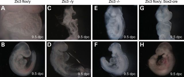
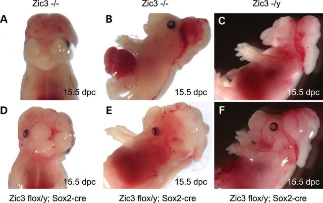
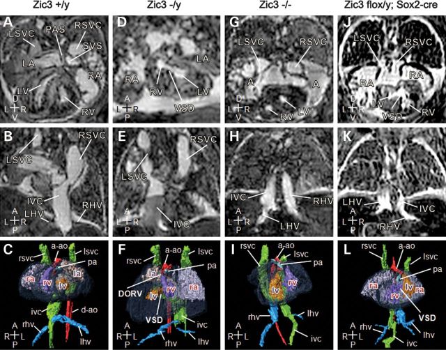
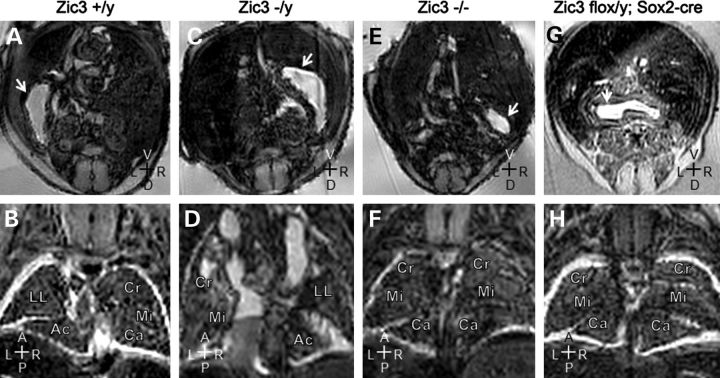
Mutations in ZIC3 cause human X-linked heterotaxy and isolated cardiovascular malformations. A mouse model with targeted deletion of Zic3 demonstrates an early role for Zic3 in gastrulation, CNS, cardiac and left-right axial development. The observation of multiple malformations in Zic3(null) mice and the relatively broad expression pattern of Zic3 suggest its important roles in multiple developmental processes. Here, we report that Zic3 is primarily required in epiblast derivatives to affect left-right patterning and its expression in epiblast is necessary for proper transcriptional control of embryonic cardiac development. However, cardiac malformations in Zic3 deficiency occur not because Zic3 is intrinsically required in the heart but rather because it functions early in the establishment of left-right body axis. In addition, we provide evidence supporting a role for Zic3 specifically in the perinodal region of the posterior lateral plate mesoderm for the establishment of laterality. These data delineate the spatial requirement of Zic3 during left-right patterning in the mammalian embryo, and provide basis for further understanding the molecular mechanisms underlying the complex interaction of Zic3 with signaling pathways involved in the early establishment of laterality.


Figures

Figure 1.
Figure 1.
Deletion of Zic3 in epiblast results in significant developmental anomalies and cardiac looping defects, similar to Zic3null embryos. Typical morphology of Zic3flox/y control (A and B), Zic3null (CF) and Zic3flox/y; Sox2-cre epiblast CKO (G and H) embryos at 9.5 dpc shows various degree of developmental defects, including head anomalies (C, E, F) and open neural tube (E), in the mutants compared with the controls (A and B). Hearts show normal dextral looping in Zic3flox/y controls (A and B), sinistral looping in Zic3null (F) and Zic3flox/y; Sox2-cre epiblast CKO (G and H) embryos, and ventral looping (C–E) in Zic3null embryos. Embryos are shown in a frontal view (A, C, E and G), right lateral position (B and D) and left lateral position (F and H), respectively.
Figure 2.
Figure 2.
Multiple laterality and CNS anomalies are seen in Zic3null (AC) and Zic3flox/y; Sox2-cre epiblast CKO (DF) embryos at 15.5 dpc. These abnormalities include exencephaly (A–F), cleft palate (A, C and D), omphalocele (B), malposition or loss of eyes (C and D), and neural tube closure defect (B and E).
Figure 3.
Figure 3.
Deletion of Zic3 in epiblast recapitulates complex cardiovascular laterality defects seen in Zic3null embryos. Magnetic resonance imaging of 15.5 dpc embryos. (AC) Transverse, coronal sections and three-dimensional reconstruction (ventral view) of a typical wild-type control heart and vasculature showing a pectinated right atrium (RA), with a systemic venous sinus (SVS), into which drains the right superior vena cava (RSVC), the left superior vena cava (LSVC) via the coronary sinus and the inferior vena cava (IVC). The left atrium (LA) is characterized by the primum atrial septum (PAS). The right ventricle (RV) is dextral to the left, and gives rise to the main pulmonary artery (pa). The left ventricle (LV), separated from the right ventricle by interventricular septum (not labeled), gives rise to the ascending aorta (a-ao), which arches to the left of the body axis. The left and right hepatic veins (LHV, RHV) join the inferior vena cava on the right of the body axis prior to entering the right atrium. (DL) Transverse, coronal sections and 3D reconstruction (ventral view) of Zic3null and Zic3flox/y; Sox2-cre epiblast CKO embryos' heart and vasculature. (D–F) Zic3−/y embryo exhibits situs inversus with heart malpositioned to the right. Atria and ventricles are inverted with ventricular septal defect (VSD). Both aorta and pulmonary artery arise from the right ventricle creating a double-outlet right ventricle (DORV). Inferior vena cava ascends on the left of the body axis. (G–I) Zic3−/− heart showing a large atrial septal defect, resulting in a common atrium (A). The heart is malpositioned to the right (dextrocardia) with inverted ventricles. Inferior vena cava ascends on the left and receives the left hepatic veins prior to entering the atrium, while the right hepatic veins enter separately. (J–L) Zic3flox/y; Sox2-cre heart showing a large atrial septal defect, leading to a common atrium, which is pectinated on each side and drains the bilateral superior vena cava. The coronary sinus is absent. These appearances indicate right atrial isomerism. Dextrocardia is also present with large ventricular septal defect (VSD). Right-sided aortic arch is noted with malposition of the great arteries and the aorta ascending anterior to the pulmonary artery. Inferior vena cava is continuous on the right and receives the right hepatic veins before draining to the atrium, while the left hepatic veins enter separately. Axis: D, dorsal; V, ventral; R, right; L, left; A, anterior; P, posterior.
Figure 4.
Figure 4.
Deletion of Zic3 in epiblast also recapitulates non-cardiovascular laterality defects seen in Zic3null embryos. Magnetic resonance imaging of 15.5 dpc embryos. (A and B) Transverse and coronal sections of a wild-type embryo showing a normal left-sided stomach (arrow), and normal pulmonary topology with cranial (Cr), middle (Mi), caudal (Ca) and accessory (Ac) lobes of the right lung and the left lung (LL). (C and D) Corresponding sections through a Zic3−/y embryo showing a right-sided stomach and inverted pulmonary topology. (E and F) Corresponding sections through a Zic3−/y embryo showing that the stomach is malpositioned to the right, and both lungs have multiple lobes suggesting right pulmonary isomerism. (G and H) Zic3flox/y; Sox2-cre embryo showing a midline stomach and right pulmonary isomerism. Axis: D, dorsal; V, ventral; R, right; L, left; A, anterior; P, posterior.
Figure 5.
Figure 5.
Whole heart transcriptional response to complete or epiblast-specfic deletion of Zic3 shows distinct gene expression patterns between Zic3 mutants and the controls at 15.5 dpc. (A) Heatmap illustrating distinct expression patterns for top differentially expressed transcripts between two groups with Q-value <0.05, false discovery rate (FDR) <0.05 and fold change larger than 1.5. Red indicates a higher expression value; blue indicates a lower expression value. (B) Proportions of significant and non-significant Gene Ontology (GO) categories for each of the two expression patterns. (C) Selection of the most important GO terms for each expression pattern. Higher fold enrichment means a GO term is more likely to be enriched in a given set of differently expressed genes. (D) Real-time PCR confirms upregulation of Nkx2.5, Cited1 and Eng, and downregulation of Tbx20 in the mutant hearts at 15.5 dpc. Four independent heart samples are used for each of the four genotype groups (WT: Zic3+/y, FLOX: Zic3flox/y, SOX2: Zic3flox/y; Sox2-cre, NULL: Zic3−/y), and each sample is assayed in triplicate and normalized to the endogenous control Gapdh. One-way ANOVA was used for statistical analyses in Qbase PLUS (Biogazelle).
Figure 6.
Figure 6.
Cardiac looping abnormalities in Zic3flox/y; T-cre embryos. Gross morphology of the Zic3flox/y control (A and B) and Zic3flox/y; T-cre embryos (CF) embryos at 9.5 dpc. Hearts show normal dextral looping (A and B), sinistral looping (C and E) and ventral looping (D and F). Embryos are shown in left lateral position (A, C and E) and right lateral position (B, D and F), respectively.
Figure 7.
Figure 7.
Laterality and complex cardiovascular abnormalities in Zic3flox/y; T-cre embryos. Magnetic resonance imaging of 15.5 dpc mutant embryos. (A and E) Transverse and coronal sections of a Zic3flox/y; T-cre embryo showing a midline stomach (arrow) and right pulmonary isomerism. (BD) Transverse, coronal sections and three-dimensional reconstruction (ventral view) of a Zic3flox/y; T-cre embryo showing a large atrial septal defect, resulting in a common atrium (A), into which drains the left and right superior vena cava (LSVC, RSVC). The heart is malpositioned to the right (dextrocardia). Inferior vena cava (IVC) is continuous on the right and receives the right hepatic veins (rhv) prior to entering the atrium, while the left hepatic veins (LHV) enter separately. (FH) Transverse, sagittal sections and three-dimensional reconstruction (right view) of a Zic3flox/y; T-cre embryo showing a normal positioned heart with a dilated common atrium (A), into which drains the left and right superior vena cava (LSVC, RSVC). Inferior vena cava (IVC) is interrupted and drains via the azygous vein (AV) to the right superior vena cava (RSVC). The hepatic veins come together and enter the atria without joining the IVC. Axis: D, dorsal; V, ventral; R, right; L, left; A, anterior; P, posterior.

Similar articles

Cited by

References

    1. Aruga J., Nagai T., Tokuyama T., Hayashizaki Y., Okazaki Y., Chapman V.M., Mikoshiba K. The mouse zic gene family. Homologues of the Drosophila pair-rule gene odd-paired. J. Biol. Chem. 1996;271:1043–1047. doi:10.1074/jbc.271.2.1043. - DOI - PubMed
    1. Aruga J., Yokota N., Hashimoto M., Furuichi T., Fukuda M., Mikoshiba K. A novel zinc finger protein, zic, is involved in neurogenesis, especially in the cell lineage of cerebellar granule cells. J. Neurochem. 1994;63:1880–1890. doi:10.1046/j.1471-4159.1994.63051880.x. - DOI - PubMed
    1. Gebbia M., Ferrero G.B., Pilia G., Bassi M.T., Aylsworth A., Penman-Splitt M., Bird L.M., Bamforth J.S., Burn J., Schlessinger D., et al. X-linked situs abnormalities result from mutations in ZIC3. Nat. Genet. 1997;17:305–308. doi:10.1038/ng1197-305. - DOI - PubMed
    1. Zhu L., Belmont J.W., Ware S.M. Genetics of human heterotaxias. Eur. J. Hum. Genet. 2006;14:17–25. - PubMed
    1. Nakata K., Nagai T., Aruga J., Mikoshiba K. Xenopus Zic3, a primary regulator both in neural and neural crest development. Proc. Natl Acad. Sci. U.S.A. 1997;94:11980–11985. doi:10.1073/pnas.94.22.11980. - DOI - PMC - PubMed

Publication types