. 2013 Jan;108(1):309.
doi: 10.1007/s00395-012-0309-x. Epub 2012 Nov 8.

Selective inhibition of Cx43 hemichannels by Gap19 and its impact on myocardial ischemia/reperfusion injury

Affiliations

Selective inhibition of Cx43 hemichannels by Gap19 and its impact on myocardial ischemia/reperfusion injury

Nan Wang et al. Basic Res Cardiol. 2013 Jan.

Abstract

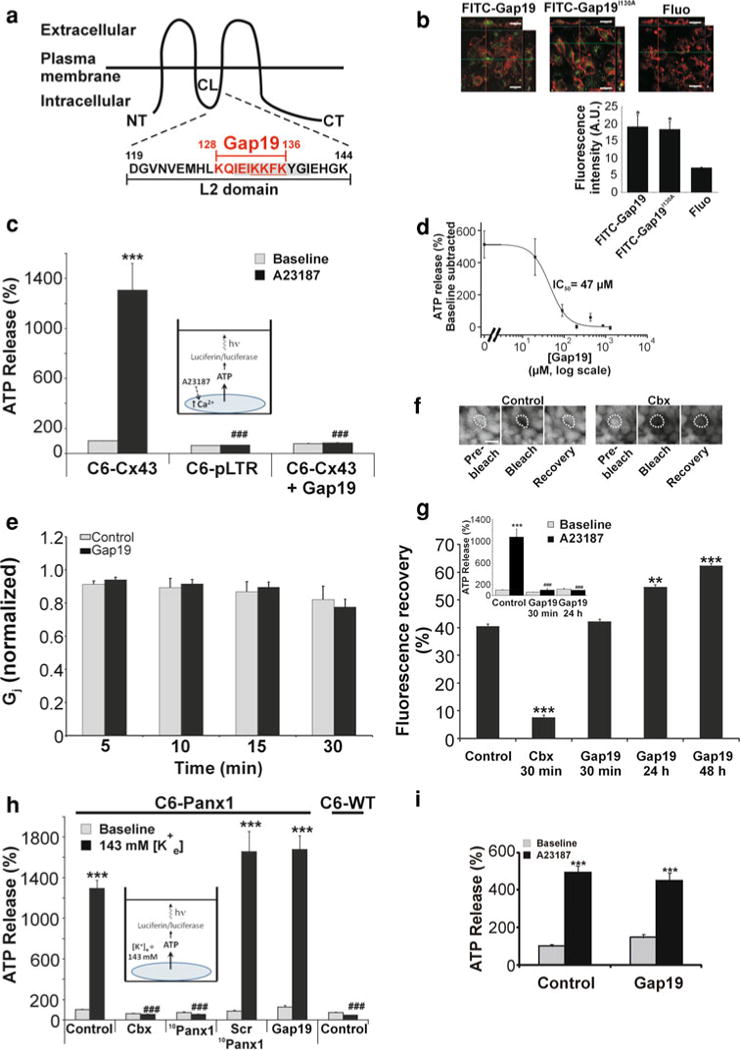
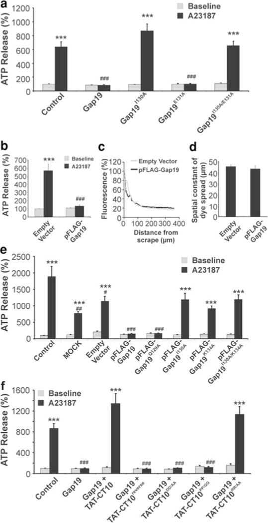
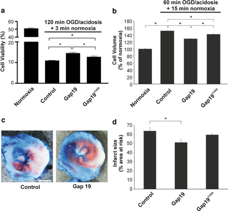
Connexin-43 (Cx43), a predominant cardiac connexin, forms gap junctions (GJs) that facilitate electrical cell-cell coupling and unapposed/nonjunctional hemichannels that provide a pathway for the exchange of ions and metabolites between cytoplasm and extracellular milieu. Uncontrolled opening of hemichannels in the plasma membrane may be deleterious for the myocardium and blocking hemichannels may confer cardioprotection by preventing ionic imbalance, cell swelling and loss of critical metabolites. Currently, all known hemichannel inhibitors also block GJ channels, thereby disturbing electrical cell-cell communication. Here we aimed to characterize a nonapeptide, called Gap19, derived from the cytoplasmic loop (CL) of Cx43 as a hemichannel blocker and examined its effect on hemichannel currents in cardiomyocytes and its influence in cardiac outcome after ischemia/reperfusion. We report that Gap 19 inhibits Cx43 hemichannels without blocking GJ channels or Cx40/pannexin-1 hemichannels. Hemichannel inhibition is due to the binding of Gap19 to the C-terminus (CT) thereby preventing intramolecular CT-CL interactions. The peptide inhibited Cx43 hemichannel unitary currents in both HeLa cells exogenously expressing Cx43 and acutely isolated pig ventricular cardiomyocytes. Treatment with Gap19 prevented metabolic inhibition-enhanced hemichannel openings, protected cardiomyocytes against volume overload and cell death following ischemia/reperfusion in vitro and modestly decreased the infarct size after myocardial ischemia/reperfusion in mice in vivo. We conclude that preventing Cx43 hemichannel opening with Gap19 confers limited protective effects against myocardial ischemia/reperfusion injury.


Conflict of interest statement

Conflict of interest None.

Figures

Fig. 1
Fig. 1
Gap19 inhibits [Ca2+]i-triggered ATP release in C6-Cx43 cells. a Topology of Cx43 and location of Gap19 in the L2 domain, part of the CL. The underlined sequence is a putative membrane translocation motif and the greyed zone is crucial for CT–CL interactions. b Confocal micrographs of C6-Cx43 cells, counter-stained for F-actin (red fluorescence), illustrating cellular uptake of fluorescein-labeled (green) Gap19 (FITC-Gap19), Gap19I130A (FITC-Gap19130A) and fluorescein only (Fluo). Scale bar is 20 μm. The bar chart below reports fluorescence intensities (A.U., arbitrary units) measured in the cells. Uptake of fluorescein-labeled peptides was significantly stronger as compared to fluorescein only (n = 4). Stars indicate significance compared to Fluo. c Inducing [Ca2+]i changes with the Ca2+ ionophore A23187 (inset shows experimental approach) triggered significant ATP release in C6-Cx43 but not in C6 cells stably transfected with the empty vector (C6-pLTR) (n = 12). Gap19 (200 μM, 30 min) strongly inhibited [Ca2+]i-triggered ATP release. d Gap19-inhibition of triggered ATP release was concentration-dependent (n = 6). e Junctional conductance (Gj) measurements in Cx43 expressing Novikoff cell pairs at different time points in the absence or presence of Gap19 (400 μM) in the recording pipette solution. Gj was normalized to the corresponding values at the beginning of the experiment. Gap19 had no effect on Gj (n = 4–16). f Representative images of a FRAP experiment in C6-Cx43 cells preloaded with CFDA. Images were acquired before photobleaching (pre-bleach), just after photobleaching the cell marked with the dotted line (bleach) and 5 min later to assess fluorescence recovery in the bleached cell. g Quantification of the fluorescence recovery 5 min after photobleaching: 30 min Gap19 (200 μM) had no influence while 24–48 h incubations promoted dye transfer (n = 5). Inset above illustrates that 24 h incubation with Gap19 inhibited ATP release equally strong as 30 min incubation (n = 12). h Exposure of C6-Panx1 cells to 143 mM [K+]e-triggered ATP release that was blocked by carbenoxolone (Cbx, 10 μM, 30 min) or 10Panx1 (200 μM, 30 min), and absent in C6-WT cells. Gap19 or Scr10Panx1 had no effect on high [K+]e-triggered ATP release (n = 12). i Gap19 (200 μM, 30 min) did not inhibit [Ca2+]i-triggered ATP release (brought about by 2 μM A23187 applied during 5 min) in HeLa-Cx40 cells (n = 6). Stars indicate statistical significance compared to the neighboring grey baseline bar (except in b); number signs mark comparisons to the black control bar; one symbol p < 0.05, two symbols p < 0.01, three symbols p < 0.001
Fig. 2
Fig. 2
Gap19 activity depends on AA 130 and 134 and is counteracted by adding CT10 peptide. a Gap19I130A (200 μM, 30 min) had no effect on [Ca2+]i-triggered ATP release in C6-Cx43 while Gap19E131A acted as Gap19. Combining the I130A/E131A modifications gave results as for Gap19I130A (n = 12). b Amino acid substitutions in the putative membrane translocation motif of Gap19 were tested using the pFLAG-Gap19 plasmid. [Ca2+]i-triggered ATP release was absent in C6-Cx43 cells transiently transfected with pFLAG-Gap19 (n = 12). c Example traces of SLDT dye (6-CF) spread experiments in C6-Cx43 cells (empty vector) and C6-Cx43 cells transfected with pFLAG-Gap19. d Quantification of the spatial constant of dye spread from SLDT experiments, demonstrating no effect of pFLAG-Gap19 on dye spread (n = 3; p = 0.6337). e pFLAG-Gap19I130A acted as Gap19I130A and did not inhibit [Ca2+]i-triggered ATP release while pFLAG-Gap19Q129A acted inhibitory. pFLAG-Gap19K134A did not inhibit [Ca2+]i-triggered ATP release; the combined I130A/K134A mutant acted as the single mutants (n = 12). f CT10 peptide counteracts Gap19 effects. In C6-Cx43 cells pre-incubated with TAT-CT10 (100 μM, 30 min), Gap19 (200 μM, 30 min together with TAT-CT10) did not inhibit ATP release while TAT-CT10reverse had no effect. The two aspartate and two proline residues in CT10 are crucial for this effect (n = 12). Stars compared to the neighboring grey baseline bar; number signs compared to the black control bar in a and f, and to the black empty vector bar in b and e
Fig. 3
Fig. 3
SPR experiments demonstrating Cx43 CT tail binding to biotin-Gap19 immobilized to a streptavidin-coated sensor chip. a Typical sensorgrams showing the association (first arrow) and dissociation (second arrow) between purified CT tail (Cx43CT, 7 μM) or purified GST (7 μM) and biotin-Gap19 or biotin-L2 immobilized to the streptavidin-coated sensor chip. The ordinate is calibrated in resonance units after correction for background binding to the control peptide (L2-reverse). b Summarized average data of experiments shown in a measured at maximal response (second arrow) (n = 3), demonstrating significantly stronger association between Cx43CT and Gap19 as compared to GST-Gap19. Cx43CT-Gap19 association was also stronger than for Cx43CT-L2. c Sensorgrams for Cx43CT-Gap19 and Cx43CT-L2 association at different concentrations of purified Cx43CT. d Summary of data shown in c, demonstrating a concentration-dependent increase in the association signal with half-maximal effect at ~2.5 μM for both Gap19 and L2 (n = 3)
Fig. 4
Fig. 4
Gap19 inhibits unitary hemichannel currents in HeLa-Cx43 cells. a Whole-cell voltage-clamp recording conditions. b ImVm plot illustrating voltage ramp experiments (−40 to +80 mV, 70 s). Unitary current activity started to appear at +50 mV (control) and was enhanced by further increasing Vm. Experiments with Gap19 or Gap19I130A in the pipette solution (400 μM) are also shown. c Typical traces of unitary currents activated by stepping Vm from −30 mV to +70 mV for 30 s. Ten consecutive runs (traces) were recorded over 7 min under control conditions (top) and when the pipette solution contained Gap19 (middle) or Gap19I130A (bottom). d Left: All-point histograms determined from each set of recordings depicted in c. Dashed vertical lines mark peaks in the histograms separated by ~220 pS. Gap19 reduced hemichannel activity as can be appreciated from the decreased number of peaks and increased frequency of the closed state. Right: Open dwell-time histograms determined from the recordings in c. Gap19 decreased the frequency of openings but had no effect on the time constant (τ) of the mono-exponential distribution of open dwell-times. Distributions with Gap19I130A were as observed in control. Data in c and d are representative for three different experiments. e Bar chart summarizing the results of integrating the current traces over time, giving the membrane charge transfer (Qm), for the different conditions applied. Gap19 significantly suppressed Qm to ~1/4 of control while Gap19I130A had no effect (n = 6 for control, 8 for Gap19 and 6 for Gap19I130A). f Gap19 inhibited Qm in a concentration-dependent manner. g Qm was not influenced by Gap19I130A unless it was applied at 1 mM concentration
Fig. 5
Fig. 5
Gap19 inhibits hemichannel unitary currents in ventricular cardiomyocytes. a Whole-cell recording performed in acutely isolated ventricular cardiomyocytes revealed a Vm dependent activation of Cx43 hemichannels (30 s voltage steps—traces representative for seven similar recordings). The all-point histogram illustration below the traces indicates a ~200 pS unitary conductance at Vm = +30 mV. In the presence of Gap19 (100 μM in the pipette), Vm steps to more positive potentials were necessary (traces representative for six similar recordings). b Example traces illustrating that unitary current activities was still present after repolarizing to −70 mV. The all-point histogram below indicates a ~200 pS unitary conductance of the unitary tail current events. c ImVm plot of open hemichannels demonstrating a reversal potential of ~0 mV and a single-channel slope conductance of ~196 pS. d Voltage dependent activation of hemichannels, demonstrating that Gap19 (100 μM) shifted the activation curve to more positive potentials. The quantity Qm/Vm was calculated by dividing the integrated unitary current activity by the corresponding Vm, and represents the integrated single-channel conductance over the 30 s voltage steps (n = 6). e Gap19 inhibition of unitary events did not influence the open hemichannel ImVm plot (~0 mV reversal potential and 204 pS single-channel slope conductance, not different from control). f Example traces recorded in a cardiomyocyte before and after exposure to MI (5 μM FCCP and 1 mM IAA). Voltage steps from −70 mV (5 s) to +40 mV (30 s) were repetitively applied. Unitary current activity was completely absent with Gap19 in the recording pipette. g Summary graph illustrating progressively increasing unitary current activity after application of MI (n = 4). Stars indicate statistical significance compared to baseline before MI induction. Recordings with Gap19 in the pipette were flat, lacking any unitary activity, also after MI induction (filled squares on the abcis). Gap19 completely suppressed MI-promoted hemichannel activity. Number signs indicate statistical significance compared to the corresponding open squares recorded without Gap19 in the pipette
Fig. 6
Fig. 6
Gap19 improves cardiomyocyte viability following ischemia/reperfusion in vitro and in vivo. a In vitro simulated ischemia/reperfusion of isolated cardiomyocytes. Gap19 (250 μM, 30 min pre-incubation and present during OGD/acidosis) improved cardiomyocyte viability after 120 min OGD/acidosis (ischemia) followed by 3 min normoxia (reperfusion) compared to control cells treated with vehicle-only; Gap19I130A had less effect (n = 6). b OGD/acidosis + normoxia caused significant swelling of cardiomyocytes compared to normoxia. Pre-incubation of cardiomyocytes with Gap19 reduced the degree of cell swelling while Gap19I130A had less effect (250 μM, 30 min) (n = 7). c In vivo experiments in mice with LAD ligation for 30 min followed by 120 min reperfusion. Images of a representative experiment illustrating a reduction of the infarct area are marked white (TTC staining). Red color indicates viable tissue and blue represents perfused tissue. Red and white zones together form the area at risk. Scale bar is 1 mm. d Summary data of experiments illustrated in c. The infarct size, relative to the area at risk, was reduced by Gap19 injected intravenously (25 mg/kg) 10 min prior to the ligation, while Gap19I130A had no significant effect (n = 11 for control, 5 for Gap19 and 8 for Gap19I130A)

References

    1. Abascal F, Zardoya R. Evolutionary analyses of gap junction protein families. Biochim Biophys Acta. 2012 doi: 10.1016/j.bbamem.2012.02.007. Epub ahead of print. - DOI - PubMed
    1. Bao X, Lee SC, Reuss L, Altenberg GA. Change in permeant size selectivity by phosphorylation of connexin 43 gap-junctional hemichannels by PKC. Proc Natl Acad Sci USA. 2007;104:4919–4924. doi: 10.1073/pnas.0603154104. - DOI - PMC - PubMed
    1. Barth K, Gentsch M, Blasche R, Pfuller A, Parshyna I, Koslowski R, Barth G, Kasper M. Distribution of caveolin-1 and connexin43 in normal and injured alveolar epithelial R3/1 cells. Histochem Cell Biol. 2005;123:239–247. doi: 10.1007/s00418-004-0727-4. - DOI - PubMed
    1. Boengler K, Buechert A, Heinen Y, Roeskes C, Hilfiker-Kleiner D, Heusch G, Schulz R. Cardioprotection by ischemic postconditioning is lost in aged and STAT3-deficient mice. Circ Res. 2008;102:131–135. doi: 10.1161/CIRCRESAHA.107.164699. - DOI - PubMed
    1. Boengler K, Dodoni G, Rodriguez-Sinovas A, Cabestrero A, Ruiz-Meana M, Gres P, Konietzka I, Lopez-Iglesias C, Garcia-Dorado D, Di LF, Heusch G, Schulz R. Connexin 43 in cardiomyocyte mitochondria and its increase by ischemic preconditioning. Cardiovasc Res. 2005;67:234–244. doi: 10.1016/j.cardiores.2005.04.014. - DOI - PubMed

Publication types

LinkOut - more resources