

doi: 10.1002/adma.201203370.
Epub 2012 Nov 26.
Functionalizing calcium phosphate biomaterials with antibacterial silver particles
Affiliations
- PMID: 23184492
- PMCID: PMC4227400
- DOI: 10.1002/adma.201203370
Item in Clipboard
Functionalizing calcium phosphate biomaterials with antibacterial silver particles
Adv Mater.
.
No abstract available
Figures
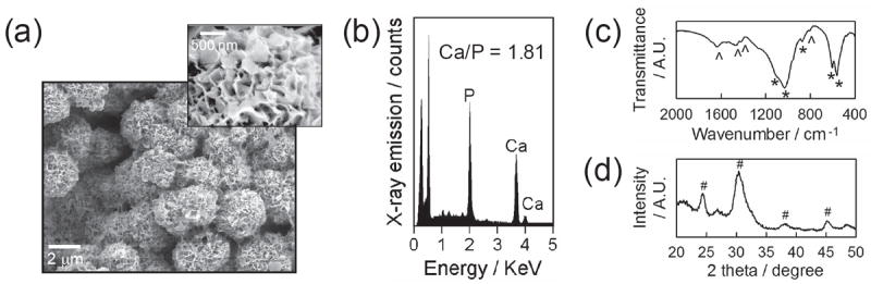
CaP coatings created on PLG films via biomimetic coating method. (a) SEM images of CaP coatings and (b) EDS, (c) FT-IR and (d) XRD spectra of CaP coatings. * and ˆ in FT-IR spectra represent characteristic peaks of phosphate and carbonate, respectively. # in XRD pattern denotes the characteristic peaks associated with hydroxyapatite.
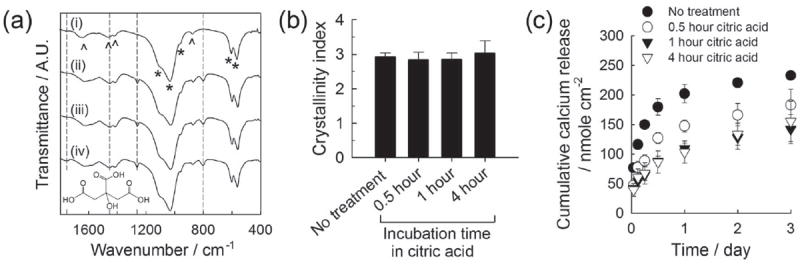
(a) FT-IR spectra of CaP coatings (i) before citric acid treatment and (ii-iv) after incubating in 5 mm citric acid solution for different time periods: (ii) 0.5, (iii) 1 and (iv) 4 hours. The structural formula of citric acid is shown on the bottom. (b) Crystallinity index calculated from FT-IR spectra based on Shemesh’s method. (c) Calcium release from CaP coatings treated differently with citric acid.
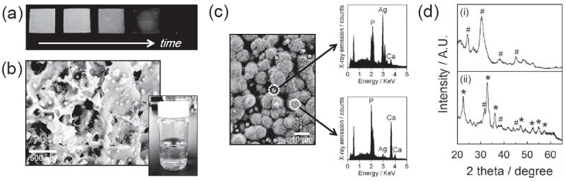
(a) Photographs of silver-incorporated, CaP-coated PLG films during the course of incubation in silver nitrate solution. The CaP-coated films were incubated in 5 mm citric acid solution for 1 hour and subsequently in 5 mm silver nitrate solution for 0, 0.5, 1, 2 and 4 hours (from left to right). (b) SEM image of nano-sized silver particles synthesized on CaP coating. Silver nanoparticles were synthesized by sequentially incubating in 5 mm citric acid solution for 1 hour and 5 mm silver nitrate solution for 0.5 hour. Lower right inset is a photograph of silver nitrate solution where a citric acid-treated CaP-coated PLG film was incubated on the bottom. (c) SEM image of micrometer-scale particles on CaP coating (left) and EDS spectra of distinct regions (right). They were created from 5 mm citric acid solution for 1 hour and 5 mm silver nitrate solution for 4 hours. (d) XRD spectra of (i) untreated CaP coating and (ii) CaP coating after silver particle formation. # and * in spectra denote the characteristic peaks from hydroxyapatite and silver phosphate or silver carbonate, respectively.
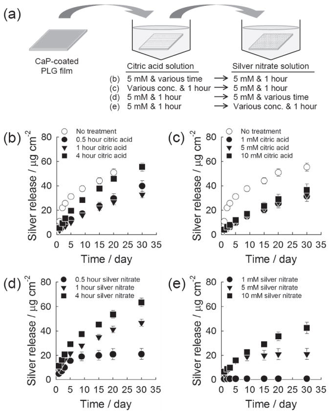
Cumulative silver release from CaP coating. (a) Schematic of experiment. (b,c) The CaP coatings were incubated (b) in 5 mm citric acid solution for different time periods or (c) in citric acid solution of different concentrations for 1 hour. The coatings were then incubated in 5 mm silver nitrate solution for 1 hour. (d,e) The CaP coatings were pretreated in 5 mm citric acid solution for 1 hour, and subsequently incubated (d) in 5 mm silver nitrate solution for different time periods or (e) in silver nitrate solution of different concentrations for 1 hour.
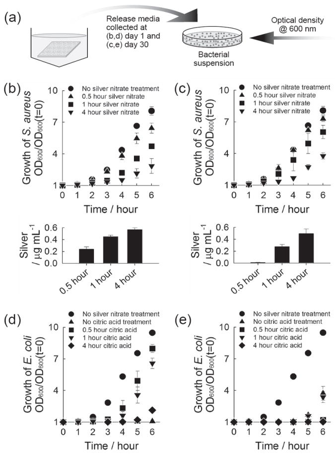
Antibacterial activity of released silver. (a) Schematic of experiment. (b-e) Growth curve of S. aureus (b,c) and E. coli (d,e) upon the addition of release media. The release media were collected from CaP coatings which were incubated sequentially in (b,c) 5 mm citric acid solution for 1 hour and 5 mm silver nitrate solution for different time periods, and (d,e) 5 mm citric acid solution for different time periods and 5 mm silver nitrate solution for 1 hour. The release media collected (b,d) at day 1 where CaP coatings were incubated for the first day and (c,e) at day 30 where the coatings were incubated from day 20 to day 30, were added to S. aureus (b,c) and E. coli (d,e). Lower plots in (b) and (c) show the concentration of silver in the bacteria solution tested.
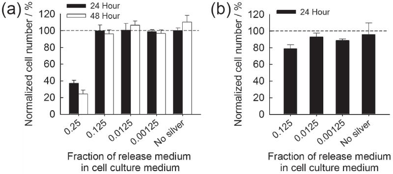
Normalized NIH 3T3 cell number when exposed to silver-containing cell culture medium. (a) Cells were seeded and cultured in silver-containing DMEM and (b) attached cells were exposed to silver-containing DMEM.
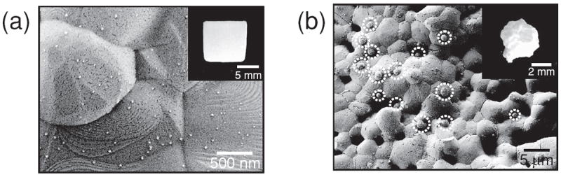
SEM images of silver particles created on sintered (a) hydroxyapatite and (b) β-tricalcium phosphate. The hydroxyapatite and β-tricalcium phosphate were incubated in 5 mm citric acid solution for 1 hour and subsequently in 5 mm silver nitrate solution for 1 hour.
References
Publication types
MeSH terms
Substances
Grants and funding
LinkOut - more resources
Full Text Sources
Other Literature Sources
Medical
