. 2013 Apr 1;591(7):1599-612.
doi: 10.1113/jphysiol.2012.245464. Epub 2012 Nov 26.

Inhibition of dendritic Ca2+ spikes by GABAB receptors in cortical pyramidal neurons is mediated by a direct Gi/o-β-subunit interaction with Cav1 channels

Affiliations

Inhibition of dendritic Ca2+ spikes by GABAB receptors in cortical pyramidal neurons is mediated by a direct Gi/o-β-subunit interaction with Cav1 channels

Enrique Pérez-Garci et al. J Physiol. .

Abstract

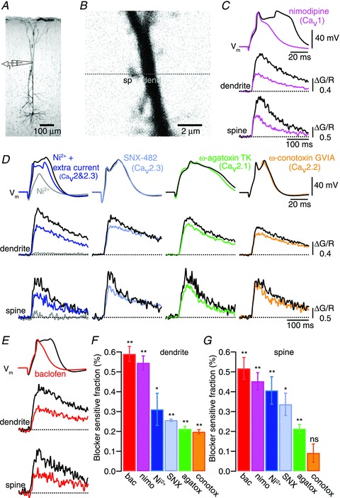
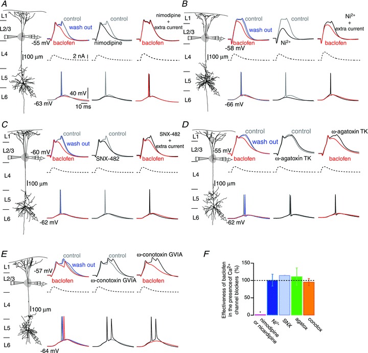
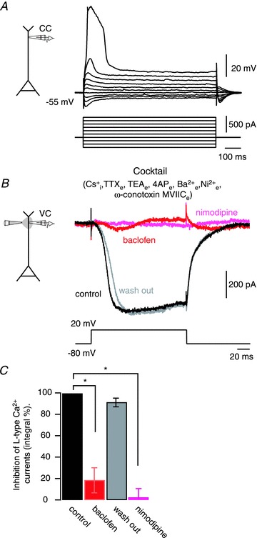
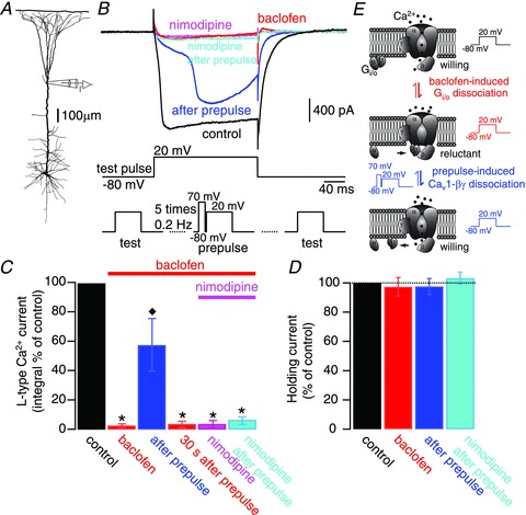
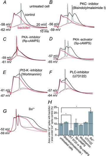
Voltage-dependent calcium channels (VDCCs) serve a wide range of physiological functions and their activity is modulated by different neurotransmitter systems. GABAergic inhibition of VDCCs in neurons has an important impact in controlling transmitter release, neuronal plasticity, gene expression and neuronal excitability. We investigated the molecular signalling mechanisms by which GABA(B) receptors inhibit calcium-mediated electrogenesis (Ca(2+) spikes) in the distal apical dendrite of cortical layer 5 pyramidal neurons. Ca(2+) spikes are the basis of coincidence detection and signal amplification of distal tuft synaptic inputs characteristic for the computational function of cortical pyramidal neurons. By combining dendritic whole-cell recordings with two-photon fluorescence Ca(2+) imaging we found that all subtypes of VDCCs were present in the Ca(2+) spike initiation zone, but that they contribute differently to the initiation and sustaining of dendritic Ca(2+) spikes. Particularly, Ca(v)1 VDCCs are the most abundant VDCC present in this dendritic compartment and they generated the sustained plateau potential characteristic for the Ca(2+) spike. Activation of GABA(B) receptors specifically inhibited Ca(v)1 channels. This inhibition of L-type Ca(2+) currents was transiently relieved by strong depolarization but did not depend on protein kinase activity. Therefore, our findings suggest a novel membrane-delimited interaction of the G(i/o)-βγ-subunit with Ca(v)1 channels identifying this mechanism as the general pathway of GABA(B) receptor-mediated inhibition of VDCCs. Furthermore, the characterization of the contribution of the different VDCCs to the generation of the Ca(2+) spike provides new insights into the molecular mechanism of dendritic computation.


Figures

Figure 1
Figure 1. Subcellular compartmentalized distribution of voltage-dependent calcium channels (VDCCs) and action of GABAB receptors
A, micrograph of a biocytin-filled L5 pyramidal neuron indicating the dendritic recording site. B, 2-photon fluorescence image (Alexa 594) of the recording site at the distal apical dendrite and a magnified view showing a spiny region in the apical tuft. Ca2+ imaging (Fluo-5) was measured by establishing a line scan (dashed line) covering a spine and its adjacent dendrite. C and D, dendritic Ca2+ spikes (top sweep) evoked by current injection via the recording pipette elicited Ca2+ transients in both the dendrite (middle panel) and in the spines (bottom panel; black sweeps). Coloured sweeps show representative examples of the action of different VDCCs blockers on the Ca2+ spikes and the resulting inhibition of Ca2+ fluorescence transients in both the dendrites and in the spines: nimodipine (10 μm, magenta traces), Ni2+ (50 μm, grey and blue sweeps, respectively, show recordings before and after injection of additional current via the recording pipette), SNX-482 (230 nm, light blue), ω-agatoxin TK (400 nm, green sweeps) and ω-conotoxin GVIA (1 μm, orange sweeps). E, baclofen added to the bath (10–30 μm) partially inhibited the Ca2+ spikes and their associated Ca2+ transients (red sweeps). F, summary bar plot showing the relative block of Ca2+ transients recorded in the dendrites for each drug tested. G, summary bar plot showing the relative block of Ca2+ transients recorded in the spines for each drug tested. Error bars indicate SEM. *P < 0.05, **P < 0.01.
Figure 2
Figure 2. Activation of dendritic GABAB receptors inhibits L-type Ca2+ conductances involved in the generation of Ca2+ spikes
AE, left columns: reconstruction of biocytin-filled L5 pyramidal neurons showing simultaneous somatic and distal (>700 μm) patch recordings, while a puff pipette expelled baclofen (50 μm) onto the apical tuft. Second columns: dendritic Ca2+ spikes (top, grey sweeps) were evoked by injecting an EPSP-shaped current waveform (middle, dashed sweeps) via the distal pipette. Dendritic spikes propagated towards the soma evoking APs (bottom, grey sweeps). Puffing baclofen on the dendrite shortened and reduced the amplitude of the Ca2+ spikes (red sweeps). The effect of baclofen was fully reversed after ceasing the local application of baclofen (blue sweeps). Third columns: control recordings and recordings obtained after bath application of different voltage-dependent calcium channels (VDCCs) blockers (black sweeps; A, nimodipne 10 μm; B, Ni2+ 50 μm; C, SNX-482, 230 nm; D, ω-agatoxin TK, 400 nm; E, ω-conotoxin GVIA, 1 μm). Fourth columns: reapplication of baclofen to the tuft in the presence of VDCCs blockers. In some cases larger current peaks were injected to the dendrite to reestablish the dendritic depolarization (see AC). F, effectiveness of baclofen estimated as the area underneath the control Ca2+ spike sensitive to the local application of baclofen. Values obtained after blockade of VDCCs are normalized to those obtained before bath application of VDCCs blockers (see SI Materials and methods). Error bars indicate SEM. *P < 0.01.
Figure 3
Figure 3. Activation of dendritic GABAB receptors inhibits dendritic L-type Ca2+ currents
A, voltage recordings performed in current-clamp mode at a distal dendritic site (700 μm) immediately after seal rupture. The internal pipette solution included 108 mm Cs+. Dendritic Ca2+ spikes occurred with depolarizing current steps (lower panel). B, a depolarizing voltage command in voltage-clamp mode was applied to the dendrite in an external medium containing TTX (1 μm), tetraethylammounium-chloride (TEA; 30 mm), 4-aminopyridine (4-AP; 5 mm) and Ba2+ (100 μm). Voltage-dependent calcium channels (VDCCs) other than Cav1 were also blocked with Ni2+ (50 μm; T- and R-type) and ω-conotoxin MVIIC (500 nm; N- and P/Q-type). Under these conditions, inward L-type Ca2+ (after leak subtraction) currents were recorded (black sweep). Local application of the GABAB agonist baclofen (50 μm) to the apical tuft inhibited the L-type Ca2+ currents (red sweep). The action of baclofen was reversed after 5 s ceasing the pressure application of baclofen (grey sweep). Subsequent bath application of nimodipine (10 μm) effectively blocked the L-type Ca2+ current (pink sweep). C, summary of the inhibitory effect of baclofen and nimodipine. Error bars indicate SEM. *P < 0.01.
Figure 4
Figure 4. GABAB inhibition of dendritic L-type Ca2+ currents is mediated by a direct Gi/o-βγ-subunit interaction
A, experimental configuration of dendritic voltage-clamp experiments. B, pharmacologically isolated dendritic L-type Ca2+ currents (black trace) were evoked by injecting a test voltage command from −80 to 20 mV (test pulse; middle trace). Bath application of baclofen (10–30 μm) inhibited the L-type Ca2+ currents (red trace). A series of 5 prepulse voltage commands from −80 mV to 70 mV (50 ms duration) preceding a depolarization from −80 mV to 20 mV (200 ms) by 60 ms (prepulse; bottom middle trace), partially and transiently relieved the L-type Ca2+ current from its inhibition (blue trace). After recovery of the GABAB-induced inhibition, nimodipine (10 μm) was supplemented to the bath (pink trace). In the presence of the Cav1 blocker, a new set of 5 prepulse voltage commands was ineffective to reverse the inhibition of L-type Ca2+ currents (cyan trace). The lower traces illustrate the sequence of test pulses and the prepulse protocol. C, summary of results. Error bars indicate SEM. *P < 0.01, relative to control; ♦P < 0.01, relative to baclofen inhibition (n = 4). D, bar graph showing the lack of variation in holding current values (at −80 mV) during the different experimental manipulations. E, sketch of the prepulse-induced relief of inhibition illustrating the interpretation of the current traces depicted in B.
Figure 5
Figure 5. GABAB receptors inhibit Ca2+ spikes independent of protein kinases
A, dendritic Ca2+ spike (black solid sweep) leading to somatic AP (black dashed sweep). Baclofen (50 μm; red sweeps) applied locally to the apical tuft induced inhibition of the Ca2+ spike. B–F, simultaneous double patch-clamp recording from the apical tuft and soma as in A, showing the baclofen-induced inhibition of Ca2+ spikes in cells pretreated with bisindolylmaleimide I (1 μm) to inhibit protein kinase C (PKC; B); Rp-cAMPS (1 mm) to inhibit cAMP-dependent protein kinases I and II (PKA; C); Sp-cAMPS (60–100 μm) to overactivate PKA (D); wortmannin (400 nm) to inhibit phosphatidylinositol 3-kinase (PI3K; E); and U73122 (1 μm) to inhibit phospholipase C (PLC; F). G, simultaneous double patch-clamp recording from the apical tuft and soma as in A, showing the baclofen-induced inhibition of Ca2+ spikes in the presence of Ba2+ (200 μm). H, summary of the inhibitory effect of baclofen (normalized to the area underneath the Ca2+ spike control) for each pharmacological condition. Error bars indicate SEM. *P < 0.05.

Similar articles

Cited by

References

    1. Almog M, Korngreen A. Characterization of voltage-gated Ca2+ conductances in layer 5 neocortical pyramidal neurons from rats. PLoS One. 2009;4:e4841. - PMC - PubMed
    1. Amico C, Marchetti C, Nobile M, Usai C. Pharmacological types of calcium channels and their modulation by baclofen in cerebellar granules. J Neurosci. 1995;15:2839–2848. - PMC - PubMed
    1. Amitai Y, Friedman A, Connors BW, Gutnick MJ. Regenerative activity in apical dendrites of pyramidal cells in neocortex. Cereb Cortex. 1993;3:26–38. - PubMed
    1. Bettler B, Kaupmann K, Mosbacher J, Gassmann M. Molecular structure and physiological functions of GABAB receptors. Physiol Rev. 2004;84:835–867. - PubMed
    1. Bloodgood BL, Sabatini BL. Nonlinear regulation of unitary synaptic signals by CaV(2.3) voltage-sensitive calcium channels located in dendritic spines. Neuron. 2007;53:249–260. - PubMed

Publication types

LinkOut - more resources