. 2013 Feb;242(2):108-21.
doi: 10.1002/dvdy.23906.

Regulation of primitive hematopoiesis by class I histone deacetylases

Affiliations

Regulation of primitive hematopoiesis by class I histone deacetylases

Rishita R Shah et al. Dev Dyn. 2013 Feb.

Abstract

Background: Histone deacetylases (HDACs) regulate multiple developmental processes and cellular functions. However, their roles in blood development have not been determined, and in Xenopus laevis a specific function for HDACs has yet to be identified. Here, we employed the class I selective HDAC inhibitor, valproic acid (VPA), to show that HDAC activity is required for primitive hematopoiesis.

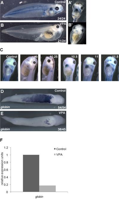
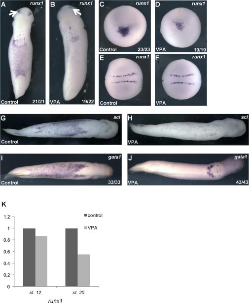
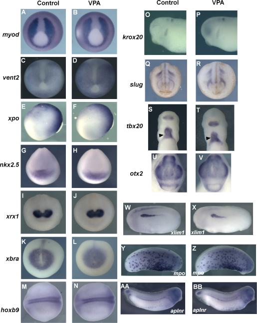
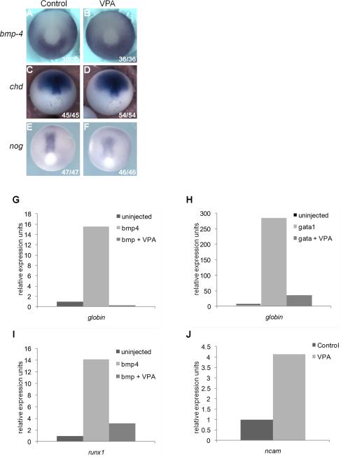
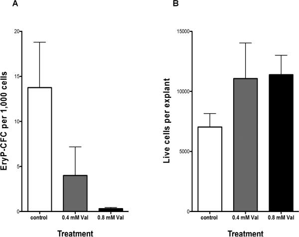
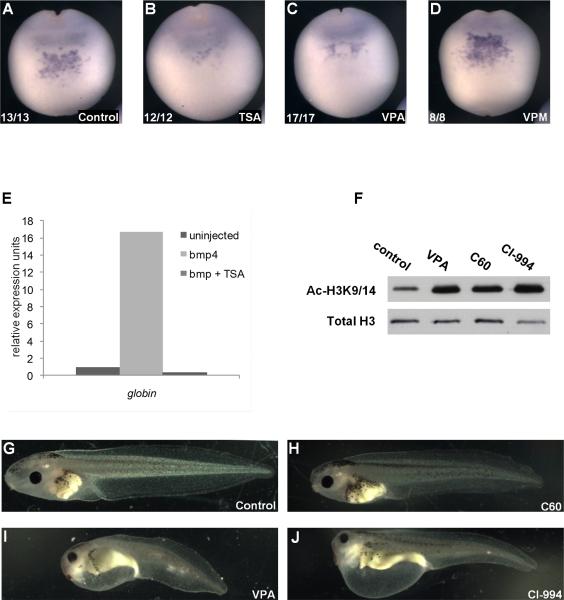
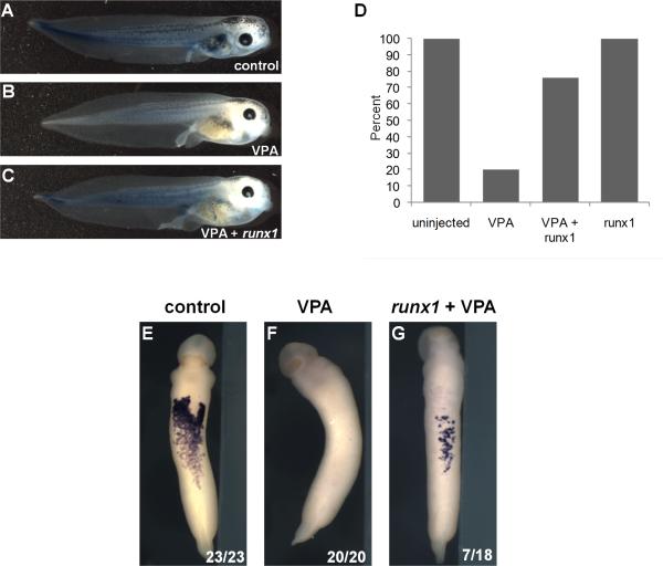
Results: VPA treatment during gastrulation resulted in a complete absence of red blood cells (RBCs) in Xenopus tadpoles, but did not affect development of other mesodermal tissues, including myeloid and endothelial lineages. These effects of VPA were mimicked by Trichostatin A (TSA), a well-established pan-HDAC inhibitor, but not by valpromide, which is structurally similar to VPA but does not inhibit HDACs. VPA also caused a marked, dose-dependent loss of primitive erythroid progenitors in mouse yolk sac explants at clinically relevant concentrations. In addition, VPA treatment inhibited erythropoietic development downstream of bmp4 and gata1 in Xenopus ectodermal explants.

Conclusions: These findings suggest an important role for class I HDACs in primitive hematopoiesis. Our work also demonstrates that specific developmental defects associated with exposure to VPA, a significant teratogen in humans, arise through inhibition of class I HDACs.


Figures

Figure 1
Figure 1. HDAC inhibition blocks primitive erythropoiesis in Xenopus laevis
A) Circulating RBCs visualized in tadpoles (stage 43) by benzidine staining (blue) for hemoglobin. RBCs are also present in pronephros (arrow) and in heart (A’, ventral view). B) Treatment of gastrulating embryos (stage 10-12) with the class I HDAC inhibitor VPA blocks blood formation, indicated by benzidine staining (B’, ventral view). C) Numbers indicate stages during gastrulation at which VPA was administered; in all conditions, VPA was removed at the end of gastrulation (stage 12). Benzidine positive RBCs are readily visible within the heart and great vessels in controls and when VPA was added at stage 10.5 or later. D,E) Expression of the erythrocyte marker alpha-T1 hemoglobin in the VBI detected by WISH in tailbud embryos (stage 31). In VPA treated embryos (E), globin expression is dramatically reduced, with residual staining restricted to the aVBI. F) Embryos were treated with control buffer or VPA during the gastrula stage and globin expression in stage 28 embryos was assessed by quantitative RT-PCR.
Figure 2
Figure 2. VPA inhibits the development of erythroid progenitors
A) Primitive erythroid marker runx1 is expressed within the VBI and olfactory placode (arrows) in control early tailbud (stage 26) embryos. B) VPA reduced runx1 expression in the VBI. C) In control late neurulae (stage 19, anterior view, with dorsal oriented to the top), runx1 is expressed in precursors of the aVBI, which lie in a ventral-anterior domain at this stage. D) VPA markedly reduces runx1 expression within presumptive hematopoietic cells in neurula stage embryos (same orientation as panel C). E) and F) Dorsal view (anterior to the left)- Expression of runx1 in Rohan Beard cells in the same embryos as (C) and (D) is not affected by VPA. G-J) Ventral views, anterior to the right. VPA treatment (H) and (J) reduces expression of erythroid markers scl (G) and (H) and gata1 (I) and (J) in the VBI of tailbud embryos (stage 31). K) runx1 expression in whole embryos is reduced with VPA treatment at late neurula stage (stage 18), as determined by qRT-PCR (data represent the mean of 4 independent experiments, normalized to ODC).
Figure 3
Figure 3. Marker analysis with VPA treatment
Embryos were treated with buffer control (A,C,E,G, I, K, M,O,Q,S,U,W, Y, AA) or VPA during stage 10-12 (B,D,F,H,J,L,N,P,R,T,V,X, Z, BB) . Whole mount in situ hybridization performed for the indicated markers shows no change in expression with VPA treatment. myod (A,B), vent2 (C,D), nkx2.5 (G,H), xrx1 (I,J), xbra (K,L), slug (Q,R), tbx20 (S,T), otx2 (U,V)- ventral view. xpo (E,F),lim (W,X), krox20 (O,P)- lateral view, anterior to the left, hoxb9 (M,N); dorsal view, anterior to the left. Also unaffected by VPA treatment were VBI derived myeloid marker mpo (Y, Z) and vascular marker aplnr (AA, BB)-lateral view, anterior to the right. myod, vent2, xpo, xbra- late gastrula. nkx2.5, xrx1, slug, hoxb9- neurula stages. lim1, krox20, mpo, aplnr, tbx20, otx2- tailbud stages.
Figure 4
Figure 4. HDAC inhibition prevents hematopoietic induction in ectodermal explants downstream of bmp4 and gata1
A-F) Expression of bmp-4 (A, B), chordin (C,D), and noggin (E,F) in late gastrula stage embryos is unaffected by VPA (B, D, F); orientation: posterior view, dorsal to the top. G-I) Quantitative RT-PCR of ectodermal explants harvested at tailbud stages (stage 28-31). G) Injection of bmp4 mRNA (2 ng) induced globin expression and VPA inhibited this effect. H) Injection gata1 mRNA (200pg) induced globin expression and VPA treatment inhibited this effect. I) Injection of bmp4 induced expression of runx1 and VPA blocked this effect. J) VPA upregulated endogenous expression of ncam in uninjected explants.
Figure 5
Figure 5. VPA inhibits primitive erythroid progenitor emergence in mouse yolk sac explants
A) EryP-CFC (mean ± SEM) per 1000 yolk sac explant cells cultured in vitro for 48 hours with 0, 0.4, and 0.8 mM VPA. B) Number of live cells (mean ± SEM) per yolk sac explant with 0, 0.4, and 0.8 mM VPA.
Figure 6
Figure 6. VPA mediated developmental defects are due to inhibition of class I HDACs
A) runx1 expression in the anterior ventral region of late neurula stage control embryos (stage 18). B, C) TSA (B) and VPA (C) suppress runx1 expression. D) The inactive VPA analog VPM does not inhibit runx1 expression. E) bmp4 mRNA (2ng) induction of globin expression in ectodermal explants is blocked by TSA (bmp+TSA). F) VPA (class I HDAC inhibitor), CI-994 (selective inhibitor of HDACs 1, 2, and 3), and C60 (IC50 for HDACs 1 and 2>>HDAC3) increase global acetylation of histone H3 at lysine 9/14 (H3K9/14Ac) at 24 hours post treatment, as demonstrated by western blotting. Bottom panel: total H3 (loading control). G-J) Lateral view of stage 45 tadpoles treated with vehicle (DMSO) control (G), C60 (H), VPA (I), and CI-994 (J) from late neurula (stage 19) to tailbud (stage 31) stages.
Figure 7
Figure 7. Runx1 expression rescues VPA-mediated hematopoietic defects
A) Benzidine staining of hemoglobin (blue) in untreated stage 43 tadpole. B) Treatment with VPA inhibits RBC development. C) Injection of runx1 mRNA partially recovered erythrocyte development in VPA treated embryos. D) Percentage of benzidine positive tadpoles for uninjected (n=20), VPA-treated (n=25), VPA-treated + runx1 (n=21), and runx1 expression alone (n=21). This experiment was repeated with 4 separate clutches of embryos and the table shows data from all 4 experiments. Intensity of benzidine stain was reduced in VPA and VPA+runx1 groups (as shown in panel C). E-G) Expression of globin was detected by WISH in all (23/23) control tadpoles, 0/20 VPA-treated tadpoles, and 7/18 VPA-treated, runx1-expressing tadpoles.

References

    1. Blythe SA, Cha SW, Tadjuidje E, Heasman J, Klein PS. beta-Catenin primes organizer gene expression by recruiting a histone H3 arginine 8 methyltransferase, Prmt2. Dev Cell. 19:220–231. - PMC - PubMed
    1. Boyes J, Byfield P, Nakatani Y, Ogryzko V. Regulation of activity of the transcription factor GATA-1 by acetylation. Nature. 1998;396:594–598. - PubMed
    1. Ciau-Uitz A, Liu F, Patient R. Genetic control of hematopoietic development in Xenopus and zebrafish. Int J Dev Biol. 54:1139–1149. - PubMed
    1. Ciau-Uitz A, Walmsley M, Patient R. Distinct origins of adult and embryonic blood in Xenopus. Cell. 2000;102:787–796. - PubMed
    1. Cunliffe VT. Histone deacetylase 1 is required to repress Notch target gene expression during zebrafish neurogenesis and to maintain the production of motoneurones in response to hedgehog signalling. Development. 2004;131:2983–2995. - PubMed

Publication types

LinkOut - more resources