

doi: 10.1002/adhm.201200115.
Epub 2012 Sep 28.
Sequence-specific crosslinking of electrospun, elastin-like protein preserves bioactivity and native-like mechanics
Affiliations
- PMID: 23184558
- PMCID: PMC3641778
- DOI: 10.1002/adhm.201200115
Item in Clipboard
Sequence-specific crosslinking of electrospun, elastin-like protein preserves bioactivity and native-like mechanics
Adv Healthc Mater.
2013 Jan.
No abstract available
Figures
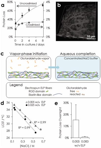
Stability of nanofibrous ELP. a) Comparison of protein loss in physiological conditions over eight days from untreated fabrics and two-stage crosslinked fabrics. b) CARS microscopy (2930 cm−1) 3D view of nanofibers after two weeks in physiological buffer. c) Schematic of two-stage crosslinking strategy. Crosslinking is initiated by exposing matrices to glutaraldehyde vapor. Complete crosslinks are formed after hydration in a concentrated sodium chloride buffer. d) LCST of ELP in varying buffer conditions. e) Comparison of overnight protein loss at physiological conditions for fabrics hydrated in varying w/v, ELP per 10× phosphate-buffered saline (PBS), during the aqueous crosslinking completion step.
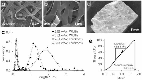
Physical characterization of electrospun ELP. a) SEM of fibers spun from aqueous solutions of a) 20%, and b) 35% w/w ELP. c) Histograms and Gaussian fits of nanofiber width and thickness (n > 100) from both solutions. d) An implantable bulk fabric in PBS, and e) a representative tensile stress-strain curve.
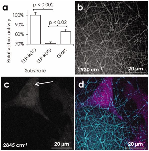
Bioactivity of the RGD ligand. a) Metabolic activity of rMSCs after 24 h in serum-free medium on ELP-RGD (8.4 mM RGD) or ELP-RDG (0 mM RGD) fabrics or glass control (n = 3). Maximal projection images obtained by CARS microscopy of rMSC after 24 h in standard conditions on electrospun ELP-RGD: b) nanofibers, c) rMSCs, d) overlay. Close contact is suggested by imprints of nanofibers on the cell membrane, white arrow.
Similar articles
-
Design and bioproduction of a recombinant multi(bio)functional elastin-like protein polymer containing cell adhesion sequences for tissue engineering purposes.J Mater Sci Mater Med. 2004 Apr;15(4):479-84. doi: 10.1023/b:jmsm.0000021124.58688.7a. J Mater Sci Mater Med. 2004. PMID: 15332621
-
Novel extracellular matrix for cell sheet recovery using genetically engineered elastin-like protein.J Biomed Mater Res B Appl Biomater. 2008 Jul;86(1):283-90. doi: 10.1002/jbm.b.31019. J Biomed Mater Res B Appl Biomater. 2008. PMID: 18161837
-
Recombinant elastin-mimetic biomaterials: Emerging applications in medicine.Adv Drug Deliv Rev. 2010 Dec 30;62(15):1468-78. doi: 10.1016/j.addr.2010.04.007. Epub 2010 May 2. Adv Drug Deliv Rev. 2010. PMID: 20441783 Free PMC article. Review.
-
Development of a collagen-like peptide polymer via end-to-end disulfide cross-linking and its application as a biomaterial.Acta Biomater. 2019 Aug;94:361-371. doi: 10.1016/j.actbio.2019.06.010. Epub 2019 Jun 11. Acta Biomater. 2019. PMID: 31200119
-
Biomimetic and bioactive nanofibrous scaffolds from electrospun composite nanofibers.Int J Nanomedicine. 2007;2(4):623-38. Int J Nanomedicine. 2007. PMID: 18203429 Free PMC article. Review.
Cited by
-
Extracellular Matrix-Mimetic Hydrogels for Treating Neural Tissue Injury: A Focus on Fibrin, Hyaluronic Acid, and Elastin-Like Polypeptide Hydrogels.Adv Healthc Mater. 2021 Nov;10(22):e2101329. doi: 10.1002/adhm.202101329. Epub 2021 Sep 8. Adv Healthc Mater. 2021. PMID: 34494398 Free PMC article. Review.
-
Hybrid elastin-like polypeptide-polyethylene glycol (ELP-PEG) hydrogels with improved transparency and independent control of matrix mechanics and cell ligand density.Biomacromolecules. 2014 Sep 8;15(9):3421-8. doi: 10.1021/bm500969d. Epub 2014 Aug 20. Biomacromolecules. 2014. PMID: 25111283 Free PMC article.
-
Immune-tolerant elastin-like polypeptides (iTEPs) and their application as CTL vaccine carriers.J Drug Target. 2016;24(4):328-39. doi: 10.3109/1061186X.2015.1077847. Epub 2015 Aug 25. J Drug Target. 2016. PMID: 26307138 Free PMC article.
-
Fibrous Scaffolds From Elastin-Based Materials.Front Bioeng Biotechnol. 2021 Jul 16;9:652384. doi: 10.3389/fbioe.2021.652384. eCollection 2021. Front Bioeng Biotechnol. 2021. PMID: 34336798 Free PMC article. Review.
-
Recombinant Resilin-Based Bioelastomers for Regenerative Medicine Applications.Adv Healthc Mater. 2016 Jan 21;5(2):266-75. doi: 10.1002/adhm.201500411. Epub 2015 Dec 3. Adv Healthc Mater. 2016. PMID: 26632334 Free PMC article.
References
Publication types
MeSH terms
Substances
Grants and funding
LinkOut - more resources
Full Text Sources
Other Literature Sources
