Mesenchymal markers on human adipose stem/progenitor cells
- PMID: 23184564
- PMCID: PMC4157311
- DOI: 10.1002/cyto.a.22227
Mesenchymal markers on human adipose stem/progenitor cells
Abstract
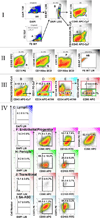
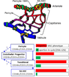
The stromal-vascular fraction (SVF) of adipose tissue is a rich source of multipotent stem cells. We and others have described three major populations of stem/progenitor cells in this fraction, all closely associated with small blood vessels: endothelial progenitor cells (EPC, CD45-/CD31+/CD34+), pericytes (CD45-/CD31-/CD146+), and supra-adventitial adipose stromal cells (SA-ASC, CD45-/CD31-/CD146-/CD34+). EPC are luminal, pericytes are adventitial, and SA-ASC surround the vessel like a sheath. The multipotency of the pericytes and SA-ASC compartments is strikingly similar to that of CD45-/CD34-/CD73+/CD105+/CD90+ bone marrow-derived mesenchymal stem cells (BM-MSC). Here, we determine the extent to which this mesenchymal pattern is expressed on the three adipose stem/progenitor populations. Eight independent adipose tissue samples were analyzed in a single tube (CD105-FITC/CD73-PE/CD146-PETXR/CD14-PECY5/CD33-PECY5/CD235A-PECY5/CD31-PECY7/CD90-APC/CD34-A700/CD45-APCCY7/DAPI). Adipose EPC were highly proliferative with (14.3 ± 2.8)% (mean ± SEM) having >2N DNA. About half (53.1 ± 7.6)% coexpressed CD73 and CD105, and (71.9 ± 7.4)% expressed CD90. Pericytes were less proliferative [(8.2 ± 3.4)% >2N DNA)] with a smaller proportion [(29.6 ± 6.9)% CD73+/CD105+, (60.5 ± 10.2)% CD90+] expressing mesenchymal associated markers. However, the CD34+ subset of CD146+ pericytes were both highly proliferative [(15.1 ± 3.6)% with >2N DNA] and of uniform mesenchymal phenotype [(93.3 ± 3.7)% CD73+/CD105+, (97.8 ± 0.7)% CD90+], suggesting transit amplifying progenitor cells. SA-ASC were the least proliferative [(3.7 ± 0.8)%>2N DNA] but were also highly mesenchymal in phenotype [(94.4 ± 3.2)% CD73+/CD105+, (95.5 ± 1.2)% CD90+]. These data imply a progenitor/progeny relationship between pericytes and SA-ASC, the most mesenchymal of SVF cells. Despite phenotypic and functional similarities to BM-MSC, SA-ASC are distinguished by CD34 expression.
Copyright © 2012 International Society for Advancement of Cytometry.
Figures


Similar articles
-
Medicinal signaling cells niche in stromal vascular fraction from lipoaspirate and microfragmented counterpart.Croat Med J. 2022 Jun 22;63(3):265-272. doi: 10.3325/cmj.2022.63.265. Croat Med J. 2022. PMID: 35722695 Free PMC article.
-
Immunophenotyping of a Stromal Vascular Fraction from Microfragmented Lipoaspirate Used in Osteoarthritis Cartilage Treatment and Its Lipoaspirate Counterpart.Genes (Basel). 2019 Jun 21;10(6):474. doi: 10.3390/genes10060474. Genes (Basel). 2019. PMID: 31234442 Free PMC article.
-
Stromal vascular progenitors in adult human adipose tissue.Cytometry A. 2010 Jan;77(1):22-30. doi: 10.1002/cyto.a.20813. Cytometry A. 2010. PMID: 19852056 Free PMC article.
-
Cell Surface Markers on Adipose-Derived Stem Cells: A Systematic Review.Curr Stem Cell Res Ther. 2017;12(6):484-492. doi: 10.2174/1574888X11666160429122133. Curr Stem Cell Res Ther. 2017. PMID: 27133085
-
Perivascular cells for regenerative medicine.J Cell Mol Med. 2012 Dec;16(12):2851-60. doi: 10.1111/j.1582-4934.2012.01617.x. J Cell Mol Med. 2012. PMID: 22882758 Free PMC article. Review.
Cited by
-
Immunocytochemistry assessment of vocal fold regeneration after cell-based implant in rabbits.Laryngoscope Investig Otolaryngol. 2024 Oct 9;9(5):e70007. doi: 10.1002/lio2.70007. eCollection 2024 Oct. Laryngoscope Investig Otolaryngol. 2024. PMID: 39386157 Free PMC article.
-
Comparison of Clinical and Imaging Outcomes of Different Doses of Adipose-Derived Stromal Vascular Fraction Cell Treatment for Knee Osteoarthritis.Cell Transplant. 2021 Jan-Dec;30:9636897211067454. doi: 10.1177/09636897211067454. Cell Transplant. 2021. PMID: 35392685 Free PMC article.
-
Utilizing Confocal Microscopy to Characterize Human and Mouse Adipose Tissue.Tissue Eng Part C Methods. 2018 Oct;24(10):566-577. doi: 10.1089/ten.TEC.2018.0154. Tissue Eng Part C Methods. 2018. PMID: 30215305 Free PMC article.
-
Cell-assisted lipotransfer in the clinical treatment of facial soft tissue deformity.Plast Surg (Oakv). 2015 Fall;23(3):199-202. doi: 10.4172/plastic-surgery.1000926. Plast Surg (Oakv). 2015. PMID: 26361629 Free PMC article. Review.
-
Phenotypic Analysis of Stromal Vascular Fraction after Mechanical Shear Reveals Stress-Induced Progenitor Populations.Plast Reconstr Surg. 2016 Aug;138(2):237e-247e. doi: 10.1097/PRS.0000000000002356. Plast Reconstr Surg. 2016. PMID: 27465185 Free PMC article.
References
-
- Zuk PA, Zhu M, Mizuno H, Huang J, Futrell JW, Katz AJ, Benhaim P, Lorenz HP, Hedrick MH. Multilineage cells from human adipose tissue: implications for cell-based therapies. Tissue Eng. 2001;7:211–228. - PubMed
Publication types
MeSH terms
Substances
Grants and funding
LinkOut - more resources
Full Text Sources
Other Literature Sources
Medical
Research Materials
Miscellaneous

