. 2013 Feb;33(3):605-21.
doi: 10.1128/MCB.01053-12. Epub 2012 Nov 26.

The C terminus of the histone chaperone Asf1 cross-links to histone H3 in yeast and promotes interaction with histones H3 and H4

Affiliations

The C terminus of the histone chaperone Asf1 cross-links to histone H3 in yeast and promotes interaction with histones H3 and H4

Briana K Dennehey et al. Mol Cell Biol. 2013 Feb.

Abstract

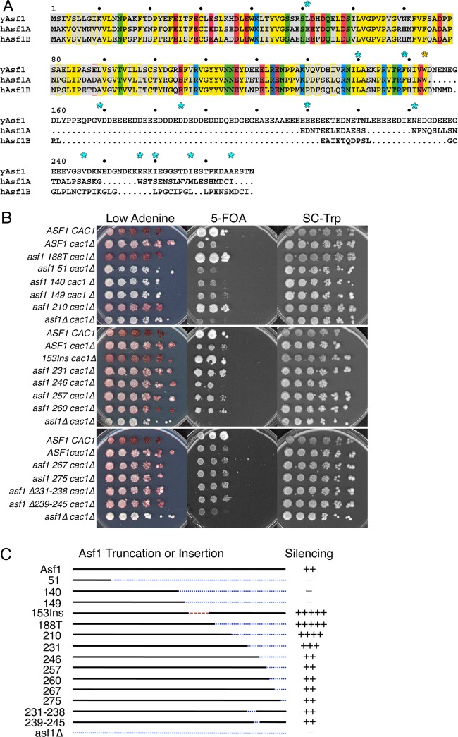
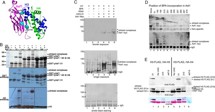
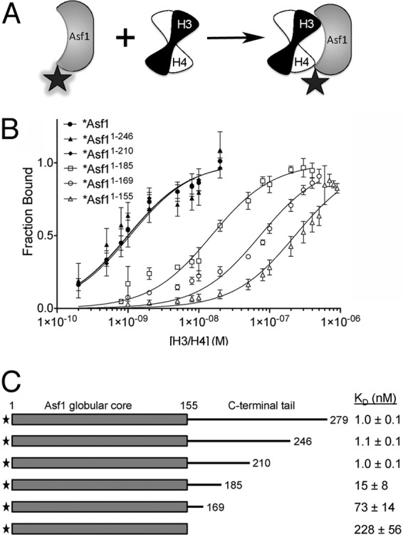
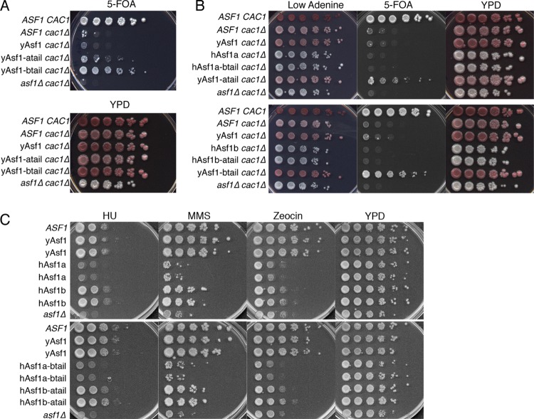
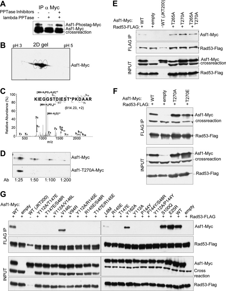
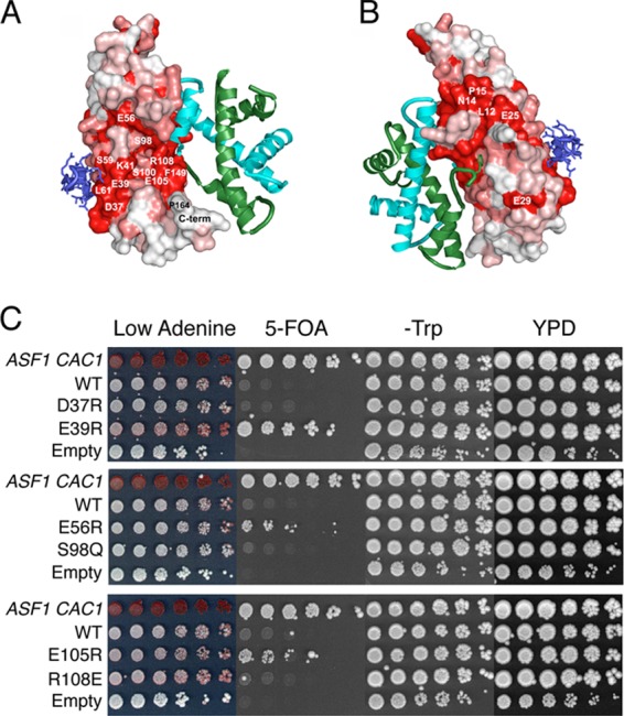
The central histone H3/H4 chaperone Asf1 comprises a highly conserved globular core and a divergent C-terminal tail. While the function and structure of the Asf1 core are well known, the function of the tail is less well understood. Here, we have explored the role of the yeast (yAsf1) and human (hAsf1a and hAsf1b) Asf1 tails in Saccharomyces cerevisiae. We show, using a photoreactive, unnatural amino acid, that Asf1 tail residue 210 cross-links to histone H3 in vivo and, further, that loss of C-terminal tail residues 211 to 279 weakens yAsf1-histone binding affinity in vitro nearly 200-fold. Via several yAsf1 C-terminal truncations and yeast-human chimeric proteins, we found that truncations at residue 210 increase transcriptional silencing and that the hAsf1a tail partially substitutes for full-length yAsf1 with respect to silencing but that full-length hAsf1b is a better overall substitute for full-length yAsf1. In addition, we show that the C-terminal tail of Asf1 is phosphorylated at T270 in yeast. Loss of this phosphorylation site does not prevent coimmunoprecipitation of yAsf1 and Rad53 from yeast extracts, whereas amino acid residue substitutions at the Asf1-histone H3/H4 interface do. Finally, we show that residue substitutions in yAsf1 near the CAF-1/HIRA interface also influence yAsf1's function in silencing.


Figures

Fig 1
Fig 1
Asf1 with an amber stop codon replacing residue 210 displays increased silencing over wild-type Asf1. (A) Alignment of human and yeast Asf1 proteins generated using multiple-sequence alignment (MSA) (61). The conserved globular core of Asf1 is shaded in gray. Conserved residues are shaded, and similar residues are colored the same, using blue (basic), red (acidic), yellow (hydrophobic), and green (polar). yAsf1 deletion endpoints later replaced with BPA are marked with cyan stars. All chimeric proteins used in this study contain the conserved core up to and including Trp153 (marked with a yellow star). (B) ROY1172, carrying an empty pRS314 vector (ASF1 CAC1), and ROY1169-based strains (asf1Δ cac1Δ), carrying either an empty pRS314 vector (asf1Δ cac1Δ) or a pRS314-Asf1-UAG-Myc plasmid encoding Asf1 proteins that are wild type or truncated at the indicated residues, were plated on the indicated media. (C) Summary of truncations and deletions with their silencing phenotypes, with minus signs indicating lack of silencing and plus signs indicating the relative strength of silencing.
Fig 2
Fig 2
Asf1 residue 210 with substitutions of BPA cross-links to histone H3. (A) Ribbon diagram of yAsf1 (aa 1 to 164) with H3 and H4 showing the proximity of yAsf1 residues Leu51, Leu140, and Phe149 (cyan) to H3. yAsf1 Trp153 is marked with an orange star. (B) Silver-stained gel (top) and Western blot analyses showing production of BPA-containing yAsf1 and cross-linking of Asf1-BPA-51 and Asf1-BPA-140 to histone H3 with UV treatment. E. coli transformed with a triple yAsf1-H3/H4 expression vector that harbors amber stop codons at position 51, 140, or 149 produced truncated yAsf1 in the absence of BPA. In the presence of BPA, full-length yAsf1 was observed. With UV treatment, cross-linked (xlinked) complexes that contain both yAsf1 and H3 appear. (C) yAsf1-Myc displays altered mobility (marked with an asterisk) only in the presence of photoactivatable artificial amino acid, BPA, the amber suppressor tRNA plasmid, and the Asf1-L51-UAG plasmid in yeast. Anti-myc immunoblot analysis of immunoprecipitated samples: in lanes 1 and 2, BKD094 (asf1Δ pRS314); in lanes 3 and 4, MCY001 (asf1Δ pRS314-Asf1-UAG-51); and in lanes 5 and 6, BKD0237 (asf1Δ pRS314-Asf1-UAG-51, pLH157-KAN). (D) Replacement of yAsf1 residues 51, 140, and 210 with BPA leads to cross-linked protein species. (Top) Anti-myc immunoblot analysis of extracts. (Bottom) Immunoprecipitated samples. The strains are BKD257 (None), BKD237 (51), BKD259 (140), BKD263 (149), BKD241 (210), BKD243 (231), BKD245 (246), BKD247 (249), BKD249 (257), BKD251 (260), BKD261 (267), and BKD255 (275). (E) yAsf1 residues 210 and 51, when replaced with BPA, cross-link to C-terminally FLAG-tagged histone H3. (Top) Immunoprecipitated yAsf1-Myc immunoblotted with anti-FLAG Ab. (Middle) Analysis of the same blot after being stripped and reprobed with anti-Myc Ab. (Bottom) Overlay of the anti-FLAG and anti-Myc Ab images. The blue/black bands indicate overlap between the Myc and FLAG signals. The HA and FLAG tags are not distinguishable by their electrophoretic mobilities. The identity of the Asf1-cross-linked central band, marked with a double asterisk, has not been established. Lanes 1 to 4, FLAG-H3 plus HA-H4; lanes 5 to 8, FLAG-H3 plus HA-H3. The strains are as follows: (lane 1) BKD382 (40), (lane 2) BKD378 (No UAG), (lane 3) BKD386 (210), (lane 4) BKD390 (asf1Δ), (lane 5) BKD379 (No UAG), (lane 6) BKD383 (51), (lane 7) BKD387 (210), and (lane 8) BKD391 (no Asf1). The yAsf1 signals seen in lanes 4 and 8 are due to interwell sample leakage.
Fig 3
Fig 3
A specific region on the yAsf1 tail enhances binding affinity to H3/H4. (A) Asf1 proteins were fluorophore labeled at a position not directly involved in binding to H3/H4 dimers. The fluorophore (shaded star) fluoresces strongly in the free Asf1 protein. However, the binding of the H3/H4 dimer to Asf1 results in quenching of this fluorescence (plain star). (B) Analysis of fluorescence-quenching data for titration of histones H3/H4 into 1 nM Alexa Fluor 532-labeled full-length yAsf1 (1 to 279) and C-terminal truncations (1-246, 1-210, 1-185, 1-169, and 1-155). The data were fitted with a ligand-depleted binding model (equation 1) or single site-binding isotherm (equation 2), depending on the final KD values. The error bars indicate standard deviations from three independent experiments. (C) Table of KD values aligned with a diagram showing yAsf1 C-terminal tail truncations demonstrating that residues 156 to 209 in the C-terminal tail of yAsf1 contribute to the binding affinity of yAsf1 for H3/H4.
Fig 4
Fig 4
Influence of hAsf1a and hAsf1b tails on the functions of the yAsf1 and hAsf1 cores. (A) Human tails enhance silencing at TELVIIL::URA3 in a cac1Δ background. Strains: ROY1172 (ASF1 CAC1), ROY1171 (ASF1 cac1Δ), BKD185 (yAsf1-myc cac1Δ), BKD198 (yAsf1-atail cac1Δ), BKD194 (yAsf1-btail cac1Δ), and ROY1169 (asf1Δ cac1Δ). (B) Influence of hAsf1a, hAsf1b, and hAsf1a/b and hAsf1b/a chimeras on silencing. (Top) ROY1172 (ASF1 CAC1), ROY1171 (ASF1 cac1Δ), BKD185 (yAsf1 cac1Δ), BKD200 (hAsf1a cac1Δ), BKD235 (hAsf1a-btail cac1Δ), BKD198 (yAsf1-atail cac1Δ), and ROY1169 (asf1Δ cac1Δ). (Bottom) ROY1172 (ASF1), ROY1171 (cac1Δ), BKD185 (yAsf cac1Δ), BKD202 (hAsf1b cac1Δ), BKD196 (hAsf1b-atail cac1Δ), BKD194 (yAsf1-btail cac1Δ), and ROY1169 (asf1Δ cac1Δ). (C) Sensitivity of hAsf1a, hAsf1b, and hAsf1a/b and hAsf11b/a chimeras to DNA-damaging and replicational-stress-inducing drugs (100 mM HU, 0.01% MMS, or 5 μg/ml zeocin). (Top) ROY1172 (ASF1), BKD187 (yAsf1), BKD188 (yAsf1), BKD135 (hAsf1a), BKD136 (hAsf1a), BKD130 (hAsf1b), BKD131 (hAsf1b), and ROY1170 (asf1Δ). (Bottom) ROY1172 (ASF1), BKD187 (yAsf1), BKD188 (yAsf1), BKD144 (hAsf1a-btail), BKD146 (hAsf1a-btail), BKD149 (hAsf1b-atail), BKD150 (hAsf1b-atail), and ROY1170 (asf1Δ).
Fig 5
Fig 5
Asf1 is phosphorylated on T270, and mutations in the Asf1-histone binding surface prevent Asf1-Rad53 coimmunoprecipitation. (A) yAsf1-Myc extracts from strain HZY1161, expressing FLAG-tagged Rad53 and containing pRS314-Asf1-Myc, were treated with and without λ phosphatase and phosphatase inhibitors. The immunoprecipitates were resolved by Mn2+ Phos-tag SDS-PAGE and analyzed by immunoblotting with anti-Myc antibodies. (B) Asf1 might be multiply phosphorylated. Shown is a 2D gel analysis of yAsf1-Myc immunoprecipitated from strain HZY1161 containing pRS314-Asf1-Myc and blotted with anti-Myc antibodies. Putative yAsf1 phosphorylation states are circled. (C) LC–MS-MS analysis of a tryptic digest of Asf1-myc immunoprecipitated from strain JKT200 carrying pRS314-Asf1-Myc. The assigned fragment ions are shown on the peptide sequence. (D) An affinity-purified antibody raised to phosphorylated yAsf1 T270 shows a signal on an immunoblot of whole-cell extracts of JKT200 with pRS314-Asf1-Myc, but not with pRS314-Asf1T270A-Myc, at dilutions from 1:50 to 1:200. (E and F) pRS314-Asf1-Myc plasmids or an empty-vector control (pRS314) was transformed into HZY1161 expressing FLAG-tagged Rad53. JKT0200 lacks ASF1 and FLAG-tagged Rad53. yAsf1-Myc was coimmunoprecipitated with Rad53-FLAG, and immoprecipitated samples were analyzed by immunoblotting. (E) Coimmunoprecipitation of WT or mutated yAsf1-Myc with Rad53-FLAG shows that mutations of yAsf1 T265 and/or T270 did not disrupt its association with Rad53. A cross-reacting protein that migrates more quickly than yAsf1-Myc (crossreaction), is detected by the anti-Myc Ab and is present in the absence of Asf1 (JKT0200). A more slowly migrating cross-reacting species is occasionally seen with the anti-FLAG Ab, and is present in the absence of Rad53-FLAG (JKT0200). (F) yAsf1 mutations to prevent or mimic phosphorylation of T270 do not abolish or enhance the association of yAsf1 and Rad53. (G) Asf1's histone binding region is important for Rad53 association. Coimmunoprecipitation of WT or mutated yAsf1-Myc with Rad53-FLAG was analyzed by immunoblotting.
Fig 6
Fig 6
Surface mutations on Asf1's conserved core display various degrees of silencing. (A and B) The Asf1-H3/H4 complex is shown docked with the B-domain peptide of HIRA (blue). Asf1 colored according to sequence conservation, with absolutely conserved residues (red), highly similar residues (pink), and the least-conserved residues (white) mapped onto the surface. (C) Wild-type strain ROY1172 (ASF1 CAC1) carrying an empty vector marked with TRP1 or ROY1169 (asf1Δ cac1Δ) carrying plasmid-borne copies of either WT or mutated Asf1 (strains BKD094, -095, -103, -104, -106, -107, -109, and -110) were serially diluted 10-fold onto the indicated media.

Similar articles

Cited by

References

    1. Luger K, Mader AW, Richmond RK, Sargent DF, Richmond TJ. 1997. Crystal structure of the nucleosome core particle at 2.8 A resolution. Nature 389: 251– 260 - PubMed
    1. Ransom M, Dennehey BK, Tyler JK. 2010. Chaperoning histones during DNA replication and repair. Cell 140: 183– 195 - PMC - PubMed
    1. Le S, Davis C, Konopka JB, Sternglanz R. 1997. Two new S-phase-specific genes from Saccharomyces cerevisiae. Yeast 13: 1029– 1042 - PubMed
    1. Singer MS, Kahana A, Wolf AJ, Meisinger LL, Peterson SE, Goggin C, Mahowald M, Gottschling DE. 1998. Identification of high-copy disruptors of telomeric silencing in Saccharomyces cerevisiae. Genetics 150: 613– 632 - PMC - PubMed
    1. Tyler JK, Adams CR, Chen SR, Kobayashi R, Kamakaka RT, Kadonaga JT. 1999. The RCAF complex mediates chromatin assembly during DNA replication and repair. Nature 402: 555– 560 - PubMed

Publication types

MeSH terms