Review
. 2012 Mar;1(2):149-158.
doi: 10.1002/adhm.201200011.

Emerging technologies for assembly of microscale hydrogels

Affiliations
Review

Emerging technologies for assembly of microscale hydrogels

Umut Atakan Gurkan et al. Adv Healthc Mater. 2012 Mar.

Abstract

Assembly of cell encapsulating building blocks (i.e., microscale hydrogels) has significant applications in areas including regenerative medicine, tissue engineering, and cell-based in vitro assays for pharmaceutical research and drug discovery. Inspired by the repeating functional units observed in native tissues and biological systems (e.g., the lobule in liver, the nephron in kidney), assembly technologies aim to generate complex tissue structures by organizing microscale building blocks. Novel assembly technologies enable fabrication of engineered tissue constructs with controlled properties including tunable microarchitectural and predefined compositional features. Recent advances in micro- and nano-scale technologies have enabled engineering of microgel based three dimensional (3D) constructs. There is a need for high-throughput and scalable methods to assemble microscale units with a complex 3D micro-architecture. Emerging assembly methods include novel technologies based on microfluidics, acoustic and magnetic fields, nanotextured surfaces, and surface tension. In this review, we survey emerging microscale hydrogel assembly methods offering rapid, scalable microgel assembly in 3D, and provide future perspectives and discuss potential applications.


Figures

Figure 1
Figure 1
Methods for directed assembly of microscale hydrogels for microphysiological 3D systems: microfluidic assembly, nano-ratchet assembly, acoustic assembly, magnetic assembly, and surface tension driven assembly.
Figure 2
Figure 2
Fabrication of microscale units for assembly: (A) Molding, (B) Continuous-flow lithography (top) and Photolithography (bottom), (C) Molecular synthesis, and (D) Folding. Reproduced with permission; A: from [209] Copyright 1999, AAAS; B: from[70] Copyright 2006, NPG; C: from [74] Copyright 2010, NPG;D: from [77] Copyright 2007, AIP.
Figure 3
Figure 3
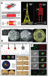
Directed assembly methods in literature for microscale hydrogels. (A) Microfluidic assembly. Reproduced with permission from[115]. Copyright 2008, NPG. (left) A schematic diagram for a microtrain (top), its cross-sectional view (middle), and design of a single microlatch component (bottom). (middle) Eiffel assembly, and (right) Skeleton assembly. (B) Assembly of microgels by acoustic fields in 2D and in 3D. Reproduced with permission from [42]. Copyright 2011, Elsevier. (left) Before and after acoustic excitation: single-layer formation (200 μm × 200 μm microgels). (right) New microgels were introduced onto a single layer to create a double-layer structure. (C) Assembly of microgels in 2D and 3D by magnetic fields. Reproduced with permission from[43]. (top) M-gels are collected from 3 different gel batches via magnetic forces to fabricate three-layer spheroids. First layer gels are stained with rhodamine-B; second layer gels are stained with FITC-dextran; third layer gels are stained with TPB (1,1,4,4-tetraphenyl-1,3-butadiene). (middle) Magnified image of the assembled single-layer 3D construct. (bottom) Images of arc- and dome-shaped constructs using a flexible surface and magnetic assembly. (D) Transport and assembly of microgels on ratchets. Reproduced with permission from [71]. Copyright 2011, AIP. (E) Surface tension driven self-assembly of microgels. Reproduced with permission from [46]. Copyright 2008, National Academy of Sciences. Directed assembly of lock-and-key-shaped microgels. Fluorescence images of cross-shaped microgels stained with FITC-dextran and rod-shaped microgels stained with Nile red. Phase-contrast and fluorescence images of lock-and-key assemblies with one, two, and three rods per cross. (bottom) Fluorescence images of assembly of microgels containing green- and red-stained cells. (Scale bars: 200 μm).

References

    1. Marga F, Neagu A, Kosztin I, Forgacs G. Birth Defects Res. C Embryo Today. 2007;81:320. - PubMed
    1. Langer R, Vacanti JP. Science. 1993;260:920. - PubMed
    1. Vunjak-Novakovic G, Tandon N, Godier A, Maidhof R, Marsano A, Martens TP, Radisic M. Tissue Eng. Part B-Rev. 2010;16:169. - PMC - PubMed
    1. Mikos AG, Herring SW, Ochareon P, Elisseeff J, Lu HH, Kandel R, Schoen FJ, Toner M, Mooney D, Atala A, Van Dyke ME, Kaplan D, Vunjak-Novakovic G. Tissue Eng. 2006;12:3307. - PMC - PubMed
    1. Davenport RJ. Science. 2005;309:84. - PubMed

Publication types

MeSH terms

LinkOut - more resources