. 2012 May;1(3):248-66.
doi: 10.1002/adhm.201200071. Epub 2012 Apr 5.

Biomaterials-based electronics: polymers and interfaces for biology and medicine

Affiliations

Biomaterials-based electronics: polymers and interfaces for biology and medicine

Meredith Muskovich et al. Adv Healthc Mater. 2012 May.

Abstract

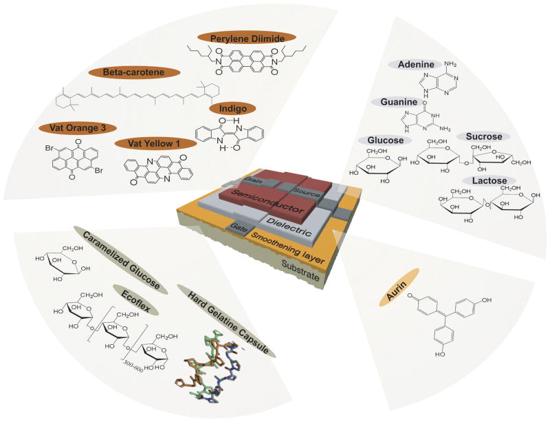
Advanced polymeric biomaterials continue to serve as a cornerstone for new medical technologies and therapies. The vast majority of these materials, both natural and synthetic, interact with biological matter in the absence of direct electronic communication. However, biological systems have evolved to synthesize and utilize naturally-derived materials for the generation and modulation of electrical potentials, voltage gradients, and ion flows. Bioelectric phenomena can be translated into potent signaling cues for intra- and inter-cellular communication. These cues can serve as a gateway to link synthetic devices with biological systems. This progress report will provide an update on advances in the application of electronically active biomaterials for use in organic electronics and bio-interfaces. Specific focus will be granted to covering technologies where natural and synthetic biological materials serve as integral components such as thin film electronics, in vitro cell culture models, and implantable medical devices. Future perspectives and emerging challenges will also be highlighted.


Figures

Figure 1
Figure 1
Naturally-derived biomaterials for organic electronic devices. Virtually all of the essential components of the organic thin film transistors can be composed of naturally-derived biomaterials. Semiconducting active layers can be fabricated from a variety of small molecule dyes and pigments. Hydrophilic biopolymers are perfectly suitable for gate dielectrics while a wide range of polymers, both natural and synthetic, are appropriate for use as flexible device substrates with additional capabilities such as compostability and bioabsorbability. Reproduced from M. Irimia-Vladu, P. A. Troshin, M. Reisinger, L. Shmygleva, Y. Kanbur, G. Schwabegger, M. Bodea, R. Schwödiauer, A. Mumyatov, J. W. Fergus, V. F. Razumov, H. Sitter, N. S. Sariciftci, S. Bauer, Advanced Functional Materials 2010, 20, 4069. Copyright Wiley-VCH Verlag GmbH & Co. KGaA. Reproduced with permission.
Figure 2
Figure 2
Melanin-like films exhibit unique optoelectronic properties. (a) The monomer used for the electrochemical synthesis of melanin-like films (5,6-dimethoxyindole-2-carboxylic acid, DMICA, left) closely resembles one of the key components of native eumelanins, 5,6-dihydroxyindole-2-carboxylic acid (DHICA, right). (b) Scanning electron micrographs of melanin-like films indicate nanophase morphology that may be useful as a structure to increase electrode surface area or promote cell adhesion. (c) Electrochromic PDMICA films are transparent when subjected to (i) negative voltages, (ii) green near 0 V, and (iii) purple above 0.5 V, which are confirmed by (d) shifts in the UV-vis spectra. Adapted with permission from L. K. Povlich, J. Le, J. Kim, D. C. Martin, Macromolecules 2010, 43, 3770. Copyright 2010. American Chemical Society.
Figure 3
Figure 3
Microfabricated organic electronic devices for precise delivery of neurotransmitters to modulate mammalian sensory function in vivo. The structure and principle of device operation are detailed at left. (a, left) The side view of the planar device used in small molecule neurotransmitter transport studies is shown. The black arrow indicates the flow of neurotransmitters from the source electrolyte, S, through the anode and over-oxidized channels, and finally out into the target electrolyte, T. (b, left), Side view showing the developmental progression from the planar device to the planar device with intermediate electrolyte (salt bridge). The white arrow indicates the flow of cations from T into the cathodic electrolyte. (c, left) The side view of the encapsulated device is shown with the arrow indicating ion flow. ( d, left) The top view of the encapsulated device, showing both electrolyte chambers and the requisite target system, T. The electrolyte reservoir tubes are 2 mm in outer diameter. The in vivo validation of the device is shown at right. (a-b, right) Briefly, the device is placed on the round window membrane of a guinea pig. The electronically activated release of neurotransmitters produces (c, right) a detectable shift in the auditory brainstem response. (d, right) The stimulated release of neurotransmitters can produce excitotic-induced damage to auditory dendrites (indicated by asterisks, ii, iv) compared to controls (i, iii). Adapted and reprinted by permission from Macmillan Publishers Ltd: Nature Materials ( D. T. Simon, S. Kurup, K. C. Larsson, R. Hori, K. Tybrandt, M. Goiny, E. W. H. Jager, M. Berggren, B. Canlon, A. Richter-Dahlfors, Nature Materials 2009, 8, 742), copyright (2009).
Figure 4
Figure 4
Elastomeric electronics for in vitro cell-device interfaces. (a) All polymer flexible microelectrode arrays with 28 microelectrodes and two reference electrodes were fabricated using a combination of soft lithography and electrodeposition for use in measuring spontaneous activity in neuronal lineages under biaxial deformation. ( b) Photograph of an organotypic hippocampal tissue slice on microelectrode arrays. Recording electrodes are indicated by circles with the electrode number indicated. Stimulating electrodes are indicated by squares. Frequency of spontaneous activity, Vrms, and Vpp before stretching (Pre), at 5% and at 10% equibiaxial strain, and after relaxation (Post); (c) Representative traces of spontaneous activity recorded on electrode 28 before stretching (Pre), at 5% and at 10% equibiaxial strain, and after relaxation (Post). The frequency of spontaneous activity is indicated. Adapted from O. Graudejus, B. Morrison, C. Goletiani, Z. Yu, S. Wagner, Advanced Functional Materials 2012, 22, 640. Copyright Wiley-VCH Verlag GmbH & Co. KGaA. Reproduced with permission.
Figure 5
Figure 5
Ashby diagram to identify ideal materials for electrically-active tissue-device interfaces for use in vitro or in vivo. The ideal material to generate reliable biotic-abiotic interfaces with soft tissue will exhibit simultaneous properties of high electrical conductivity and high mechanical compliance (dashed border). This optimal materials domain is currently inaccessible by virtue of hydrogels, polymers, or composites (unlabeled intermediate regions).
Figure 6
Figure 6
Highly conformal conducting polymer electrodes for in vivo recordings. (a) Schematic of the all polymer electrode arrays used in conjunction with silicon-based probes for in vivo cortical recording and (b) photograph showing the implantation. (c) Representative recordings from 25 electrodes in the polymer array and from 10 electrodes in the silicon probe measured from the surface and interior of the cortex, respectively. (d) Time-frequency (TF) analysis of the signals recorded by electrodes (black frames, x-axis: time, 10 min; y-axis: frequency, 0.1–50 Hz; color coding: power (dB) and their cross-spectrum coherences (open boxes, same axes as TF plots, color coding: coherence). Adapted from D. Khodagholy, T. Doublet, M. Gurfinkel, P. Quilichini, E. Ismailova, P. Leleux, T. Herve, S. Sanaur, C. Bernard, G. G. Malliaras, Advanced Materials 2011, 23, H268. Copyright Wiley-VCH Verlag GmbH & Co. KGaA. Reproduced with permission.
Figure 7
Figure 7
Biodegradable organic thin film transistors. (a) A combination of bio-inspired small molecule semiconductors and synthetic bioerodible/bioexcretable polymers can be assembled into (b) organic thin film transistor geometries. (c) These devices exhibit transfer characteristics, hole mobilities, and Ion-Ioff ratios that are suitable for a variety of simple operations that would be useful when integrated into temporary medical implants. Adapted from C. J. Bettinger, Z. Bao, Advanced Materials 2010, 22, 651. Copyright Wiley-VCH Verlag GmbH & Co. KGaA. Reproduced with permission.

References

    1. Levin M. Bio Essays. 2012;34:205.
    1. Zhao M, Song B, Pu J, Wada T, Reid B, Tai G, Wang F, Guo A, Walczysko P, Gu Y, Sasaki T, Suzuki A, Forrester JV, Bourne HR, Devreotes PN, McCaig CD, Penninger JM. Nature. 2006;442:457. - PubMed
    1. Irimia-Vladu M, Troshin PA, Reisinger M, Shmygleva L, Kanbur Y, Schwabegger G, Bodea M, Schwödiauer R, Mumyatov A, Fergus JW, Razumov VF, Sitter H, Sariciftci NS, Bauer S. Advanced Functional Materials. 2010;20:4017.
    1. Ito S. Pigment Cell Research. 2003;16:230. - PubMed
    1. Tran ML, Powell BJ, Meredith P. Biophysical Journal. 2006;90:743. - PMC - PubMed

Publication types

LinkOut - more resources