. 2013 Jan 11;288(2):1397-408.
doi: 10.1074/jbc.M112.396309. Epub 2012 Nov 26.

Identification of a His-Asp-Cys catalytic triad essential for function of the Rho inactivation domain (RID) of Vibrio cholerae MARTX toxin

Affiliations

Identification of a His-Asp-Cys catalytic triad essential for function of the Rho inactivation domain (RID) of Vibrio cholerae MARTX toxin

Sebastian Ahrens et al. J Biol Chem. .

Abstract

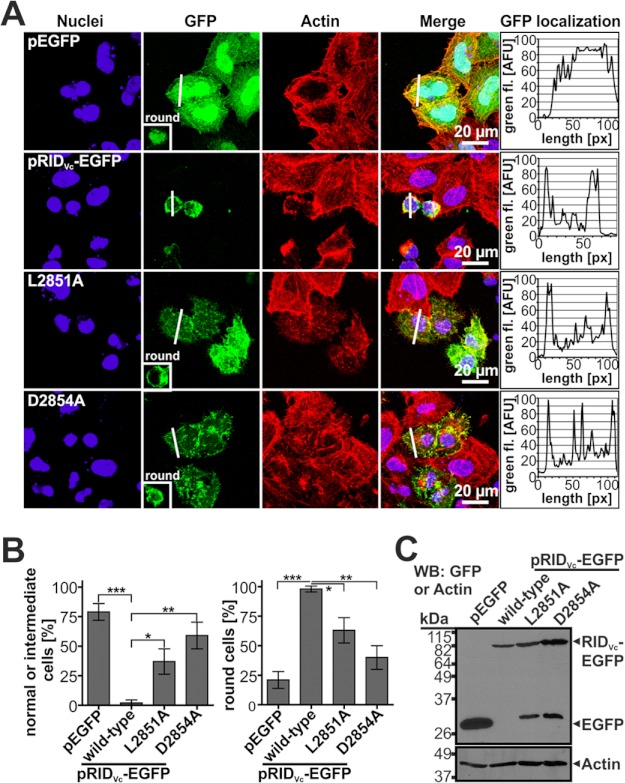
Vibrio cholerae is the causative agent of the severe diarrheal disease cholera. For V. cholerae to colonize the intestinal epithelium, accessory toxins such as the multifunctional autoprocessing repeats-in-toxin (MARTX(Vc)) toxin are required. MARTX toxins are composite toxins comprised of arrayed effector domains that carry out distinct functions inside the host cell. Among the three effector domains of MARTX(Vc) is the Rho inactivation domain (RID(Vc)) known to cause cell rounding through inactivation of small RhoGTPases. Using alanine scanning mutagenesis in the activity subdomain of RID(Vc), four residues, His-2782, Leu-2851, Asp-2854, and Cys-3022, were identified as impacting RID(Vc) function in depolymerization of the actin cytoskeleton and inactivation of RhoA. Tyr-2807 and Tyr-3015 were identified as important potentially for forming the active structure for substrate contact but are not involved in catalysis or post translational modifications. Finally, V. cholerae strains modified to carry a catalytically inactive RID(Vc) show that the rate and efficiency of MARTX(Vc) actin cross-linking activity does not depend on a functional RID(Vc), demonstrating that these domains function independently in actin depolymerization. Overall, our results indicate a His-Asp-Cys catalytic triad is essential for function of the RID effector domain family shared by MARTX toxins produced by many Gram-negative bacteria.


Figures

FIGURE 1.
FIGURE 1.
Alignment of RID effector domains from various bacterial species show high similarity in N- and C-terminal regions. A, shown is a diagram of the RIDVc effector from the CPD cleavage sites 2434 to 3085 in MARTXVc. Location of the 4HBM membrane localization domain is shown. Numbering is based on annotation of the rtxA gene as determined by Lin et al. (35). Shaded regions represent regions of the AD with >30% identity across the 10 aligned RID proteins. B, shown is ClustalW alignment of the AD of 10 known or predicted RID effectors. Gray shading indicates 70% identical residues, whereas boxed residues denote the six residues identified in this study as defective for RhoA inactivation and cell rounding when changed to Ala. Asterisks label residues that were targeted for alanine mutagenesis but did not cause a defect in cell rounding. AD sequences in alignment are ordered top to bottom by percent similarity to RIDVc and are based on deduced protein sequences from publically available genome sequences at GenBankTM or UniprotKB of V. cholerae (Vcho, AAD21057.1); V. vulnificus (Vvul, Q8D6P9); V. anguillarum (Vang, B1NY97); Vibrio caribbenthicus (Vcar, E3BQK8); V. nigripulchritudo (Vnig, F0V1C5); P. mirabilis (Pmir1 and Pmir2, B4F0Y5); Photorhabudus luminescens subsp. laumondii (Plum, 2801318); Xenorhabdus bovienii (Xbov, D3UXB8), and Xenorhabdus nematophila (Xnem, D3VAK8). Large gaps in sequence for Pmir1 and Vnig were removed to improve alignment where indicated.
FIGURE 2.
FIGURE 2.
The activity domain of RIDVc is the smallest functional unit to induce cell rounding. HeLa cells were transient-transfected with pEGFP-N3 (A), pRIDVc-EGFP wild-type (pKS113) (B), pΔMD-RIDVc-EGFP (pSA41) (C), and pΔCTD-RIDVc-EGFP (pSA42) (D) fusion proteins as indicated. E, shown is Western blotting (WB) analysis of EGFP fusion-protein expression from transfected HeLa cells. Detection of actin was used as the loading control.
FIGURE 3.
FIGURE 3.
RIDVc mutants H2782A and C3022A prevent cell rounding in HeLa cells. A, HeLa cells were transfected with pRIDVc-EGFP (pKS113) expressing wild-type or mutant RIDVc fusion proteins as indicated. Cells were stained with rhodamine-phalloidin and DAPI. Histograms in the right panel display green fluorescence in arbitrary units (AFU) of a cross-section (white bar, length in pixels ([px])) indicating membrane localization. Insets represent the phenotype of round cells for RIDVc mutants and pEGFP-N3 control. B, shown is quantification of cell morphology based on a total of 100–300 EGFP-positive cells. Data represent the mean and S.D. of three independent transfections. C, shown is Western blotting analysis of EGFP fusion-protein expression from transfected HeLa cells. Detection of actin was used as loading control. ***, p ≤ 0.0001.
FIGURE 4.
FIGURE 4.
RIDVc mutants L2851A and D2854A cause intermediate cell rounding in HeLa cells. A, HeLa cells were transfected with pRIDVc-EGFP (pKS113) expressing wild-type or mutant RIDVc-EGFP fusion proteins as indicated. Cells were stained with rhodamine-phalloidin and DAPI. Histograms in the right panel display green fluorescence in arbitrary units (AFU) of a cross section (white bar, length in pixels ([px])) indicating membrane localization. Insets represent the phenotype of round cells for RIDVc mutants and pEGFP-N3 control. B, shown is quantification of cell morphology based on a total of 100–300 EGFP-positive cells. Data represent the mean and S.D. of three independent transfections. Intermediate phenotype refers to cells that are shrunken but normal in shape compared with normally sized and shaped pEPGF-N3-transfected cells. C, shown is Western blotting (WB) analysis of EGFP fusion-protein expression from transfected HeLa cells. Detection of actin was used as loading control. ***, p ≤ 0.0001; **, p ≤ 0.001; *, p ≤ 0.01.
FIGURE 5.
FIGURE 5.
YA but not YF RIDVc mutants prevent cell rounding. A, HeLa cells expressing pRIDVc-EGFP (pKS113) wild-type or Tyr-mutant RIDVc-EGFP fusion proteins as indicated were stained with rhodamine-phalloidin and DAPI. Insets represent round cells observed in transfections with YA RIDVc-EGFP mutants or pEGFP-N3 control. B, Western blotting (WB) analysis of EGFP fusion-protein expression from transfected HeLa cells is shown. Detection of actin was used as loading control.
FIGURE 6.
FIGURE 6.
Point mutants do not alter the overall structure of RIDVc. A, shown is SDS-PAGE of 2 μg of purified recombinant LFNRIDVc wild-type or mutant proteins. B–D, shown are CD spectra of 0.5 μm LFN fusion proteins. The spectra shown were averaged from five separate readings for two independent samples.
FIGURE 7.
FIGURE 7.
RIDVc alanine point mutants prevent inactivation of RhoA. HeLa cells treated with 28 nm PA plus 12 nm of the indicated LFN fusion proteins for 4 h were stained with rhodamine-phalloidin and DAPI and visualized by confocal microscopy (A) or lysed and assayed for RhoA-GTP (A490) using RhoA G-LISATM assays (B–D). Western blot detection of total recovered RhoA from cell lysates is shown below the bar diagrams. ***, p ≤ 0.0001; **, p ≤ 0.001; *, p ≤ 0.01; n.s., not significant.
FIGURE 8.
FIGURE 8.
H2782A and C3022A prevent cell rounding and RhoA inactivation in the context of V. cholerae infections. HeLa cells treated with V. cholerae CCO5 (rtxA+Δacd), KFV92 (ΔrtxA), SAHV2 (CCO5rtxAH2782A), or BGV1 (CCO5rtxAC3022A) or mock-treated with PBS were stained with rhodamine-phalloidin and DAPI and visualized by confocal microscopy (A) or lysed and assayed for RhoA-GTP (A490) using RhoA G-LISATM assays (B). Western blot (WB) detection of total recovered RhoA from cell lysates is shown below the bar diagrams. ***, p ≤ 0.0001.
FIGURE 9.
FIGURE 9.
Actin cross-linking efficiency of MARTXVc does not depend on functional RIDVc. HeLa cells treated with the indicated V. cholerae strains KFV119 (rtxA+), KFV92 (ΔrtxA), and SAHV1 (KFV119rtxAH2782A) or mock-treated with PBS were sampled at different time points and analyzed by Western blotting using monoclonal anti-actin antibody to monitor actin cross-linking and anti-α-tubulin antibody as loading control. M, actin monomer (42 kDa); D, actin dimer (84 kDa); T, actin trimer (126 kDa); Q, actin quatramer (168 kDa); MM, actin multimers (>180 kDa).

References

    1. Ma A. T., McAuley S., Pukatzki S., Mekalanos J. J. (2009) Translocation of a Vibrio cholerae type VI secretion effector requires bacterial endocytosis by host cells. Cell Host. Microbe 5, 234–243 - PMC - PubMed
    1. Olivier V., Queen J., Satchell K. J. (2009) Successful small intestine colonization of adult mice by Vibrio cholerae requires ketamine anesthesia and accessory toxins. PLoS One 4, e7352. - PMC - PubMed
    1. Queen J., Satchell K. J. (2012) Neutrophils are essential for containment of Vibrio cholerae to the intestine during the proinflammatory phase of infection. Infect Immun. 80, 2905–2913 - PMC - PubMed
    1. Satchell K. J. (2011) Structure and function of MARTX toxins and other large repetitive RTX proteins. Annu. Rev. Microbiol. 65, 71–90 - PubMed
    1. Egerer M., Satchell K. J. (2010) Inositol hexakisphosphate-induced autoprocessing of large bacterial protein toxins. PLoS Pathog. 6, e1000942. - PMC - PubMed

Publication types

LinkOut - more resources