. 2012 Dec 11;109(50):E3395-404.
doi: 10.1073/pnas.1217982109. Epub 2012 Nov 26.

Stromal epigenetic dysregulation is sufficient to initiate mouse prostate cancer via paracrine Wnt signaling

Affiliations

Stromal epigenetic dysregulation is sufficient to initiate mouse prostate cancer via paracrine Wnt signaling

Yang Zong et al. Proc Natl Acad Sci U S A. .

Abstract

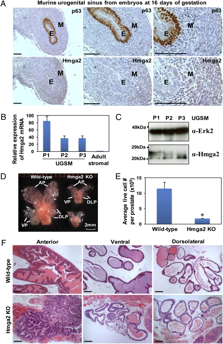
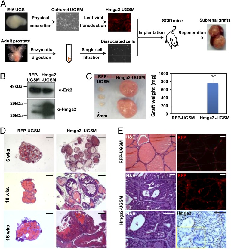
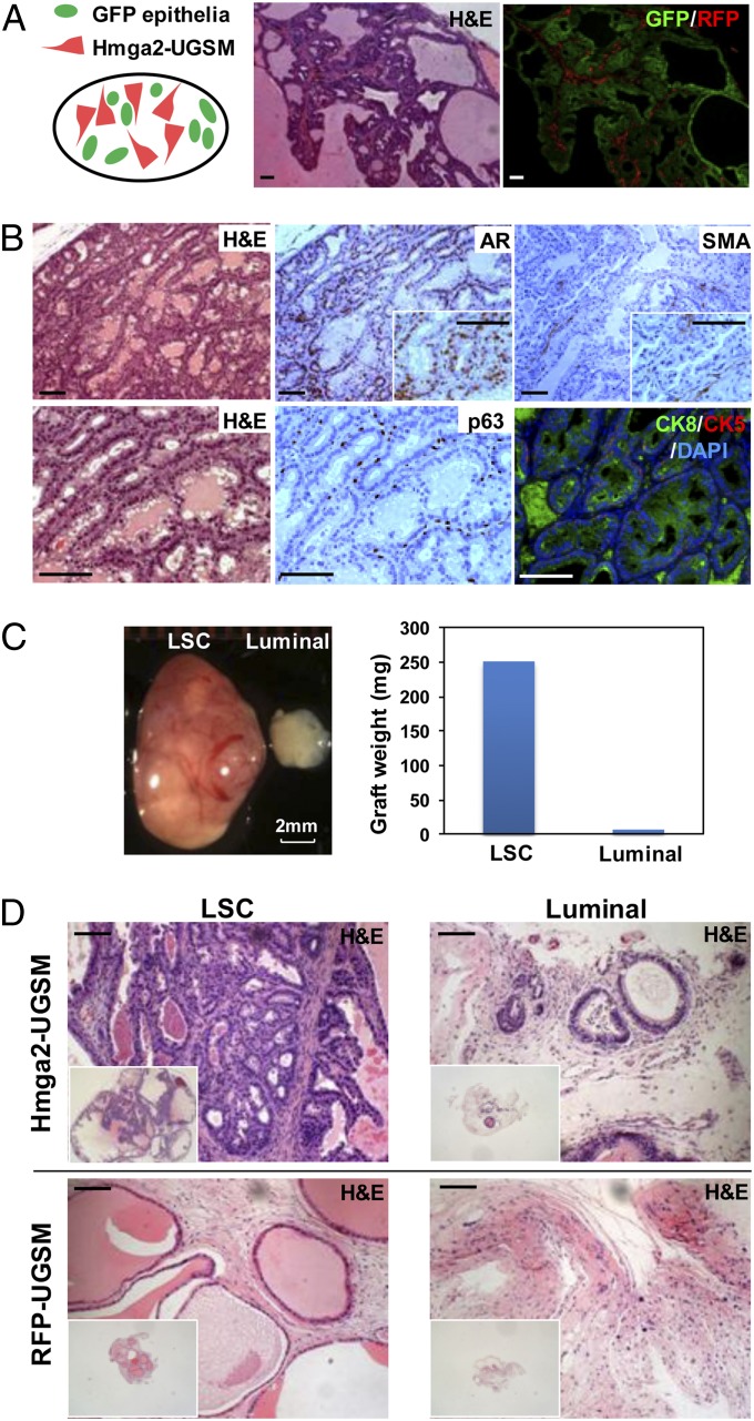
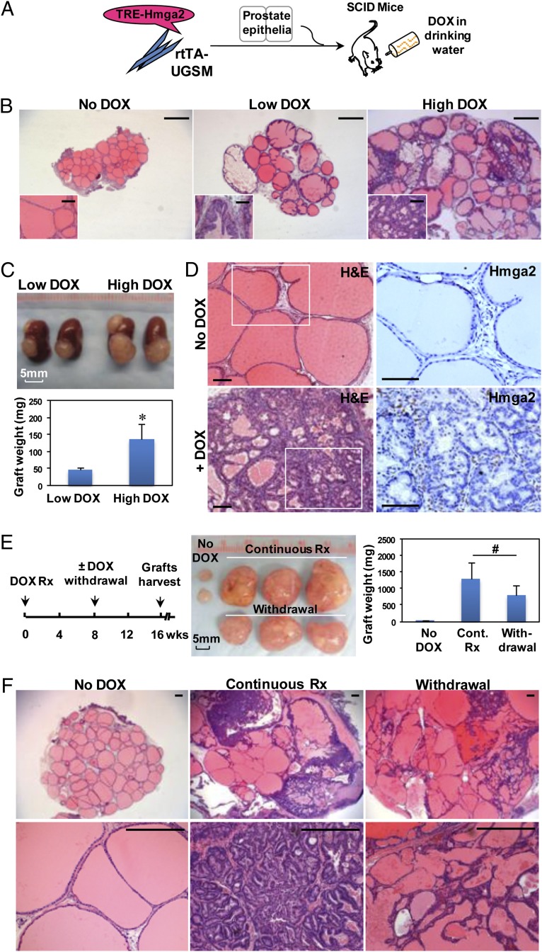
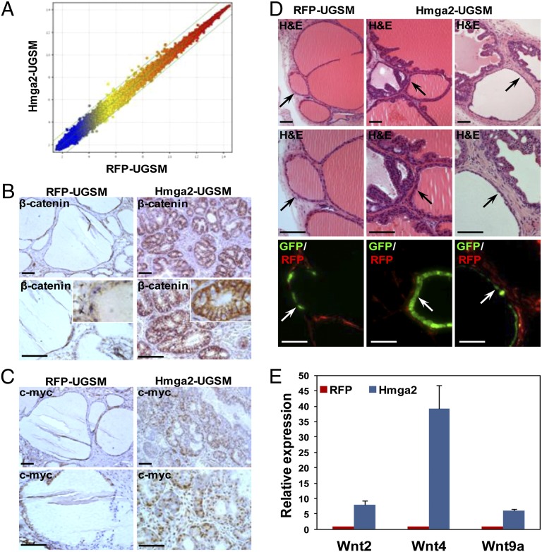
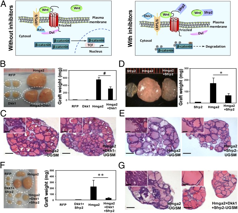
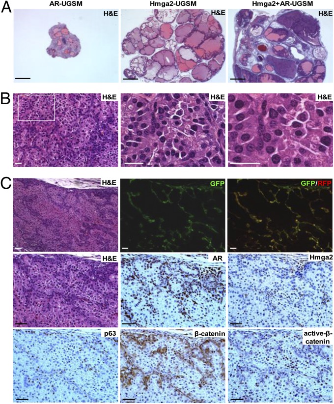
Carcinomas most often result from the stepwise acquisition of genetic alterations within the epithelial compartment. The surrounding stroma can also play an important role in cancer initiation and progression. Given the rare frequencies of genetic events identified in cancer-associated stroma, it is likely that epigenetic changes in the tumor microenvironment could contribute to its tumor-promoting activity. We use Hmga2 (High-mobility group AT-hook 2) an epigenetic regulator, to modify prostate stromal cells, and demonstrate that perturbation of the microenvironment by stromal-specific overexpression of this chromatin remodeling protein alone is sufficient to induce dramatic hyperplasia and multifocal prostatic intraepithelial neoplasia lesions from adjacent naïve epithelial cells. Importantly, we find that this effect is predominantly mediated by increased Wnt/β-catenin signaling. Enhancement of Hmga2-induced paracrine signaling by overexpression of androgen receptor in the stroma drives frank murine prostate adenocarcinoma in the adjacent epithelial tissues. Our findings provide compelling evidence for the critical contribution of epigenetic changes in stromal cells to multifocal tumorigenesis.


Conflict of interest statement

The authors declare no conflict of interest.

Figures

Fig. 1.
Fig. 1.
Temporospatial expression of Hmga2 in murine developing UGS mesenchyme. (A) IHC analysis of E16 murine UGS with p63 and Hmga2 antibodies. E, epithelia; M, mesenchyme. (Scale bars, 100 µm.) (B) Relative expression of Hmga2 mRNA in cultured UGSM cells with different passage determined by quantitative PCR, with comparison with that in FACS-sorted adult prostate stromal (Lin-CD49f-Sca-1+) cells. Data are presented as mean ± SD after normalization with Gapdh expression. (C) Immunoblotting of UGSM cells with anti-Hmga2 and Erk2 as a loading control. (D) Representative photograph of prostate glands from 11-wk-old wild-type and Hmga2 knockout (KO) mice. AP, anterior prostate; VP, ventral prostate; DLP, dorsolateral prostate. (E) Comparison of live cells in dissociated prostate glands from wild-type and Hmga2 KO mice, determined by trypan blue exclusion and represented as mean ± SD. *P < 0.05. (F) Histological analysis of different lobes of prostate from Hmga2 KO mice and the aged-matched normal control. H&E staining. (Scale bars, 100 µm.)
Fig. 2.
Fig. 2.
Overexpression of Hmga2 in the stroma leads to the formation of multifocal high-grade PIN lesions. (A) Schematic design for the dissociated prostate regeneration assay. Dissociated prostate cells from wild-type adult mice were combined with lentiviral-transduced UGSM cells that were originally derived from E16 mouse UGS mesenchyme and engrafted into the subrenal capsules of male immunodeficient mice for in vivo regeneration. (B) Western blotting of Hmga2-transduced UGSM cells. (C) Representative prostate grafts derived from wild-type adult murine prostate cells and Hmga2-UGSM cells, compared with control grafts. Data are presented as mean ± SD. **P < 0.01. (D) Histological sections of prostate regenerated tissues harvested at the indicated time points after engraftment. H&E staining. (Scale bars, 1 mm.) (E) Hmga2 staining with corresponding H&E and fluorescent (RFP) images of prostate regenerated tissue sections. (Scale bars, 100 µm for main images, 50 µm for Inset.)
Fig. 3.
Fig. 3.
Hmga2-UGSM cells induced PIN lesions are of adult prostate epithelial origin, and prostate basal stem cells preferentially respond to the growth-promoting effects of Hmga2-modified stroma. (A) Representative histological section and corresponding fluorescent photo of prostate grafts derived from Hmga2-UGSM cells and adult prostate cells from GFP transgenic mice. (B) IHC analysis of Hmga2-UGSM grafts with the indicated antibodies. (Scale bars, 100 µm.) (C) Representative photograph and weight measurement of Hmga2-UGSM grafts derived from prostate stem cells-enriched (LSC) basal cells or luminal epithelial cells. (D) H&E staining of Hmga2-UGSM grafts and RFP-UGSM grafts derived from LSC (basal-enriched) cells or luminal epithelial cells. (Scale bars, 100 µm.)
Fig. 4.
Fig. 4.
Devolvement and maintenance of PIN lesions are dependent on continuous expression of Hmga2 in the stroma. (A) Diagram of the approach used to assess the requirement of stromal Hmga2 for the maintenance of PIN lesions. The rtTA-expressing UGSM cells were infected with DOX-inducible Hmga2 lentivirus (TRE-Hmga2). The in vivo regeneration assay was performed using dissociated adult normal prostate cells in the absence or presence of systemic administration of DOX. TRE, tetracycline responsive element; rtTA, reverse tetracycline-controlled transactivator. (B) Histology of prostate grafts with inducible expression of stromal Hmga2 from recipient mice treated for 8 wk with two concentrations of DOX (Low DOX: 0.4 mg/mL; High DOX: 2 mg/mL). H&E staining. (Scale bars, 1 mm for main images, 100 µm for Insets.) (C) Photograph and weight measurement of representative subrenal grafts from recipient mice treated with two concentrations of DOX. Data are presented as mean ± SD. *P < 0.05. (D) Hmga2 staining of prostate regenerated tissues from DOX-uninduced mice or mice treated with 2 mg/mL DOX for 8 wk. (Scale bars, 100 µm.) (E and F) Gross appearance, graft weight, and H&E staining of prostate regenerated tissues from recipients in various treatment conditions as indicated. Data are presented as mean ± SD. #P > 0.05. (Scale bars, 300 µm.)
Fig. 5.
Fig. 5.
Augmented Wnt/β-catenin signaling in murine PIN lesions induced by Hmga2-modified stromal cells. (A) Scatter plot for expression ratios of the global gene profiles between Hmga2-UGSM cells and control cells. Each dot represents the average fold change for individual genes summarized from three independent microarrays. (B and C) IHC analysis of prostate grafts with β-catenin and c-Myc antibodies. (Scale bars, 100 µm.) (D) Histological sections (H&E staining) and corresponding merged fluorescent images of regenerated tissues derived from Cre-transduced prostate cells from β-cateninfl/fl mice. Arrows indicate the Cre/GFP lentiviral transduced prostate tubules. (Scale bars, 100 µm.) (E) Quantitative PCR analyses of Wnt ligands differentially expressed between Hmga2-UGSM cells and RFP-UGSM control samples. Data are presented as mean ± SD after normalization with β-actin.
Fig. 6.
Fig. 6.
Forced expression of two Wnt inhibitors in the stroma significantly suppresses Hmga2-UGSM induced PIN lesions. (A) Schematic representation of Wnt signaling pathway in the absence or presence of Dkk1 and Sfrp2. (B and C) Gross appearance, weight, and H&E staining of prostate grafts with stromal overexpression of Dkk1 and Hmga2. Data are presented as mean ± SD. #P > 0.05. (D and E) Representative photograph and histological sections of regenerated tissues derived from Hmga2 or Hmga2+Sfrp2-UGSM cells. Data are presented as mean ± SD. *P < 0.05. (F and G) Macroscopic overview, weight, and H&E staining of prostate grafts with stromal co-overexpression of Dkk1, Sfrp2, and Hmga2. Data are expressed as mean ± SD. **P < 0.01. (Scale bars, 1 mm for main images, 200 µm for Insets.)
Fig. 7.
Fig. 7.
Combined expression of Hmga2 and AR in the stroma results in the development of invasive prostate cancer. (A) Representative histological sections of prostate grafts with stromal coexpression of Hmga2 and AR. (Scale bars, 1 mm.) (B) H&E staining of tissue recombinants consisting of Hmga2+AR-UGSM and wild-type prostate cells. (Scale bars, 25 µm.) (C) IHC analysis of prostate grafts derived from Hmga2+AR-UGSM cells with various antibodies as indicated, with corresponding H&E staining and fluorescent images. (Scale bars, 50 µm.)

Similar articles

Cited by

References

    1. Siegel R, Naishadham D, Jemal A. Cancer statistics, 2012. CA Cancer J Clin. 2012;62(1):10–29. - PubMed
    1. Andreoiu M, Cheng L. Multifocal prostate cancer: Biologic, prognostic, and therapeutic implications. Hum Pathol. 2010;41(6):781–793. - PubMed
    1. Slaughter DP, Southwick HW, Smejkal W. Field cancerization in oral stratified squamous epithelium; clinical implications of multicentric origin. Cancer. 1953;6(5):963–968. - PubMed
    1. Bhowmick NA, et al. TGF-beta signaling in fibroblasts modulates the oncogenic potential of adjacent epithelia. Science. 2004;303(5659):848–851. - PubMed
    1. Memarzadeh S, et al. Enhanced paracrine FGF10 expression promotes formation of multifocal prostate adenocarcinoma and an increase in epithelial androgen receptor. Cancer Cell. 2007;12(6):572–585. - PMC - PubMed

Publication types

MeSH terms

LinkOut - more resources