. 2012 Dec 11;109(50):20407-12.
doi: 10.1073/pnas.1209565109. Epub 2012 Nov 26.

Alternative bacterial two-component small heat shock protein systems

Affiliations

Alternative bacterial two-component small heat shock protein systems

Alexander Bepperling et al. Proc Natl Acad Sci U S A. .

Abstract

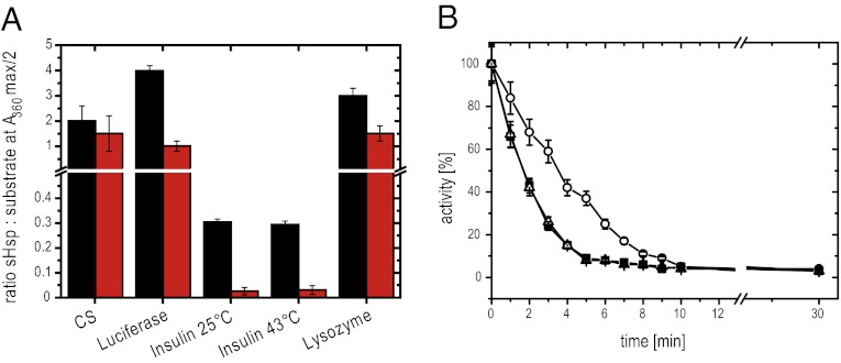
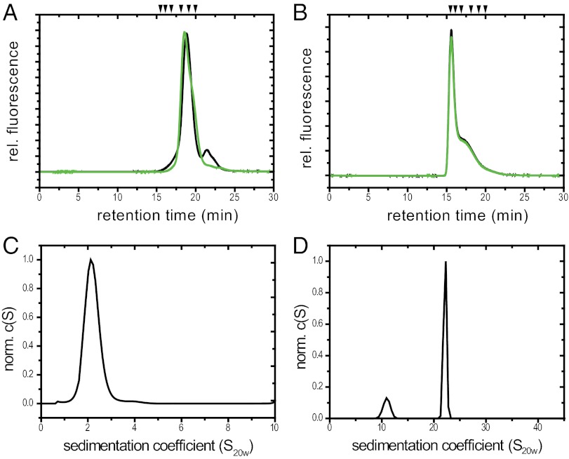
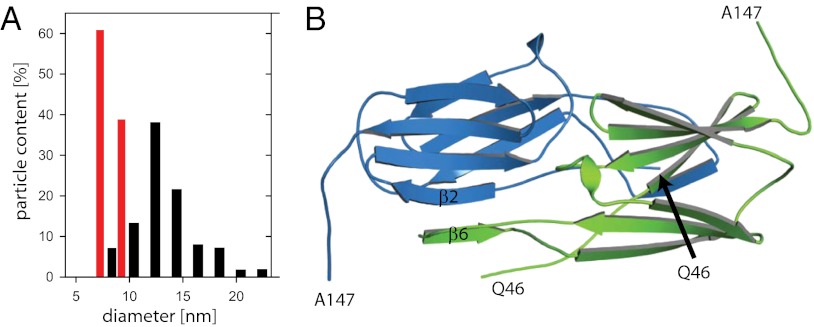
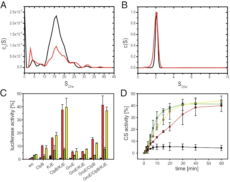
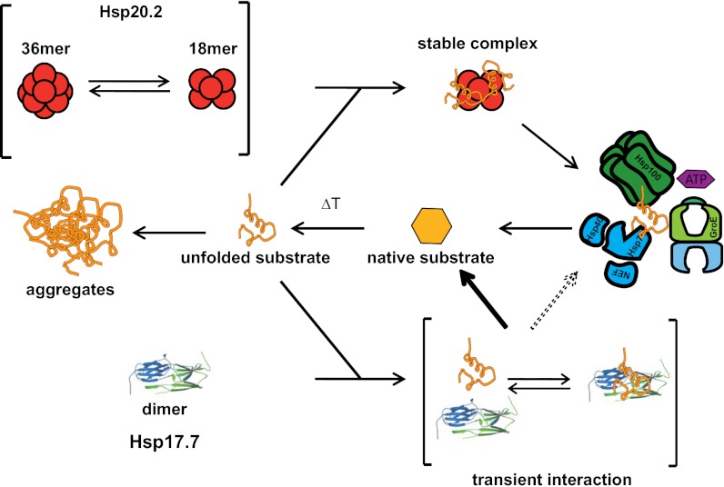
Small heat shock proteins (sHsps) are molecular chaperones that prevent the aggregation of nonnative proteins. The sHsps investigated to date mostly form large, oligomeric complexes. The typical bacterial scenario seemed to be a two-component sHsps system of two homologous sHsps, such as the Escherichia coli sHsps IbpA and IbpB. With a view to expand our knowledge on bacterial sHsps, we analyzed the sHsp system of the bacterium Deinococcus radiodurans, which is resistant against various stress conditions. D. radiodurans encodes two sHsps, termed Hsp17.7 and Hsp20.2. Surprisingly, Hsp17.7 forms only chaperone active dimers, although its crystal structure reveals the typical α-crystallin fold. In contrast, Hsp20.2 is predominantly a 36mer that dissociates into smaller oligomeric assemblies that bind substrate proteins stably. Whereas Hsp20.2 cooperates with the ATP-dependent bacterial chaperones in their refolding, Hsp17.7 keeps substrates in a refolding-competent state by transient interactions. In summary, we show that these two sHsps are strikingly different in their quaternary structures and chaperone properties, defining a second type of bacterial two-component sHsp system.


Conflict of interest statement

The authors declare no conflict of interest.

Figures

Fig. 1.
Fig. 1.
Comparison of chaperone activity of Hsp17.7 and Hsp20.2. (A) Influence of Hsp17.7 (black) and Hsp20.2 (red) on the thermal aggregation of different substrate proteins. The ratio of sHsp to substrate at half-maximum suppression of aggregation is displayed. Error bars represent the SD in three independent assays. (B) Influence of Hsp17.7 and Hsp20.2 on the thermal inactivation of CS. Aliquots from a thermostated solution (43 °C) of 75 nM CS (filled squares) and 75 nM CS in the presence of 600 nM Hsp17.7 (open circles) or 600 nM Hsp20.2 (open triangles) were extracted at the indicated time points, and the enzymatic activity was determined (37). Mean values of three independent assays and the respective SD are indicated.
Fig. 2.
Fig. 2.
Quaternary structure of Hsp17.7 and Hsp 20.2. (A and B) SEC-HPLC was performed using a TosoHaas TSK 4000 PW column as described in Materials and Methods. Hsp17.7 (A) and Hsp20.2 (B) at a concentration of 1.5 mg/mL were separated at 25 °C (green) and at 43 °C (black). Arrows indicate calibration markers as described in Materials and Methods. (C and D) Analytical ultracentrifugation sedimentation analysis. Normalized c(S) distribution of Hsp17.7 at 1.0 mg/mL (C) and Hsp20.2 at 1.5 mg/mL (D).
Fig. 3.
Fig. 3.
Structure of Hsp20.2 and Hsp17.7. (A) Size distributions (circumscribing diameters, in nm) of Hsp20.2 oligomers (black) and Hsp17.7 (red), based on electron microscopic images from negatively stained particles. (B) Crystal structure of Hsp17.7 (Protein Data Bank ID code 4FEI). Ribbon diagram of the Hsp17.7 dimer. The monomers are colored in green and blue. The N-terminal 45 and the C-terminal 19 amino acids are not shown as they are structurally distorted.
Fig. 4.
Fig. 4.
Analysis of substrate binding and release of Hsp17.7 and Hsp20.2. (A) Multisignal sedimentation velocity analysis of substrate binding to Hsp20.2. ck(S) distribution of 10 µM lysozyme + 1 mM Tris(2-carboxyethyl)phosphine + 20 µM Hsp20.2 calculated from parallel scans with absorbance and interference optics. The contribution of Hsp20.2 (black) and lysozyme (red) to the total signal was deconvoluted using the multiwavelength discrete/continuous distribution analysis of SEDPHAT. (B) c(S) distribution of 1 µM Hsp17.7-Alexa 488 (black) and 10 µM Hsp17.7-Alexa 488 + 100 µM Hsp20.2 (red). (C) Effects of Hsp17.7 and Hsp20.2 on the refolding efficiency of heat-denatured luciferase by the KJE, ClpB, and GroE chaperone machineries. Luciferase (80 nM) was incubated at 43 °C for 10 min in the absence (black) of sHsps or in the presence of 600 nM Hsp20.2 (red), 600 nM Hsp17.7 (green), or 600 nM of Hsp20.2 and Hsp17.7 (yellow). After complete inactivation, the samples were shifted to 25 °C and subsequently DnaK/DnaJ/GrpE (KJE, 0.6, 1.2, and 0.6 µM), ClpB (600 nM), and GroE (600 nM GroEL and GroES) were added in different combinations as indicated. Luciferase activity was assayed after 60 min in comparison with 80 nM untreated luciferase (100% value). Error bars represent the SD in three independent assays. (D) CS was denatured for 1 h in 6 M GdmCl and diluted 200-fold to a final concentration of 300 nM in a solution containing DnaK/DnaJ/GrpE (2, 2, and 0.5 µM), 2 mM ATP, and 3 mM MgCl2 (black) supplemented with 1.2 µM Hsp17.7 (green), 1.2 µM Hsp20.2 (red), or a mixture of 0.6 µM Hsp17.7 and 0.6 µM Hsp20.2 (yellow). Aliquots were withdrawn at the indicated time points, and the enzymatic activity was determined (37). Mean values of three independent assays and the respective SDs are indicated.
Fig. 5.
Fig. 5.
Model of the D. radiodurans two-component sHsp system. Dimeric Hsp17.7 and the ensemble of 36 and 18meric Hsp20.2 form the D. radiodurans two-component sHsp system. Stress conditions, like heat shock, lead to the unfolding of substrate proteins (orange). Unfolding proteins are stably trapped by Hsp20.2 18mers or transiently bound by Hsp17.7. Hsp104 and Hsp70 or GroE can reactivate the Hsp20.2-bound substrate proteins in an energy-dependent reaction or refold the substrate proteins spontaneously released from Hsp17.7. This indicates that Hsp20.2 and Hsp17.7 work in parallel and independently of each other.

References

    1. McHaourab HS, Godar JA, Stewart PL. Structure and mechanism of protein stability sensors: Chaperone activity of small heat shock proteins. Biochemistry. 2009;48(18):3828–3837. - PMC - PubMed
    1. Haslbeck M, Franzmann T, Weinfurtner D, Buchner J. Some like it hot: The structure and function of small heat-shock proteins. Nat Struct Mol Biol. 2005;12(10):842–846. - PubMed
    1. Liberek K, Lewandowska A, Zietkiewicz S. Chaperones in control of protein disaggregation. EMBO J. 2008;27(2):328–335. - PMC - PubMed
    1. Basha E, O'Neill H, Vierling E. Small heat shock proteins and alpha-crystallins: dynamic proteins with flexible functions. Trends Biochem Sci. 2012;37(3):106–117. - PMC - PubMed
    1. Richter K, Haslbeck M, Buchner J. The heat shock response: Life on the verge of death. Mol Cell. 2010;40(2):253–266. - PubMed

Publication types

MeSH terms

Associated data

LinkOut - more resources