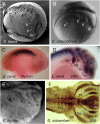
Variation in the schedules of somite and neural development in frogs
- PMID: 23184997
- PMCID: PMC3528599
- DOI: 10.1073/pnas.1219307110
Variation in the schedules of somite and neural development in frogs
Abstract
The timing of notochord, somite, and neural development was analyzed in the embryos of six different frog species, which have been divided into two groups, according to their developmental speed. Rapid developing species investigated were Xenopus laevis (Pipidae), Engystomops coloradorum, and Engystomops randi (Leiuperidae). The slow developers were Epipedobates machalilla and Epipedobates tricolor (Dendrobatidae) and Gastrotheca riobambae (Hemiphractidae). Blastopore closure, notochord formation, somite development, neural tube closure, and the formation of cranial neural crest cell-streams were detected by light and scanning electron microscopy and by immuno-histochemical detection of somite and neural crest marker proteins. The data were analyzed using event pairing to determine common developmental aspects and their relationship to life-history traits. In embryos of rapidly developing frogs, elongation of the notochord occurred earlier relative to the time point of blastopore closure in comparison with slowly developing species. The development of cranial neural crest cell-streams relative to somite formation is accelerated in rapidly developing frogs, and it is delayed in slowly developing frogs. The timing of neural tube closure seemed to be temporally uncoupled with somite formation. We propose that these changes are achieved through differential timing of developmental modules that begin with the elongation of the notochord during gastrulation in the rapidly developing species. The differences might be related to the necessity of developing a free-living tadpole quickly in rapid developers.
Conflict of interest statement
The authors declare no conflict of interest.
Figures


Similar articles
-
A comparative analysis of frog early development.Proc Natl Acad Sci U S A. 2007 Jul 17;104(29):11882-8. doi: 10.1073/pnas.0705092104. Epub 2007 Jul 2. Proc Natl Acad Sci U S A. 2007. PMID: 17606898 Free PMC article.
-
Development of the dendrobatid frog Colostethus machalilla.Int J Dev Biol. 2004 Sep;48(7):663-70. doi: 10.1387/ijdb.041861ed. Int J Dev Biol. 2004. PMID: 15470639
-
Comparison of Lim1 expression in embryos of frogs with different modes of reproduction.Int J Dev Biol. 2010;54(1):195-202. doi: 10.1387/ijdb.092870mv. Int J Dev Biol. 2010. PMID: 19876816
-
The extraordinary biology and development of marsupial frogs (Hemiphractidae) in comparison with fish, mammals, birds, amphibians and other animals.Mech Dev. 2018 Dec;154:2-11. doi: 10.1016/j.mod.2017.12.002. Epub 2018 Jan 3. Mech Dev. 2018. PMID: 29305906 Review.
-
Embryogenesis of Marsupial Frogs (Hemiphractidae), and the Changes that Accompany Terrestrial Development in Frogs.Results Probl Cell Differ. 2019;68:379-418. doi: 10.1007/978-3-030-23459-1_16. Results Probl Cell Differ. 2019. PMID: 31598865 Review.
Cited by
-
Accurate staging of chick embryonic tissues via deep learning of salient features.Development. 2023 Nov 15;150(22):dev202068. doi: 10.1242/dev.202068. Epub 2023 Nov 16. Development. 2023. PMID: 37830145 Free PMC article.
-
Heterokairy: a significant form of developmental plasticity?Biol Lett. 2016 Sep;12(9):20160509. doi: 10.1098/rsbl.2016.0509. Biol Lett. 2016. PMID: 27624796 Free PMC article. Review.
References
-
- Keller R, Shook D. In: Gastrulation from Cells to Embryo. Stern CD, editor. Cold Spring Harbor, NY: Cold Spring Harbor Lab Press; 2004. pp. 171–203.
-
- Keller R, Davidson LA, Shook DR. How we are shaped: The biomechanics of gastrulation. Differentiation. 2003;71(3):171–205. - PubMed
-
- Wallingford JB, Harland RM. Xenopus Dishevelled signaling regulates both neural and mesodermal convergent extension: Parallel forces elongating the body axis. Development. 2001;128(13):2581–2592. - PubMed
-
- Wallingford JB, Fraser SE, Harland RM. Convergent extension: The molecular control of polarized cell movement during embryonic development. Dev Cell. 2002;2(6):695–706. - PubMed
Publication types
MeSH terms
LinkOut - more resources
Full Text Sources

