Review
. 2012 Nov 26;199(5):723-34.
doi: 10.1083/jcb.201208152.

The cell biology of disease: lysosomal storage disorders: the cellular impact of lysosomal dysfunction

Affiliations
Review

The cell biology of disease: lysosomal storage disorders: the cellular impact of lysosomal dysfunction

Frances M Platt et al. J Cell Biol. .

Abstract

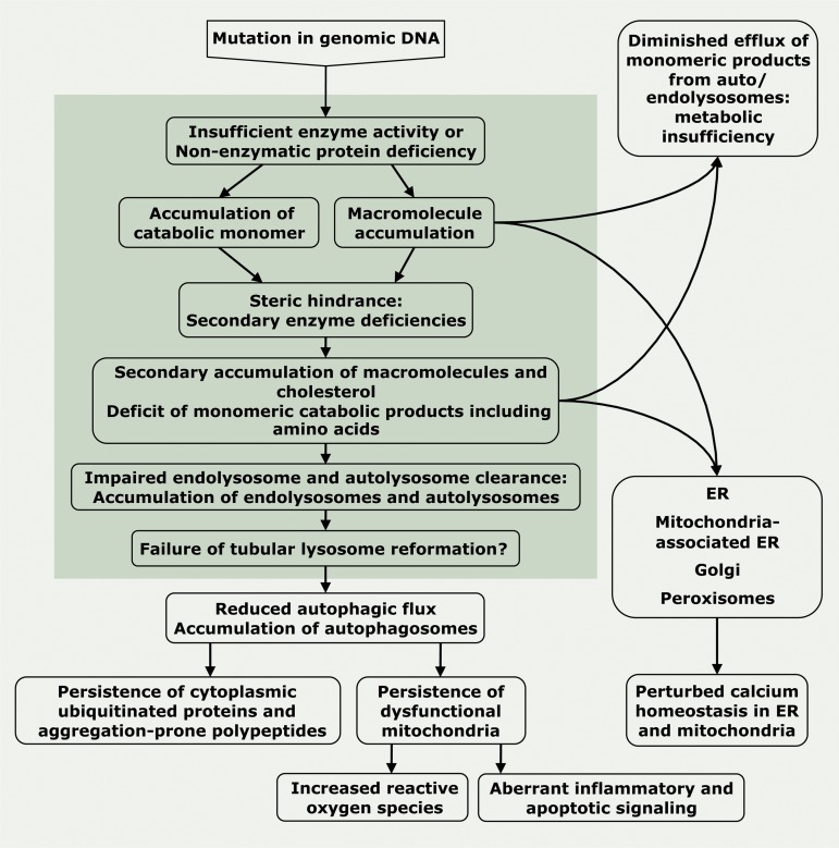
Lysosomal storage diseases (LSDs) are a family of disorders that result from inherited gene mutations that perturb lysosomal homeostasis. LSDs mainly stem from deficiencies in lysosomal enzymes, but also in some non-enzymatic lysosomal proteins, which lead to abnormal storage of macromolecular substrates. Valuable insights into lysosome functions have emerged from research into these diseases. In addition to primary lysosomal dysfunction, cellular pathways associated with other membrane-bound organelles are perturbed in these disorders. Through selective examples, we illustrate why the term "cellular storage disorders" may be a more appropriate description of these diseases and discuss therapies that can alleviate storage and restore normal cellular function.


Figures

Figure 1.
Figure 1.
Lysosomes as catabolic centers of the cell. Lysosomes utilize four distinct pathways for the degradation of cellular material. (A) Macroautophagy begins with the formation of isolation membranes that sequester regions of the cytosol that include denatured proteins, lipids, carbohydrates, and old/damaged organelles into encapsulated vesicles known as autophagosomes. The dynamic kinetics of autophagosome production and clearance by lysosomes is known as autophagic flux. (B) Endosomal degradation by lysosomes predominantly targets late endosomes/multivesicular bodies. Fusion between late endosomes and lysosomes can occur by (i) full fusion/degradation or (ii) kiss-and-run content mixing, where transient endosomal docking occurs. (C) Microautophagy involves the pinocytosis of cytosolic regions surrounding lysosomes. (D) Chaperone-mediated autophagy (CMA) selectively targets proteins with a KFERQ motif for delivery to lysosomes using Hsc-70 as its chaperone and LAMP-2A as its receptor.
Figure 2.
Figure 2.
Subtypes of storage organelles accumulate in LSDs. In different LSDs, cells display a unique spectrum of dysfunctional organelles depending on the specific lysosomal enzyme or non-enzymatic protein affected. (A) In primary LSDs, deficiencies in degradative enzymes prevent the clearance of autophagic and endocytic substrates, resulting in the accumulation of (i) autolysosomes (LC3-II (+), LAMP-1 (+)), (ii) endolysosomes (CI-MPR (+), LAMP-1 (+)), and (iii), in the case of certain lipase deficiencies, lipid-rich multilamellar bodies (CI-MPR (+), LAMP-1 (+)). (B) In a secondary storage disease such as Niemann-Pick type C1, lysosomal enzyme function remains intact, but impaired heterotypic fusion of autophagic and endocytic organelles with lysosomes results in the accumulation of (iv) autophagosomes (LC3-II (+), LAMP-1 (−)), (v) late endosomes (CI-MPR (+), active cathepsin D (−)), and (vi) endosome-derived multilamellar bodies (lipid-rich, CI-MPR (+), active cathepsin D (−)). Note: many primary storage diseases also accumulate organelles seen in secondary storage diseases (see text).
Figure 3.
Figure 3.
Summary of organelles affected in LSDs. Also shown are selective examples of LSDs. See Table 1 and main text for details.
Figure 4.
Figure 4.
Hypothetical cascade of events in LSD pathology. How gene mutations in lysosomal enzymes and non-enzymatic lysosomal proteins could lead to LSDs. Endo/autolysosomal events are confined to the darker shaded background, whereas processes taking place in the cytoplasm that affect autophagosomes, the ER, Golgi, peroxisomes, and mitochondria are on the lighter background. Processes depicted have been observed in a number of LSDs but do not necessarily apply to all LSDs.

References

    1. Angelini C., Semplicini C. 2012. Enzyme replacement therapy for Pompe disease. Curr. Neurol. Neurosci. Rep. 12:70–75 10.1007/s11910-011-0236-5 - DOI - PubMed
    1. Aqul A., Liu B., Ramirez C.M., Pieper A.A., Estill S.J., Burns D.K., Liu B., Repa J.J., Turley S.D., Dietschy J.M. 2011. Unesterified cholesterol accumulation in late endosomes/lysosomes causes neurodegeneration and is prevented by driving cholesterol export from this compartment. J. Neurosci. 31:9404–9413 10.1523/JNEUROSCI.1317-11.2011 - DOI - PMC - PubMed
    1. Balch W.E., Morimoto R.I., Dillin A., Kelly J.W. 2008. Adapting proteostasis for disease intervention. Science. 319:916–919 10.1126/science.1141448 - DOI - PubMed
    1. Ballabio A. 2009. Disease pathogenesis explained by basic science: lysosomal storage diseases as autophagocytic disorders. Int. J. Clin. Pharmacol. Ther. 47(Suppl 1):S34–S38 - PubMed
    1. Ballabio A., Gieselmann V. 2009. Lysosomal disorders: from storage to cellular damage. Biochim. Biophys. Acta. 1793:684–696 10.1016/j.bbamcr.2008.12.001 - DOI - PubMed