

DNA abasic site-selective enhancement of sanguinarine fluorescence with a large emission shift
- PMID: 23185252
- PMCID: PMC3502418
- DOI: 10.1371/journal.pone.0048251
DNA abasic site-selective enhancement of sanguinarine fluorescence with a large emission shift
Abstract
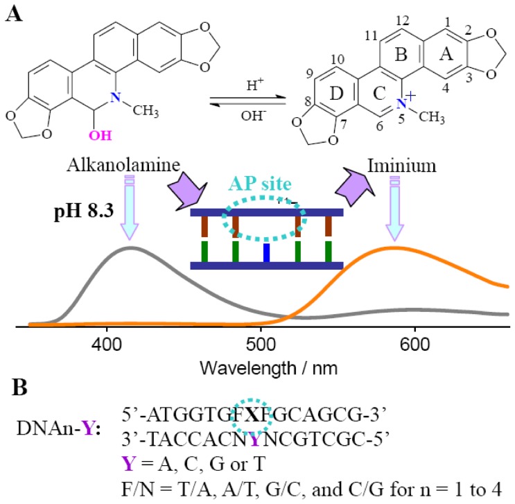
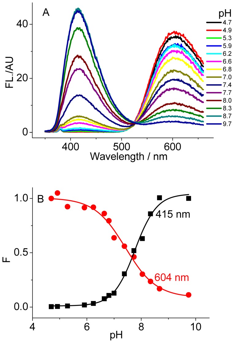
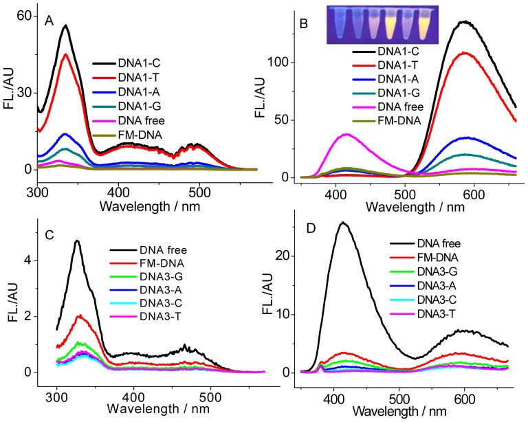
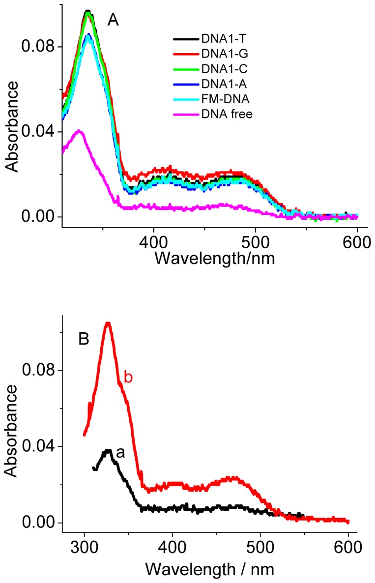
Small molecules that can specifically bind to a DNA abasic site (AP site) have received much attention due to their importance in DNA lesion identification, drug discovery, and sensor design. Herein, the AP site binding behavior of sanguinarine (SG), a natural alkaloid, was investigated. In aqueous solution, SG has a short-wavelength alkanolamine emission band and a long-wavelength iminium emission band. At pH 8.3, SG experiences a fluorescence quenching for both bands upon binding to fully matched DNAs without the AP site, while the presence of the AP site induces a strong SG binding and the observed fluorescence enhancement for the iminium band are highly dependent on the nucleobases flanking the AP site, while the alkanolamine band is always quenched. The bases opposite the AP site also exert some modifications on the SG's emission behavior. It was found that the observed quenching for DNAs with Gs and Cs flanking the AP site is most likely caused by electron transfer between the AP site-bound excited-state SG and the nearby Gs. However, the flanking As and Ts that are not easily oxidized favor the enhanced emission. This AP site-selective enhancement of SG fluorescence accompanies a band conversion in the dominate emission from the alkanolamine to iminium band thus with a large emission shift of about 170 nm. Absorption spectra, steady-state and transient-state fluorescence, DNA melting, and electrolyte experiments confirm that the AP site binding of SG occurs and the stacking interaction with the nearby base pairs is likely to prevent the converted SG iminium form from contacting with water that is thus emissive when the AP site neighbors are bases other than guanines. We expect that this fluorophore would be developed as a promising AP site binder having a large emission shift.
Conflict of interest statement
Figures

Similar articles
-
Simultaneous fluorescence light-up and selective multicolor nucleobase recognition based on sequence-dependent strong binding of berberine to DNA abasic site.Org Biomol Chem. 2012 Apr 28;10(16):3300-7. doi: 10.1039/c2ob00028h. Epub 2012 Mar 13. Org Biomol Chem. 2012. PMID: 22410866
-
Structural and thermodynamic studies on the interaction of iminium and alkanolamine forms of sanguinarine with hemoglobin.J Phys Chem B. 2014 Apr 10;118(14):3771-84. doi: 10.1021/jp409764z. Epub 2014 Mar 27. J Phys Chem B. 2014. PMID: 24635139
-
Interaction of the anticancer plant alkaloid sanguinarine with bovine serum albumin.PLoS One. 2011 Apr 6;6(4):e18333. doi: 10.1371/journal.pone.0018333. PLoS One. 2011. PMID: 21494677 Free PMC article.
-
Sanguinarine and Its Role in Chronic Diseases.Adv Exp Med Biol. 2016;928:155-172. doi: 10.1007/978-3-319-41334-1_7. Adv Exp Med Biol. 2016. PMID: 27671816 Review.
-
Antimicrobial action of sanguinarine.J Clin Dent. 1989 Spring;1(4):96-101. J Clin Dent. 1989. PMID: 2700895 Review.
Cited by
-
Shape-selective recognition of DNA abasic sites by metallohelices: inhibition of human AP endonuclease 1.Nucleic Acids Res. 2015 Jun 23;43(11):5297-306. doi: 10.1093/nar/gkv438. Epub 2015 May 4. Nucleic Acids Res. 2015. PMID: 25940617 Free PMC article.
-
Target-switched triplex nanotweezer and synergic fluorophore translocation for highly selective melamine assay.Mikrochim Acta. 2018 Dec 19;186(1):42. doi: 10.1007/s00604-018-3134-6. Mikrochim Acta. 2018. PMID: 30569196
-
Selective lighting up of epiberberine alkaloid fluorescence by fluorophore-switching aptamer and stoichiometric targeting of human telomeric DNA G-quadruplex multimer.Anal Chem. 2015 Jan 6;87(1):730-7. doi: 10.1021/ac503730j. Epub 2014 Dec 9. Anal Chem. 2015. PMID: 25429435 Free PMC article.
-
8-Styryl-substituted coralyne derivatives as DNA binding fluorescent probes.RSC Adv. 2017 Feb 8;7(18):10660-10667. doi: 10.1039/c6ra27684a. RSC Adv. 2017. PMID: 28496973 Free PMC article.
References
-
- Marnett LJ, Pastaras JP (2001) Endogenous DNA damage and mutation. Trends Genet 17: 214–221. - PubMed
-
- Iyer RR, Pluciennik A, Burdett V, Modrich PK (2006) DNA mismatch repair: functions and mechanisms. Chem Rev 106: 302–323. - PubMed
-
- Benner K, Granzhan A, Ihmels H, Viola G (2007) Targeting Abasic Sites in DNA by Aminoalkyl-Substituted Carboxamidoacridizinium Derivatives and Acridizinium–Adenine Conjugates. Eur J Org Chem 2007: 4721–4730.
Publication types
MeSH terms
Substances
LinkOut - more resources
Full Text Sources
Research Materials
