

Targeting of CRMP-2 to the primary cilium is modulated by GSK-3β
- PMID: 23185275
- PMCID: PMC3504062
- DOI: 10.1371/journal.pone.0048773
Targeting of CRMP-2 to the primary cilium is modulated by GSK-3β
Abstract
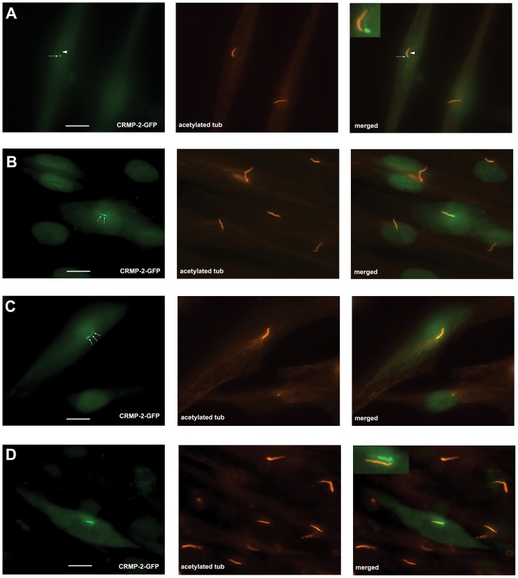
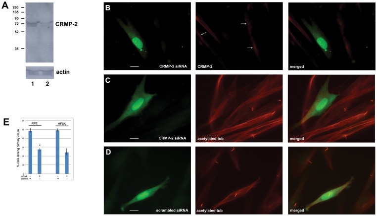
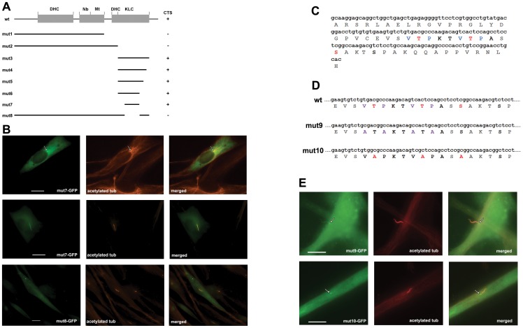
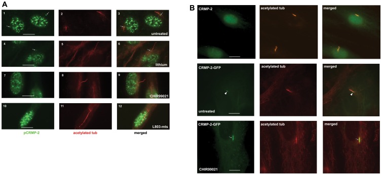
CRMP-2 plays a pivotal role in promoting axon formation, neurite outgrowth and elongation in neuronal cells. CRMP-2's role in other cells is unknown. Our preliminary results showed CRMP-2 expression in cilia of fibroblasts. To localize CRMP-2, define its role and study the regulation of CRMP-2's expression in cilia we carried out the following experiments. We find that in fibroblasts CRMP-2 localizes to the centrosome and is associated with the basal body and -at a low level- is present in primary cilia. Phosphorylated pCRMP-2 can only be detected at the basal body. RNAi knockdown of CRMP-2 interfered with primary cilium assembly demonstrating a critical requirement for CRMP-2. Deletion analysis of CRMP-2 identified a 51 amino acid sequence in the C-terminus that is required for targeting to the basal body and primary cilium. This domain contains GSK-3β phosphorylation sites as well as two repeats of the VxPx motif, previously identified as a cilium targeting signal in other primary cilium proteins. To our surprise, mutation of the CRMP-2 VxPx motifs did not eliminate primary cilium targeting. Instead, mutation of the GSK-3β phosphorylation sites abolished CRMP-2 targeting to the primary cilium without affecting basal body localization. Treatment of cells with lithium, a potent GSK-3β inhibitor, or with two specific GSK-3β inhibitors (the L803-mts peptide inhibitor and CHIR99021) resulted in cilium elongation and decreased basal body levels of pCRMP-2 as well as increased levels of total CRMP-2 at the primary cilium. In summary, we identified CRMP-2 as a protein critically involved in primary cilia formation. To our knowledge this is the first demonstration of modulation of primary cilium targeting by GSK-3β.
Conflict of interest statement
Figures


Similar articles
-
GSK-3beta regulates phosphorylation of CRMP-2 and neuronal polarity.Cell. 2005 Jan 14;120(1):137-49. doi: 10.1016/j.cell.2004.11.012. Cell. 2005. PMID: 15652488
-
Role of the Akt/GSK-3β/CRMP-2 pathway in axon degeneration of dopaminergic neurons resulting from MPP+ toxicity.Brain Res. 2015 Mar 30;1602:9-19. doi: 10.1016/j.brainres.2014.08.030. Epub 2014 Aug 21. Brain Res. 2015. PMID: 25150591
-
GSK-3 mediates the okadaic acid-induced modification of collapsin response mediator protein-2 in human SK-N-SH neuroblastoma cells.J Cell Biochem. 2008 Apr 15;103(6):1833-48. doi: 10.1002/jcb.21575. J Cell Biochem. 2008. PMID: 17902168
-
[Molecular mechanisms of neuronal polarity].Nihon Shinkei Seishin Yakurigaku Zasshi. 2005 Aug;25(4):169-74. Nihon Shinkei Seishin Yakurigaku Zasshi. 2005. PMID: 16190365 Review. Japanese.
-
Molecular mechanisms of axon specification and neuronal disorders.Ann N Y Acad Sci. 2006 Nov;1086:116-25. doi: 10.1196/annals.1377.013. Ann N Y Acad Sci. 2006. PMID: 17185510 Review.
Cited by
-
Ouabain affects cell migration via Na,K-ATPase-p130cas and via nucleus-centrosome association.PLoS One. 2017 Aug 17;12(8):e0183343. doi: 10.1371/journal.pone.0183343. eCollection 2017. PLoS One. 2017. PMID: 28817661 Free PMC article.
-
Purinergic A2b Receptor Activation by Extracellular Cues Affects Positioning of the Centrosome and Nucleus and Causes Reduced Cell Migration.J Biol Chem. 2016 Jul 15;291(29):15388-403. doi: 10.1074/jbc.M116.721241. Epub 2016 May 13. J Biol Chem. 2016. PMID: 27226580 Free PMC article.
-
Lithium-induced ciliary lengthening sparks Arp2/3 complex-dependent endocytosis.Mol Biol Cell. 2023 Apr 1;34(4):ar26. doi: 10.1091/mbc.E22-06-0219. Epub 2023 Feb 8. Mol Biol Cell. 2023. PMID: 36753380 Free PMC article.
-
Reduction of ciliary length through pharmacologic or genetic inhibition of CDK5 attenuates polycystic kidney disease in a model of nephronophthisis.Hum Mol Genet. 2016 Jun 1;25(11):2245-2255. doi: 10.1093/hmg/ddw093. Epub 2016 Apr 5. Hum Mol Genet. 2016. PMID: 27053712 Free PMC article.
-
Loss of Arf4 causes severe degeneration of the exocrine pancreas but not cystic kidney disease or retinal degeneration.PLoS Genet. 2017 Apr 14;13(4):e1006740. doi: 10.1371/journal.pgen.1006740. eCollection 2017 Apr. PLoS Genet. 2017. PMID: 28410364 Free PMC article.
References
-
- Brailov I, Bancila M, Brisorgueil MJ, Miquel MC, Hamon M, et al. (2000) Localization of 5-HT(6) receptors at the plasma membrane of neuronal cilia in the rat brain. Brain Res 872: 271–275. - PubMed
-
- Handel M, Schulz S, Stanarius A, Schreff M, Erdtmann-Vourliotis M, et al. (1999) Selective targeting of somatostatin receptor 3 to neuronal cilia. Neuroscience 89: 909–926. - PubMed
-
- Pazour GJ, San Agustin JT, Follit JA, Rosenbaum JL, Witman GB (2002) Polycystin-2 localizes to kidney cilia and the ciliary level is elevated in orpk mice with polycystic kidney disease. Curr Biol 12: R378–R380. - PubMed
-
- Rohatgi R, Milenkovic L, Scott MP (2007) Patched1 regulates hedgehog signaling at the primary cilium. Science 317: 372–376. - PubMed
-
- Schneider L, Clement CA, Teilmann SC, Pazour GJ, Hoffmann EK, et al. (2005) PDGFRalphaalpha signaling is regulated through the primary cilium in fibroblasts. Curr Biol 15: 1861–1866. - PubMed
Publication types
MeSH terms
Substances
Grants and funding
LinkOut - more resources
Full Text Sources
Miscellaneous
