. 2012;7(11):e49349.
doi: 10.1371/journal.pone.0049349. Epub 2012 Nov 21.

Autoproteolysis and intramolecular dissociation of Yersinia YscU precedes secretion of its C-terminal polypeptide YscU(CC)

Affiliations

Autoproteolysis and intramolecular dissociation of Yersinia YscU precedes secretion of its C-terminal polypeptide YscU(CC)

Stefan Frost et al. PLoS One. 2012.

Abstract

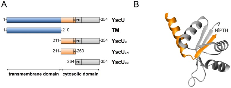
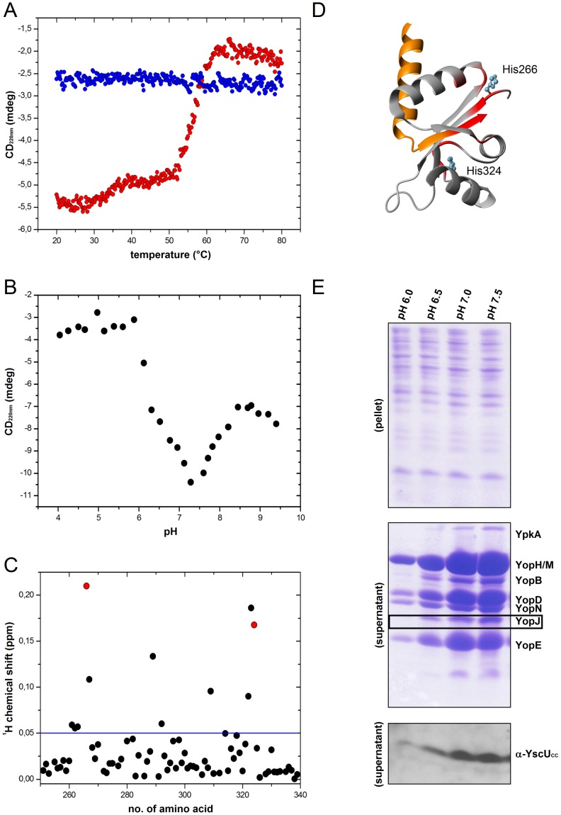
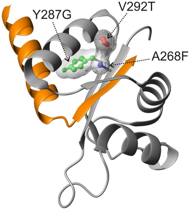
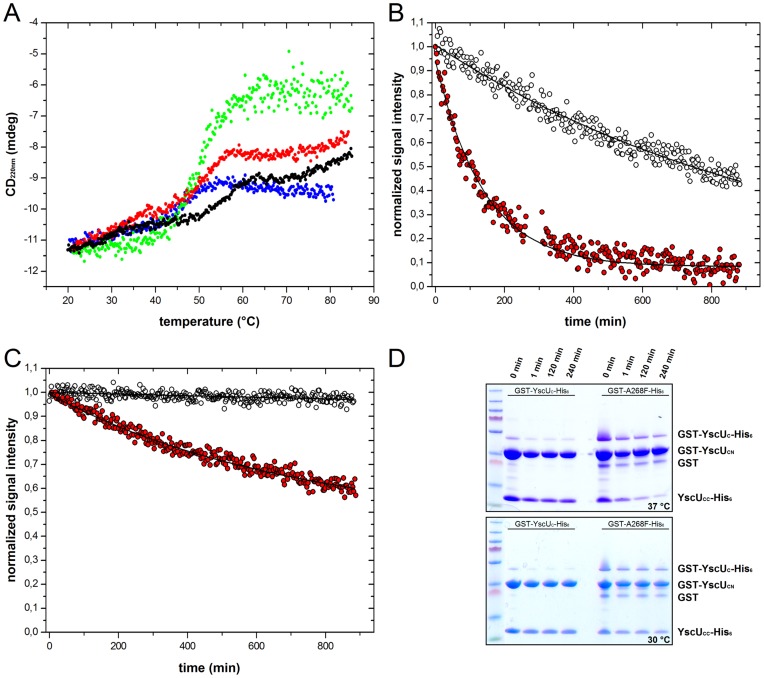
Type III secretion system mediated secretion and translocation of Yop-effector proteins across the eukaryotic target cell membrane by pathogenic Yersinia is highly organized and is dependent on a switching event from secretion of early structural substrates to late effector substrates (Yops). Substrate switching can be mimicked in vitro by modulating the calcium levels in the growth medium. YscU that is essential for regulation of this switch undergoes autoproteolysis at a conserved N↑PTH motif, resulting in a 10 kDa C-terminal polypeptide fragment denoted YscU(CC). Here we show that depletion of calcium induces intramolecular dissociation of YscU(CC) from YscU followed by secretion of the YscU(CC) polypeptide. Thus, YscU(CC) behaved in vivo as a Yop protein with respect to secretion properties. Further, destabilized yscU mutants displayed increased rates of dissociation of YscU(CC)in vitro resulting in enhanced Yop secretion in vivo at 30°C relative to the wild-type strain.These findings provide strong support to the relevance of YscU(CC) dissociation for Yop secretion. We propose that YscU(CC) orchestrates a block in the secretion channel that is eliminated by calcium depletion. Further, the striking homology between different members of the YscU/FlhB family suggests that this protein family possess regulatory functions also in other bacteria using comparable mechanisms.


Conflict of interest statement

Competing Interests: The authors have declared that no competing interests exist.

Figures

Figure 1
Figure 1. Domain structure of YscU and crystallographic structure of YscUC.
(A) Schematic domain structure of the integral membrane protein, YscU of Y. pseudotuberculosis. The full-length protein contains 354 amino acid residues. The N-terminal 210 residues constitute four transmembrane helices (TM). The cytosolic domain of YscU (YscUC) undergoes autoproteolytic cleavage at the N↑PTH-motif (amino acids 263–266), which leaves an N-terminal cytoplasmic polypeptide, denoted YscUCN, and a C-terminal polypeptide, denoted YscUCC. (B) Ribbon drawing of the cleaved cytosolic domain YscUC (2JLI.PDB) from Y. pestis . YscUCN and YscUCC resulting from cleavage at the N↑PTH motif are colored in orange and grey, respectively.
Figure 2
Figure 2. In vitro dissociation of YscUC.
(A) 1H-15N HSQC spectra of YscUC at 20°C, pH 7.4, before (blue contours) and after (red contours) incubation at 60°C for 10 min. Only resonances that corresponded to YscUCN were visible after the thermal treatment. (B) Thermal up- and down-scans of YscUC at pH 7.4 monitored with CD spectroscopy at 220 nm in the absence of calcium. (C) Thermal signatures of P264A, a non-cleavable mutant, at pH 7.4 in the absence of calcium. Up- and down scans of YscUC and P264A are shown in red and blue circles, respectively.
Figure 3
Figure 3. Calcium effects on YscUC stability and Yop secretion.
(A) Thermal up- and down-scans of YscUC at pH 7.4 monitored with CD spectroscopy at 220 nm in the presence of 2.5 mM calcium. Up- and down scans of YscUC are shown in red and blue circles, respectively. (B) Titration of calcium to YscUC monitored with CD spectroscopy at 220 nm. The calcium binding isotherm to YscUC was fit to a one-site binding model (red line). The resulting Kd was 800 µM. (C) Western Blot analysis of YopB, YopD, and YopE in wild-type Y. pseudotuberculosis. The bacteria were grown for 2 h at 26°C and shifted to 37°C for 3 h (temperature shift for induction of Yop secretion) with varying concentrations of free calcium. “Pellet” indicates intracellular proteins; “supernatant” denotes secreted proteins. The LB growth medium was initially supplemented with 1 mM EGTA to complex residual calcium content (approximately 500 µM); thereafter, calcium was added to set the indicated concentrations of free calcium.
Figure 4
Figure 4. In vivo dissociation and secretion of YscUCC in different Yersinia strains.
Calcium dependent regulation of Yop and YscUCC secretion in wild-type Y. pseudotuberculosis, in a ΔyscC mutant and ΔyscN mutant strain without and with in trans complementation of YscUCC. Bacteria transformed with empty pBADmycHis B (pBAD), or pBAD with one additional yscUCC copy (pBAD(YscUCC)), were grown for 2 h at 26°C and 3 h at 37°C in calcium depleted (−) or calcium supplemented (+) medium. The expression of yscUCC was induced by addition of arabinose. Yop secretion is coupled to the secretion of YscUCC in all analysed Yersinia strains and required a functional T3SS. Secreted Yops visualized on Coomassie stained PAGE gels; YscUCC visualized on immunoblots with anti-YscUCC peptide antibodies. “pellet” indicates intracellular proteins; “supernatant” denotes secreted proteins. The YopJ protein (black box) was subjected to densitometric analysis for quantification of secretion levels (see Table 2).
Figure 5
Figure 5. pH-dependencies of YscUC dissociation in vitro and Yop/YscUCC secretion in vivo.
(A) Thermal up- and down-scans of YscUC at pH 6.0, monitored with CD spectroscopy at 220 nm in the absence of calcium. The thermal signature of YscUC displayed one large-amplitude transition at 55°C. Up- and down scans of YscUC are shown in red and blue circles, respectively. (B) The pH-dependency of the YscUC monitored with CD spectroscopy at 220 nm. (C) Chemical shift perturbations of YscUC quantified from 1H-15N HSQC spectra, in response to a pH-shift from 7.4 to 6.0, displayed against the primary sequence. The blue line indicates the threshold value (0.05 ppm) used in Figure 5D. The chemical shift perturbation of the two histidines at positions 266 and 324 are shown in red. (D) Structural distributions of residues that show significant chemical shift perturbations in response to a pH-shift from 7.4 to 6.0 are shown in red on the YscUC structure (2JLI.PDB). The YscUCN and YscUCC fragments are colored orange and gray, respectively. The two histidine residues (266 and 324) in the folded part of YscUC are indicated. (E) Coomassie stained gels show Yop secretion under different pH conditions. (top panel) “pellet” indicates intracellular proteins; (middle panel) “supernatant” denotes secreted proteins. The YopJ protein (black box) was subjected to densitometric analysis for quantification of secretion levels (see Table 2). (bottom panel) The pH-dependency of YscUCC secretion was visualized on immunoblots with anti-YscUCC peptide antibodies.
Figure 6
Figure 6. YscUC suppressor mutations are buried in the structure.
The spatial locations of single mutations in YscUC that suppressed the non-secreting ΔyscP phenotype in vivo are shown on the YscUC structure of Y. pestis (2JLI.PDB). All positions are either fully or partially buried in the protein structure. YscUCN and YscUCC polypeptides are colored orange and gray, respectively.
Figure 7
Figure 7. YscUC suppressor mutant stabilities and dissociation kinetics.
(A) Thermal induced unfolding of YscUC and suppressor mutants. Dissociation temperatures (T diss) of single suppressor mutants at pH 7.4 were quantified with the CD signal at 220 nm and a scan rate of 1°C/min; YscUC (black), A268F (blue), Y287G (green), and V292T (red). All suppressor mutants are destabilized compared to wild-type YscUC. T diss values are summarized in Table 3. (B), (C) Dissociation kinetics quantified as dissociation life-times (τdiss) of YscUC (black) and V292T (red) at pH 7.4 followed with NMR-spectroscopy at (B) 37°C and (C) 30°C, respectively. Primary NMR data for (B) is shown in Figure S6. Solid lines correspond to fits of the experimental data to single exponential decays. (D) Time dependent GST-pulldown experiments show the dissociation of wild-type YscUC and the suppressor mutant A268F at 30°C and 37°C after varying incubation times. The suppressor mutant A268F displayed pronounced dissociation of YscUCC-His6 at 37°C and moderate dissociation at 30°C; wild-type YscUC displayed no dissociation of YscUCC-His6 at 37°C or at 30°C over the observed time period. Note! Dissociation of YscUCC is manifested as disappearance of YscUCC-His6 over time since the dissociation is irreversible and YscUCC-His6 cannot bind itself to the used resin.
Figure 8
Figure 8. Yersinia strains with destabilized yscU suppressor mutants secrete YscUCC and Yops at lower temperatures (30°C) than wild-type.
Coomassie stained analysis of Yop secretion in Y. pseudotuberculosis incubated at 30°C. Bacteria expressing either wild-type yscU or one of the suppressor mutants, A268F or V292T. “pellet” indicates intracellular proteins; “supernatant” denotes secreted proteins. Secretion of YscUCC was analyzed on immunoblots with anti-YscUCC peptide antibodies. Yersinia harboring yscU suppressor mutants showed strongly elevated secretion of Yops after cultivation at 30°C.

Similar articles

Cited by

References

    1. Hills GM, Spurr ED (1952) The effect of temperature on the nutritional requirements of Pasteurella pestis. J Gen Microbiol 6: 64–73. - PubMed
    1. Higuchi K, Kupferberg LL, Smith JL (1959) Studies on the nutrition and physiology of Pasteurella pestis. III. Effects of calcium ions on the growth of virulent and avirulent strains of Pasteurella pestis. J Bacteriol 77: 317–321. - PMC - PubMed
    1. Bölin I, Portnoy DA, Wolf-Watz H (1985) Expression of the temperature-inducible outer membrane proteins of yersiniae. Infect Immun 48: 234–240. - PMC - PubMed
    1. Heesemann J, Algermissen B, Laufs R (1984) Genetically manipulated virulence of Yersinia enterocolitica. Infect Immun 46: 105–110. - PMC - PubMed
    1. Portnoy DA, Falkow S (1981) Virulence-associated plasmids from Yersinia enterocolitica and Yersinia pestis. J Bacteriol 148: 877–883. - PMC - PubMed

Publication types

MeSH terms