. 2012;7(11):e49566.
doi: 10.1371/journal.pone.0049566. Epub 2012 Nov 21.

Ezrin interacts with the SARS coronavirus Spike protein and restrains infection at the entry stage

Affiliations

Ezrin interacts with the SARS coronavirus Spike protein and restrains infection at the entry stage

Jean Kaoru Millet et al. PLoS One. 2012.

Abstract

Background: Entry of Severe Acute Respiratory Syndrome coronavirus (SARS-CoV) and its envelope fusion with host cell membrane are controlled by a series of complex molecular mechanisms, largely dependent on the viral envelope glycoprotein Spike (S). There are still many unknowns on the implication of cellular factors that regulate the entry process.

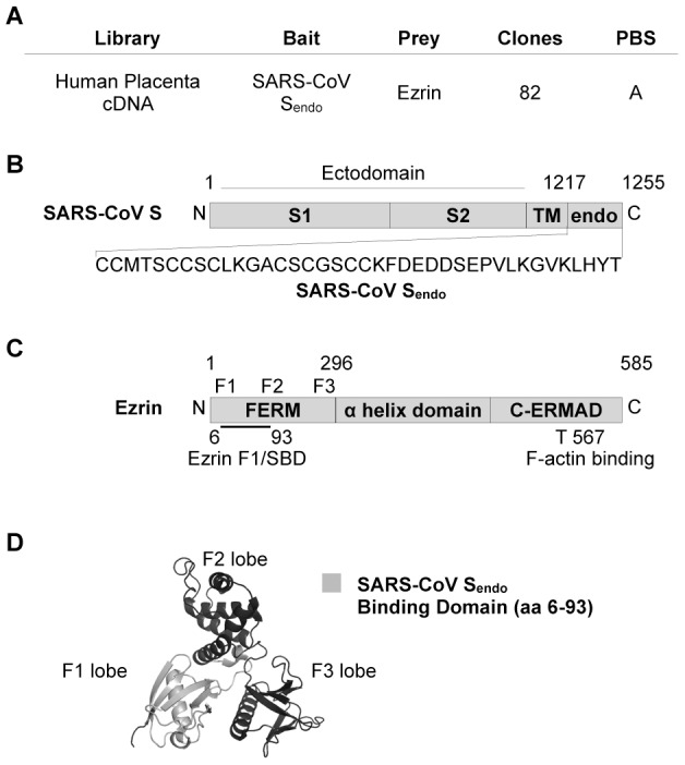
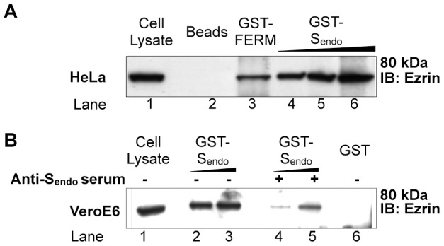
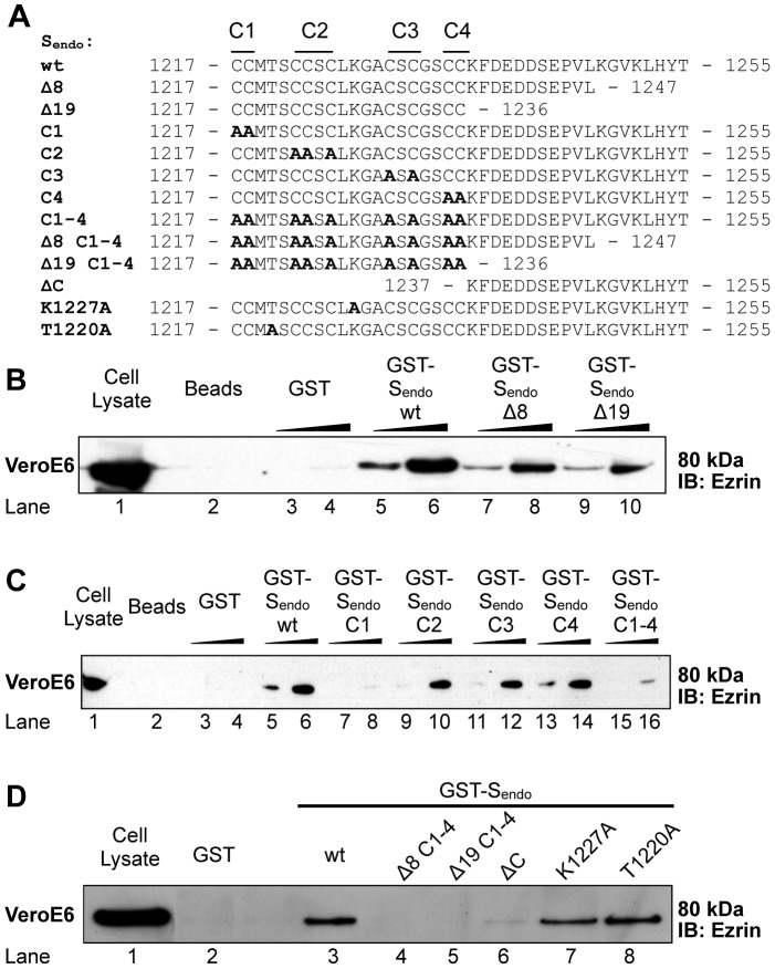
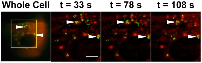
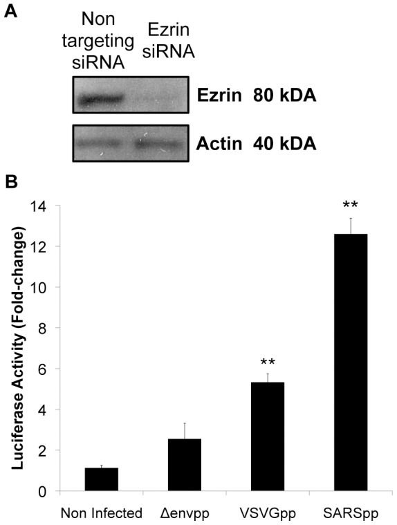
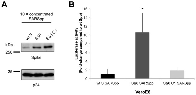
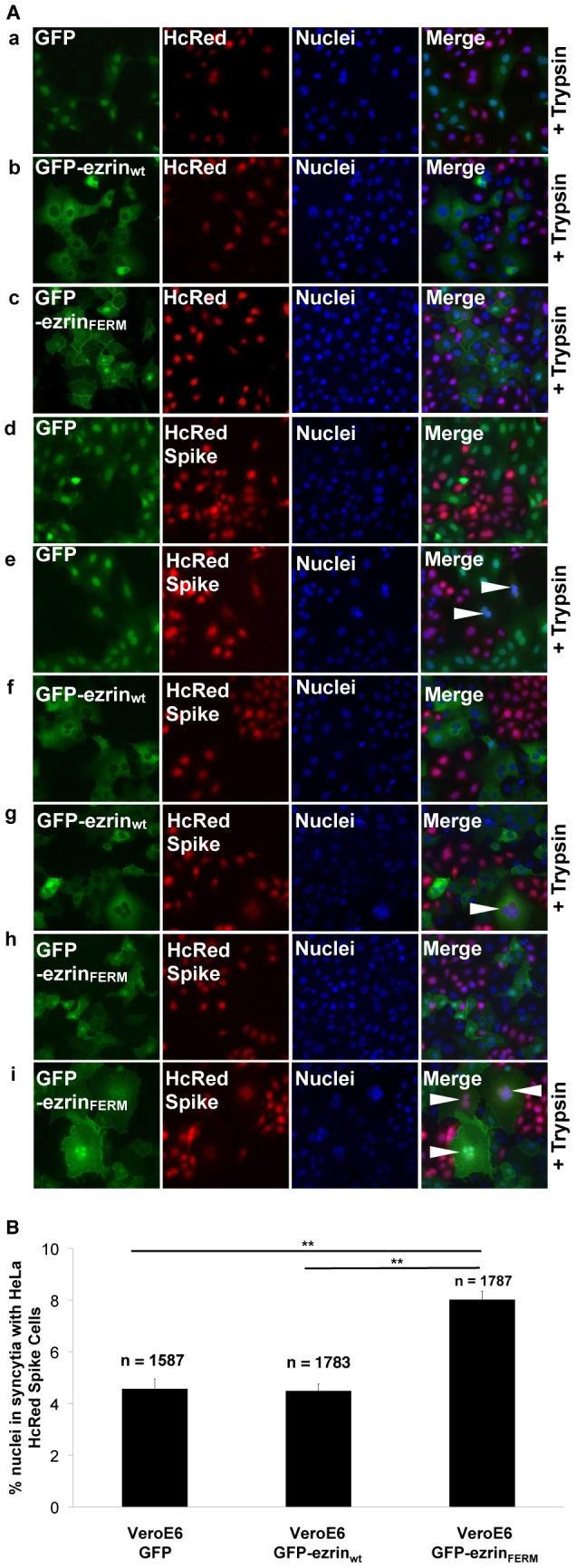
Methodology/principal findings: We performed a yeast two-hybrid screen using as bait the carboxy-terminal endodomain of S, which faces the cytosol during and after opening of the fusion pore at early stages of the virus life cycle. Here we show that the ezrin membrane-actin linker interacts with S endodomain through the F1 lobe of its FERM domain and that both the eight carboxy-terminal amino-acids and a membrane-proximal cysteine cluster of S endodomain are important for this interaction in vitro. Interestingly, we found that ezrin is present at the site of entry of S-pseudotyped lentiviral particles in Vero E6 cells. Targeting ezrin function by small interfering RNA increased S-mediated entry of pseudotyped particles in epithelial cells. Furthermore, deletion of the eight carboxy-terminal amino acids of S enhanced S-pseudotyped particles infection. Expression of the ezrin dominant negative FERM domain enhanced cell susceptibility to infection by SARS-CoV and S-pseudotyped particles and potentiated S-dependent membrane fusion.

Conclusions/significance: Ezrin interacts with SARS-CoV S endodomain and limits virus entry and fusion. Our data present a novel mechanism involving a cellular factor in the regulation of S-dependent early events of infection.


Conflict of interest statement

Competing Interests: The authors have declared that no competing interests exist.

Figures

Figure 1
Figure 1. The F1 lobe of the ezrin FERM domain binds to the SARS-CoV S endodomain.
A. Table summarizing the yeast two-hybrid screening results for ezrin - S endodomain interaction. B. Schematic representation of the SARS-CoV S endodomain sequence (not drawn to scale). S1: Subunit 1. S2: Subunit 2. TM: transmembrane domain. endo: endodomain. C. Schematic representation of ezrin (not drawn to scale). The bold line represents the common sequence of the 82 cDNAs corresponding to ezrin found in the yeast two-hybrid screening. The domain was named ezrin F1/Spike Binding Domain (SBD). F1, F2, and F3 represent approximate regions corresponding to the three lobes of the FERM domain. T567: threonine 567. D. Representation of the three-dimensional crystal structure of the ezrin FERM domain, showing the three distinct globular lobes F1, F2, and F3. The region in light grey represents amino-acids of the F1/SBD. The FERM domain crystal structure for ezrin (PDB ID: 1NI2) was downloaded from the Protein Data Bank (http://www.pdb.org). 3-D rendering of the ezrin FERM domain was performed using MacPyMol software (DeLano Scientific).
Figure 2
Figure 2. In vitro confirmation of the interaction between SARS-CoV S endodomain and ezrin.
A. The endodomain of S pulls down ezrin from HeLa cell lysate. Cell lysate was incubated with Glutathione-Sepharose beads either uncoupled (lane 2) or coupled with the fusion protein GST-FERM (positive control, lane 3) or increasing amounts of GST-Sendo (lanes 4–6). B. The endodomain of S pulls down ezrin from Vero E6 cell lysate. Similarly, cell lysate was incubated with Glutathione-Sepharose beads bound either to GST fused to Sendo in absence (lanes 2–3) or presence (lanes 4–5) of rabbit serum that recognizes the endodomain of S, or to GST alone (lane 6). For both A. and B. 10 µL of cell lysate was used as input control which represents 1.6% and 4% of the volume used for the pull down for A. and B., respectively. IB: Immunoblot. Results shown are representative of two independent experiments.
Figure 3
Figure 3. Characterization of interactions determinants of S endodomain binding to ezrin.
A. Sequences of the wild type (wt) and mutated S endodomain used in the GST-pull down analysis. Twelve GST fusion proteins were produced with mutations in the S endodomain. Δ indicates truncations and C indicates cysteine to alanine mutations (in bold) of cysteine clusters (1 to 4). B. & C. Effect of truncations or cysteine to alanine mutations. Vero E6 lysate was incubated with Glutathione-Sepharose beads coupled or not with GST, GST-Sendo wt (B. & C.), GST-Sendo Δ8, and GST-Sendo Δ19(B.), GST-Sendo C1, GST-Sendo C2, GST-Sendo C3, GST-Sendo C4, and GST-Sendo C1-4 (C.) using 1 or 5 µg of GST fusion proteins. D. Effects of cysteine to alanine mutations and truncations or point mutations (K1227A and T1220A). Vero E6 lysate was incubated with Glutathione-Sepharose beads coupled with either GST, GST-Sendo wt, GST-Sendo Δ8 C1-4, and GST-Sendo Δ19 C1-4, GST-Sendo ΔC, GST-Sendo K1227A, GST-Sendo T1220A using 1 µg of each GST fusion protein. 5 µL of cell lysate was used as input control for B. & C. (8% of volume used in each pull down); 10 µL of cell lysate was used as input control for D. (5% of volume used in each pull down). IB: Immunoblot. Results shown are representative of two independent experiments.
Figure 4
Figure 4. Ezrin accumulates at sites of entry of SARS-CoV S pseudotyped particles.
Vero E6 cells stably expressing RFP-ezrin were inoculated with SARS-CoV S-pseudotyped lentiviral particles harboring a GFP-tagged Vpr protein (SARSpp GFP-Vpr) on ice for 30 minutes. Unbound particles were washed. Internalization of particles was induced by placing the culture dish in a 37°C 5% CO2 chamber. At 30 minutes post temperature switch (t = 0), cells were analyzed by TIRFM. Time-lapse images were acquired every 3 seconds. The whole cell (first frame) is shown on the left panel. The region shown for time-lapse images is indicated by a square. 3 frames at t = 33, 78 and 108 seconds out of a total of 50 frames are shown and correspond to the times after the start of image acquisitions. The movie of this image sequence is shown as supporting material (Supporting movie S1). Scale bar = 40 µm.
Figure 5
Figure 5. Knock down of ezrin by siRNA increases entry of SARS-CoV S pseudotyped particles.
A. Ezrin expression knock down by siRNA. HeLa-F5 cells stably expressing the SARS-CoV receptor ACE2 were transfected twice with ezrin or non-targeting siRNAs and knock down efficiency was estimated by Western blot analysis of ezrin and actin content of cell lysates. B. Luciferase activity fold change analysis. HeLa-F5 cells treated with siRNAs as indicated above were infected with pseudotyped lentiviral particles harboring the VSV G or SARS-CoV S viral envelope glycoproteins. Δenvpp indicates lentiviral particles without any viral surface glycoprotein. The fold change in luciferase activity was calculated using the non-targeting siRNA condition as negative control. Experiments were performed in triplicates and the results of this experiment are representative of at least three independent experiments. ** indicates a value of p<0.001 in two-tailed t-tests.
Figure 6
Figure 6. S C-terminal mutations that decrease ezrin interaction enhance transduction by pseudotyped particles in Vero E6 cells.
A. Generation of lentiviral pseudotyped particles harboring wild-type (wt) or mutated (Δ8 and Δ8 C1) SARS-CoV Spike proteins. A Western blot assay was performed on concentrated SARSpp particles where the Spike protein and the lentiviral backbone protein p24 were probed. Estimation of protein quantities was performed using densitometry analysis. B. Entry of wt and mutated SARS-CoV S pseudotyped particles. Vero E6 cells were infected by wt S SARS-CoV pseudotyped particles, along with mutated SARSpp (SΔ8 SARSpp and SΔ8 C1 SARSpp). Results are expressed as fold-change in luciferase activity compared to the wt S SARSpp. The results are averages of triplicates and are representative of at least three independent experiments. * indicates a value of p<0.05 in a two-tailed t-test.
Figure 7
Figure 7. Expression of the FERM domain of ezrin increases Vero E6 cell susceptibility to SARS-CoV infection.
A. Flow cytometry analysis (a.) and subcellular localization (b.) of the wt or FERM domain GFP-ezrin in clonal Vero E6 stable cell lines. (b.) Arrows indicate enrichments of wt or FERM ezrin. Scale bars: 20 µm. B. Time course of SARS-CoV replication in Vero E6 stable cell lines expressing wt or FERM GFP-ezrin. Vero E6, Vero E6 GFP-ezrinwt, Vero E6 GFP-ezrinFERM were infected with SARS-CoV (strain HK39849) at M.O.I. 5. At 3, 6 and 24 hours post infection, SARS-CoV N RNA levels were measured using qRT-PCR with 18S rRNA normalization. For each condition the average of two measurements of two independent wells was calculated. C. SARS-CoV infection rates in Vero E6, Vero E6 GFP-ezrinwt and Vero E6 GFP-ezrinFERM cell lines. Cells were infected with SARS-CoV (strain HK39849) at M.O.I. of 5. 24 h post-infection, cells were immunolabeled for SARS-CoV S. (a.), scale bars: 100 µm. (b.) for each cell line, images of ten random microscopy fields were acquired and analyzed for total number of cells (n; DAPI or Phalloidin AMCA staining) and SARS-CoV S positive cells (TRITC staining) using Imaris 6.3 software. ** indicates a value of p<0.001 in two-tailed t-tests.
Figure 8
Figure 8. Expression of the FERM domain of ezrin enhances entry of SARS-CoV S pp.
A. Vero E6, Vero E6 GFP-ezrinwt and Vero E6 GFP-ezrinFERM clonal cells were infected with SARSpp and incubated for 72 hours and the activity of luciferase was measured (results correspond to average of triplicates). B. (a.) Flow cytometry analysis of polyclonal Vero E6 stable cells expressing GFP-ezrinFERM. Lentiviral vector transductions enabled Vero E6 cells to express irrelevant HcRed or GFP-ezrinFERM proteins. MFI: Mean Fluorescence Intensity. (b.) Polyclonal Vero E6 cells stably expressing HcRed (negative control) or GFP-ezrinFERM were infected with SARSpp, incubated for 72 hours, and the activity of luciferase was measured. The results of this experiment are representative of at least three independent experiments. * indicates a value of p<0.05 in two-tailed t-tests.
Figure 9
Figure 9. Effect of wt or FERM ezrin expression on S-mediated cell-cell fusion.
A. Microscopy analysis of syncytia formation induced by activated SARS-CoV S. Vero E6 GFP, Vero E6 GFP-ezrinwt and Vero E6 GFP-ezrinFERM cells were overlaid on HeLa cells stably expressing HcRed alone or HcRed and SARS-CoV S. SARS-CoV S was activated or not with trypsin for 15 minutes (+Trypsin). 18 hours later, cells were fixed, nuclei stained with DAPI and analyzed by microscopy for syncytia. Arrows indicate syncytia. B. Quantification of syncytia. For the conditions in which HeLa HcRed Spike cells (+Trypsin) were incubated with Vero E6 GFP or Vero E6 GFP-ezrinwt or Vero E6 GFP-ezrinFERM cells, 10 random microscopy fields were analyzed for total number of nuclei (DAPI) and number of nuclei in multi-nucleated cells (DAPI/HcRed/GFP). Results are representative of three independent experiments. ** indicates a value of p<0.001 in two-tailed t-tests.

Similar articles

Cited by

References

    1. Woo PC, Lau SK, Yip CC, Huang Y, Yuen KY (2009) More and More Coronaviruses: Human Coronavirus HKU1. Viruses 1: 57–71. - PMC - PubMed
    1. Woo PC, Lau SK, Huang Y, Yuen KY (2009) Coronavirus diversity, phylogeny and interspecies jumping. Exp Biol Med (Maywood) 234: 1117–1127. - PubMed
    1. Wise AG, Kiupel M, Maes RK (2006) Molecular characterization of a novel coronavirus associated with epizootic catarrhal enteritis (ECE) in ferrets. Virology 349: 164–174. - PMC - PubMed
    1. Lau SK, Woo PC, Yip CC, Fan RY, Huang Y, et al. (2012) Isolation and characterization of a novel Betacoronavirus subgroup A coronavirus, rabbit coronavirus HKU14, from domestic rabbits. J Virol 86: 5481–5496. - PMC - PubMed
    1. Cavanagh D (2007) Coronavirus avian infectious bronchitis virus. Vet Res 38: 281–297. - PubMed

Publication types

MeSH terms