

Duodenum clamping trauma induces significant postoperative intraperitoneal adhesions on a rat model
- PMID: 23185403
- PMCID: PMC3502250
- DOI: 10.1371/journal.pone.0049673
Duodenum clamping trauma induces significant postoperative intraperitoneal adhesions on a rat model
Abstract
Objective: The purpose of this study was to investigate the histological and morphological changes in the first two postoperative weeks on a rat intraperitoneal adhesion model induced by duodenum clamping trauma.
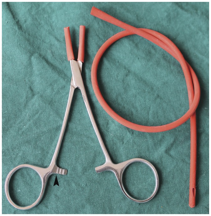
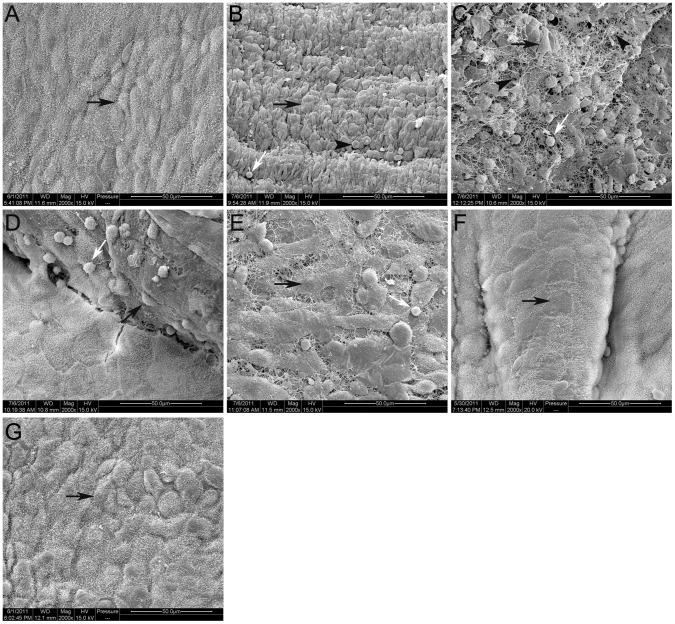
Method: The rat model of postoperative intraperitoneal adhesions was established in 48 male Wistar rats by laparotomy, followed by the duodenum clamping trauma. Rats were sacrificed respectively on 1(st), 3(rd), 5(th), 7(th) and 14(th) day after the operation. The control rats were sacrificed immediately after the operation (0 day). Then the intraperitoneal adhesions were assessed macroscopically. Histopathology and immunohistochemistry were performed to evaluate the fibrosis, inflammatory responses, neovascularization, and cells infiltration in adhesion tissues. In addition, the changes of the mesothelium covering the surgical sites were examined by scanning electron microscopy.
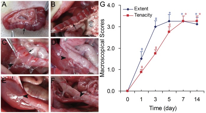
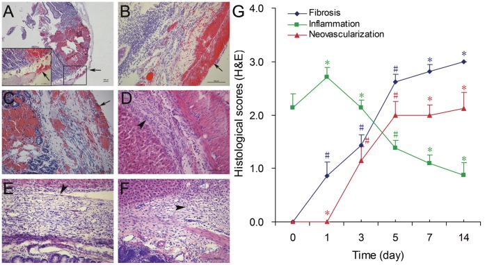
Results: Our study revealed that duodenum clamping trauma induced by mosquito hemostat can result in significant postoperative intraperitoneal adhesions formation. The extent and tenacity of intraperitoneal adhesions reached their peaks on 3(rd) and 5(th) days, respectively. Histopathological examination showed that all rats developed inflammatory responses at the clamped sites of duodenum, which was most prominent on 1(st) day; the scores of fibrosis and vascular proliferation increased slowly from 3(rd) to 5(th) day. Myofibroblasts proliferated significantly in the adhesion tissues from 3(rd) day, which were examined by immunohistochemical method. And the mesothelium covering the surgical sites and the adhesion tissues healed on 7(th) day.
Conclusion: This study suggests that clamping trauma to the duodenum can result in significant postoperative intraperitoneal adhesions formation, which represents an ideal rat model for intraperitoneal adhesions research and prevention. And myofibroblasts may play an important role in the forming process of intraperitoneal adhesions.
Conflict of interest statement
Figures

Similar articles
-
Effect of intraabdominal administration of Allium sativum (garlic) oil on postoperative peritoneal adhesion.Eur J Obstet Gynecol Reprod Biol. 2014 Jun;177:44-7. doi: 10.1016/j.ejogrb.2014.03.018. Epub 2014 Apr 13. Eur J Obstet Gynecol Reprod Biol. 2014. PMID: 24793933
-
A semiquantitative rat model for intraperitoneal postoperative adhesion formation.Gynecol Obstet Invest. 1994;37(2):99-105. doi: 10.1159/000292534. Gynecol Obstet Invest. 1994. PMID: 8150379
-
The preventive effect of Rofecoxib in postoperative intraperitoneal adhesions.Acta Chir Belg. 2004 Feb;104(1):97-100. doi: 10.1080/00015458.2003.11978403. Acta Chir Belg. 2004. PMID: 15053473
-
Prevention of peritoneal adhesions by intraperitoneal administration of vitamin E and human amniotic membrane.Int J Surg. 2009 Dec;7(6):561-5. doi: 10.1016/j.ijsu.2009.09.007. Epub 2009 Sep 30. Int J Surg. 2009. PMID: 19800036
-
Postsurgical Adhesions: Is There Any Prophylactic Strategy Really Working?J Clin Med. 2023 Jun 8;12(12):3931. doi: 10.3390/jcm12123931. J Clin Med. 2023. PMID: 37373626 Free PMC article. Review.
Cited by
-
Administration of Intravenous Inf liximab for Prevention of Peritoneal Adhesions Formation in Rats.Bull Emerg Trauma. 2015 Jul;3(3):97-103. Bull Emerg Trauma. 2015. PMID: 27162911 Free PMC article.
References
-
- Liakakos T, Thomakos N, Fine PM, Dervenis C, Young RL (2001) Peritoneal adhesions: Etiology, pathophysiology, and clinical significance - Recent advances in prevention and management. Digestive Surgery 18: 260–273. - PubMed
-
- Hellebrekers BW, Kooistra T (2011) Pathogenesis of postoperative adhesion formation. Br J Surg 98: 1503–1516. - PubMed
-
- Ellis H, Moran BJ, Thompson JN, Parker MC, Wilson MS, et al. (1999) Adhesion-related hospital readmissions after abdominal and pelvic surgery: a retrospective cohort study. Lancet 353: 1476–1480. - PubMed
-
- Lauder CI, Garcea G, Strickland A, Maddern GJ (2010) Abdominal adhesion prevention: still a sticky subject? Dig Surg 27: 347–358. - PubMed
Publication types
MeSH terms
LinkOut - more resources
Full Text Sources
Medical
Miscellaneous
