. 2012;7(11):e49763.
doi: 10.1371/journal.pone.0049763. Epub 2012 Nov 19.

Novel MeCP2 isoform-specific antibody reveals the endogenous MeCP2E1 expression in murine brain, primary neurons and astrocytes

Affiliations

Novel MeCP2 isoform-specific antibody reveals the endogenous MeCP2E1 expression in murine brain, primary neurons and astrocytes

Robby M Zachariah et al. PLoS One. 2012.

Abstract

Rett Syndrome (RTT) is a severe neurological disorder in young females, and is caused by mutations in the X-linked MECP2 gene. MECP2/Mecp2 gene encodes for two protein isoforms; MeCP2E1 and MeCP2E2 that are identical except for the N-terminus region of the protein. In brain, MECP2E1 transcripts are 10X higher, and MeCP2E1 is suggested to be the relevant isoform for RTT. However, due to the unavailability of MeCP2 isoform-specific antibodies, the endogenous expression pattern of MeCP2E1 is unknown. To gain insight into the expression of MeCP2E1 in brain, we have developed an anti-MeCP2E1 antibody and validated its specificity in cells exogenously expressing individual MeCP2 isoforms. This antibody does not show any cross-reactivity with MeCP2E2 and detects endogenous MeCP2E1 in mice brain, with no signal in Mecp2(tm1.1Bird) y/- null mice. Additionally, we show the endogenous MeCP2E1 expression throughout different brain regions in adult mice, and demonstrate its highest expression in the brain cortex. Our results also indicate that MeCP2E1 is highly expressed in primary neurons, as compared to primary astrocytes. This is the first report of the endogenous MeCP2E1 expression at the protein levels, providing novel avenues for understanding different aspects of MeCP2 function.


Conflict of interest statement

Competing Interests: The authors have declared that no competing interests exist.

Figures

Figure 1
Figure 1. Validation of the newly developed anti-MeCP2E1 antibody.
A) Schematics of MeCP2 isoforms with known functional domains. The difference in the initial amino acids of the N-terminus is highlighted. B) Schematics of the previously reported MECP2E1 (Retro-EF1α-E1) and MECP2E2 (Retro-EF1α-E2) retroviral vectors that were used for transfections (C-D) and transductions (E). C) Western blot experiments with Phoenix cell extracts from control non-transfected (NT), MECP2E1 transfected (E1-T), MECP2E2 transfected (E2-T), and MECP2E1 with peptide competition. Anti-MYC labelling was used as a positive control and ACTIN was used as a loading control. D) Western blot experiments with Phoenix cell extracts from non-transfected cells (NT), and MECP2E1 transfected cells (E1-T), probed with the anti-MeCP2E1 antibody after pre-incubation with increasing concentrations of peptide (0%, 0.1%, 1%, and 5%, of peptide as compared to the amount of antibody used). E) Immunofluorescence staining of NIH3T3 cells transduced with MECP2E1 (top row) or MECP2E2 (bottom row), with the anti-MeCP2E1 and an anti-C-MYC antibody are shown. DAPI signals are shown in blue. Note that the signals in both transduced cells are detectable with anti-C-MYC, but only transduced cells with MECP2E1 show positive signals when incubated with the anti-MeCP2E1 antibody. Scale bars represent 10 µm. MBD: methyl binding domain, ID: intervening domain, TRD: transcriptional repression domain, CTD: C-terminal domain.
Figure 2
Figure 2. Anti-MeCP2E1 validation by immunohistochemistry and MeCP2E1 detection in adult mouse hippocampus.
A) A tiled image of MeCP2 immunolabelling in the adult mice hippocampus (A1) is shown. A higher magnification of MeCP2 immunolabelling (A2, red) in hippocampus CA1 region of adult mouse is shown, as well as the merged signals with DAPI signals (blue) in the nuclei (A3). As a negative control for A2–A3, the absence of MeCP2 immunolabelling is shown (A4, red) in the hippocampus CA1 region of Mecp2tm1.1Bird y/− mouse where DAPI (blue) labelling in nuclei is present and it is shown in the merged image (A5). B) A tiled image of MeCP2E1 immunolabelling in the hippocampus (B1) of adult mice brain. Higher magnification of MeCP2E1 immunolabelling (B2, red) in the hippocampus CA1 region of adult mouse brain, and the merge image with DAPI signals (blue) in the nuclei (B3). The absence of MeCP2E1 immunolabelling (B4) in hippocampus CA1 region of adult mouse brain where DAPI signals (blue) in the nuclei is shown in the merged image (B5). C) Confocal images of MeCP2 (red) immunolabelling in adult mice hippocampus in the CA1 region (C1), shown to overlap with DAPI (blue) nuclear labelling (C2). Signal intensity profile analysis of C2 in two white circles (shown by white arrows), shows enriched MeCP2 signals localized at the DAPI-rich nuclear regions of cells within the hippocampus CA1 (C3). D) Confocal images of MeCP2E1 (red) in wild type adult mouse brain in the hippocampus CA1 region (D1), and overlap with DAPI (blue) nuclear labelling (D2). Signal intensity profile analysis indicates enriched MeCP2E1 (D3) localized at the DAPI-rich regions of hippocampus CA1 nuclei. Scale bars: A2–A5, B2–B5 = 20 µm; A1, B1 = 200 µm.
Figure 3
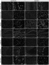
Figure 3. Expression of total MeCP2 and MeCP2E1 in adult murine brain.
Expression of total MeCP2 and MeCP2E1, respectively in olfactory bulb (A1, H1), cerebral cortex (B1, I1), striatum (C1, J1), dentate gyrus of hippocampus (D1, K1), thalamus (E1, L1), cerebellum (F1, M1) and brain stem (G1, N1). Higher magnification images for total MeCP2 (A2–G2) and MeCP2E1 (H2–N2) shows their nuclear expression pattern within the various brain regions. Scale bars: A1–G1; H1–N1 = 80 µm, A2–G2; H2–N2 = 20 µm.
Figure 4
Figure 4. Differential expression of total MeCP2 and MeCP2E1 in adult murine brain regions.
Quantification of total MeCP2 (A) and MeCP2E1 (B) in total cell extracts from the wild type Mecp2tm1.1Bird y/+ mice whole brain (Brain-WT), olfactory bulb, striatum, cerebral cortex, hippocampus, thalamus, brain stem and cerebellum. Mecp2tm1.1Bird y/− mice whole brain (Brain-Null) was included as a negative control. Equal loading of protein lysates was verified by probing the same membrane with ACTIN (N = 3±SEM).
Figure 5
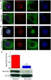
Figure 5. Expression of total MeCP2 and MeCP2E1 in primary neurons and astrocytes. A
) Expression of total MeCP2 in embryonic primary cortical neurons and astrocytes are detected by immunofluorescence labelling. Cells were labelled with β-III tubulin (β TUB III) and GFAP to mark neurons and astrocytes, respectively. B) Expression of MeCP2E1 in primary cortical neurons and astrocytes. Cells were labelled with NEUN and GFAP to mark neurons and astrocytes, respectively. Scale bars represent 5 µm. C) Western blot analysis of MeCP2E1 levels in primary cortical neurons and astrocytes. The graph depicts the quantification of MeCP2E1 in neurons and astrocytes, p<0.01 (N = 2±SEM).

References

    1. Meehan RR, Lewis JD, Bird AP (1992) Characterization of MeCP2, a vertebrate DNA binding protein with affinity for methylated DNA. Nucleic Acids Res 20: 5085–5092. - PMC - PubMed
    1. Amir RE, Van den Veyver IB, Wan M, Tran CQ, Francke U, et al. (1999) Rett syndrome is caused by mutations in X-linked MECP2, encoding methyl-CpG-binding protein 2. Nat Genet 23: 185–188. - PubMed
    1. Hagberg B (1985) Rett’s syndrome: prevalence and impact on progressive severe mental retardation in girls. Acta Paediatr Scand 74: 405–408. - PubMed
    1. Zachariah RM, Rastegar M (2012) Linking Epigenetics to Human Disease and Rett Syndrome: The Emerging Novel and Challenging Concepts in MeCP2 Research,. Neural Plasticity 2012: 10. - PMC - PubMed
    1. Chahrour M, Zoghbi HY (2007) The story of Rett syndrome: from clinic to neurobiology. Neuron 56: 422–437. - PubMed

Publication types