

Histone deacetylase (HDAC) 1 controls the expression of beta defensin 1 in human lung epithelial cells
- PMID: 23185513
- PMCID: PMC3502185
- DOI: 10.1371/journal.pone.0050000
Histone deacetylase (HDAC) 1 controls the expression of beta defensin 1 in human lung epithelial cells
Abstract
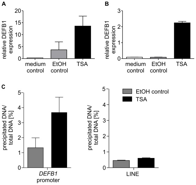
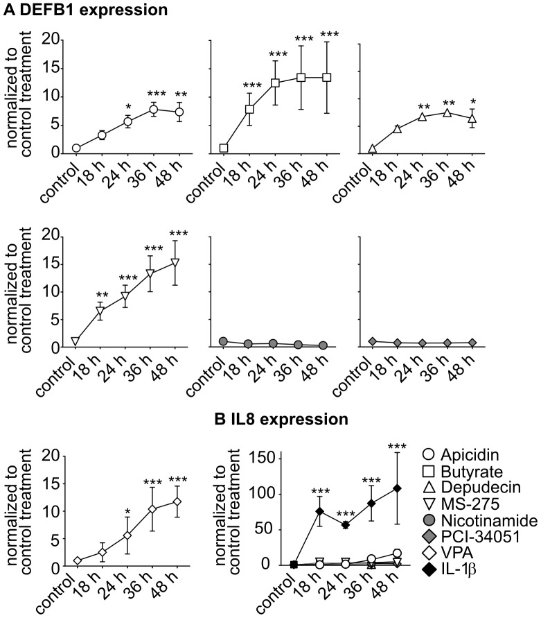
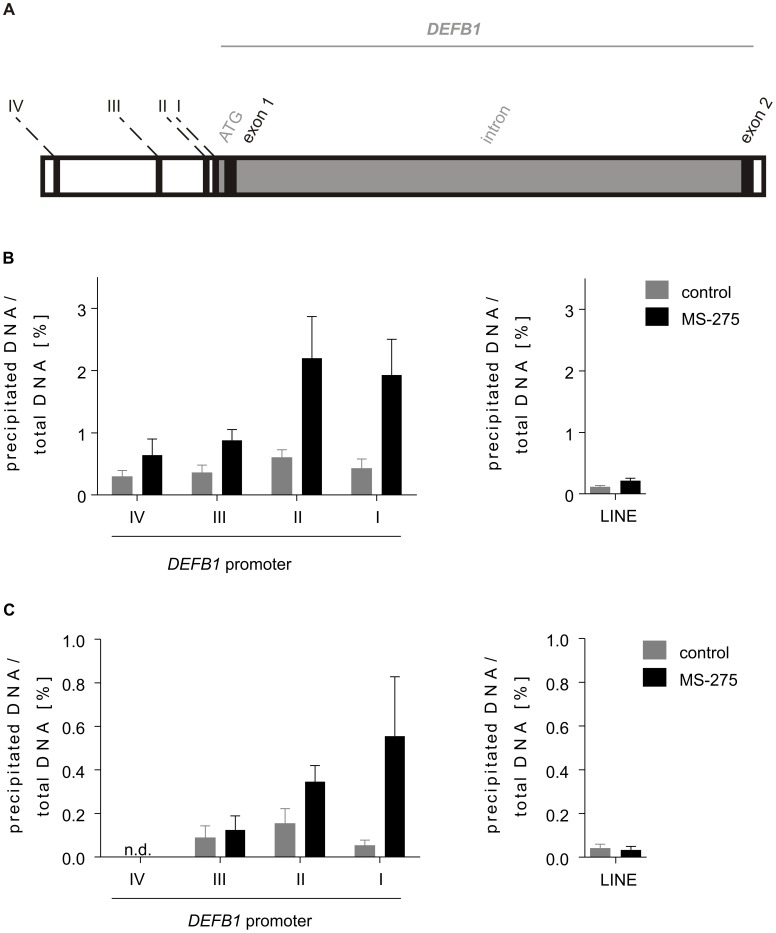
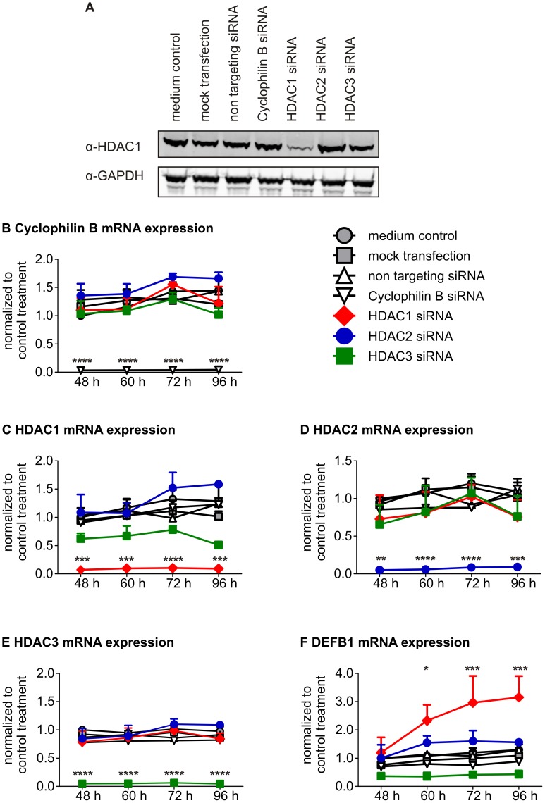
Deregulation of the expression human beta defensin 1 (DEFB1), an antimicrobial peptide, has been implicated in the pathogenesis of COPD and asthma. Since the molecular mechanisms that regulate DEFB1 gene expression are widely unknown, the epigenetic processes involved in the regulation of the constitutive expression of DEFB1 in lung epithelial cells (A549) were investigated. The data demonstrate that histone deacetylases (HDACs) participate in the regulation of DEFB1 gene expression. Inhibition of the class I HDACs, HDACs 1-3, increases DEFB1 gene expression in A549 cells. Chromatin immunoprecipitation (ChIP) assays revealed that the inhibition of the class I HDACs also results in modifications of the chromatin at the DEFB1 promoter. Histone modifications, histone H3 acetylation and H3K4 trimethylation, that are associated with transcriptional activation, were found to increase after inhibition of HDACs 1-3. Finally, RNAi knockdown experiments identified HDAC1 as the sole HDAC responsible for maintaining the constitutive level of DEFB1 transcription. Taken together, our data reveal epigenetic mechanisms which are the basis of the maintenance of the constitutive gene expression of human beta defensin 1.
Conflict of interest statement
Figures
Similar articles
-
Increased expression of beta-defensin 1 (DEFB1) in chronic obstructive pulmonary disease.PLoS One. 2011;6(7):e21898. doi: 10.1371/journal.pone.0021898. Epub 2011 Jul 19. PLoS One. 2011. PMID: 21818276 Free PMC article.
-
Trichostatin A induces 5-lipoxygenase promoter activity and mRNA expression via inhibition of histone deacetylase 2 and 3.J Cell Mol Med. 2012 Jul;16(7):1461-73. doi: 10.1111/j.1582-4934.2011.01420.x. J Cell Mol Med. 2012. PMID: 21883892 Free PMC article.
-
Histone deacetylase 1 and p300 can directly associate with chromatin and compete for binding in a mutually exclusive manner.PLoS One. 2014 Apr 10;9(4):e94523. doi: 10.1371/journal.pone.0094523. eCollection 2014. PLoS One. 2014. PMID: 24722339 Free PMC article.
-
Epigenetic regulation of airway inflammation.Curr Opin Immunol. 2007 Dec;19(6):694-700. doi: 10.1016/j.coi.2007.07.016. Epub 2007 Aug 27. Curr Opin Immunol. 2007. PMID: 17720468 Review.
-
Histone deacetylases and their inhibitors: new implications for asthma and chronic respiratory conditions.Curr Opin Allergy Clin Immunol. 2014 Feb;14(1):44-8. doi: 10.1097/ACI.0000000000000029. Curr Opin Allergy Clin Immunol. 2014. PMID: 24322009 Review.
Cited by
-
Association between Genetic Polymorphisms in DEFB1 and Susceptibility to Digestive Diseases.Med Sci Monit. 2015 Aug 2;21:2240-50. doi: 10.12659/MSM.893453. Med Sci Monit. 2015. PMID: 26232989 Free PMC article.
-
Identification of a Neisseria gonorrhoeae Histone Deacetylase: Epigenetic Impact on Host Gene Expression.Pathogens. 2020 Feb 18;9(2):132. doi: 10.3390/pathogens9020132. Pathogens. 2020. PMID: 32085531 Free PMC article.
-
Histone deacetylase (HDAC) inhibitors- based drugs are effective to control Mycobacterium tuberculosis infection and promote the sensibility for rifampicin in MDR strain.Mem Inst Oswaldo Cruz. 2023 Dec 22;118:e230143. doi: 10.1590/0074-02760230143. eCollection 2023. Mem Inst Oswaldo Cruz. 2023. PMID: 38126492 Free PMC article.
-
Integrating Proteomes for Lung Tissues and Lavage Reveals Pathways That Link Responses in Allergen-Challenged Mice.ACS Omega. 2021 Jan 5;6(2):1171-1189. doi: 10.1021/acsomega.0c04269. eCollection 2021 Jan 19. ACS Omega. 2021. PMID: 33490776 Free PMC article.
-
Repurposing HDAC inhibitors to enhance ribonuclease 4 and 7 expression and reduce urinary tract infection.Proc Natl Acad Sci U S A. 2023 Jan 24;120(4):e2213363120. doi: 10.1073/pnas.2213363120. Epub 2023 Jan 18. Proc Natl Acad Sci U S A. 2023. PMID: 36652479 Free PMC article.
References
-
- Zasloff M (2002) Antimicrobial peptides of multicellular organisms. Nature 415: 389–395. - PubMed
-
- Yang D, Chertov O, Bykovskaia SN, Chen Q, Buffo MJ, et al. (1999) Beta-defensins: linking innate and adaptive immunity through dendritic and T cell CCR6. Science 286: 525–528. - PubMed
-
- Murphy CJ, Foster BA, Mannis MJ, Selsted ME, Reid TW (1993) Defensins are mitogenic for epithelial cells and fibroblasts. J Cell Physiol 155: 408–413. - PubMed
-
- Bensch KW, Raida M, Magert HJ, Schulz-Knappe P, Forssmann WG (1995) hBD-1: a novel beta-defensin from human plasma. FEBS Lett 368: 331–335. - PubMed
-
- Goldman MJ, Anderson GM, Stolzenberg ED, Kari UP, Zasloff M, et al. (1997) Human beta-defensin-1 is a salt-sensitive antibiotic in lung that is inactivated in cystic fibrosis. Cell 88: 553–560. - PubMed
Publication types
MeSH terms
Substances
LinkOut - more resources
Full Text Sources
Miscellaneous
