. 2012;7(11):e50052.
doi: 10.1371/journal.pone.0050052. Epub 2012 Nov 20.

Lipocalin 2 regulates inflammation during pulmonary mycobacterial infections

Affiliations

Lipocalin 2 regulates inflammation during pulmonary mycobacterial infections

Lokesh Guglani et al. PLoS One. 2012.

Abstract

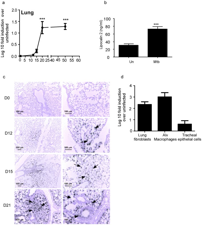
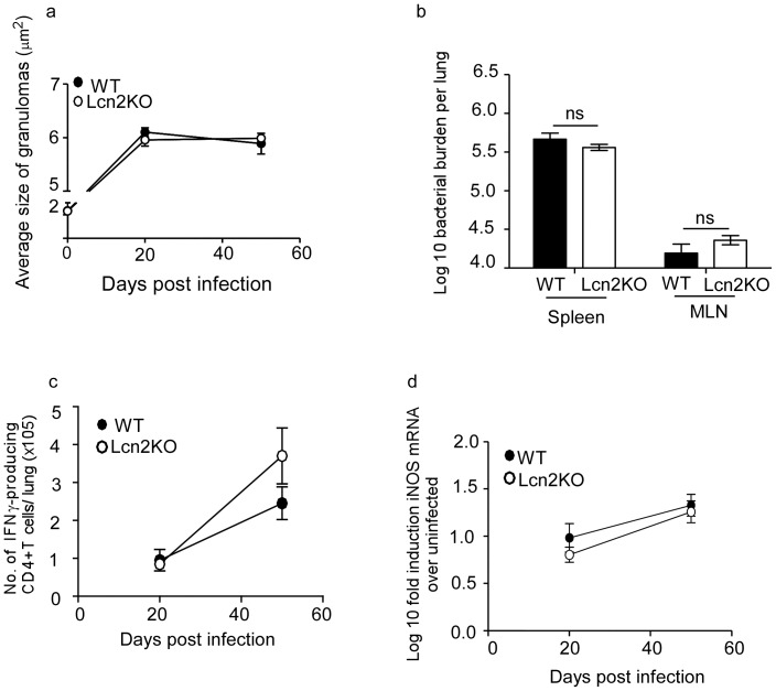
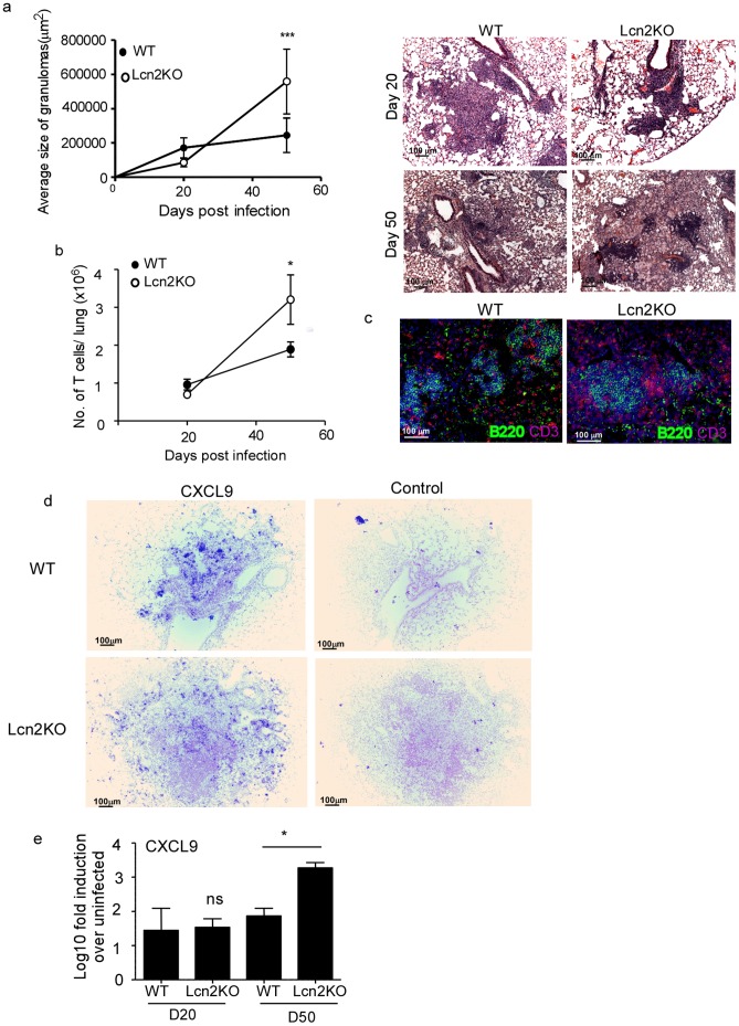
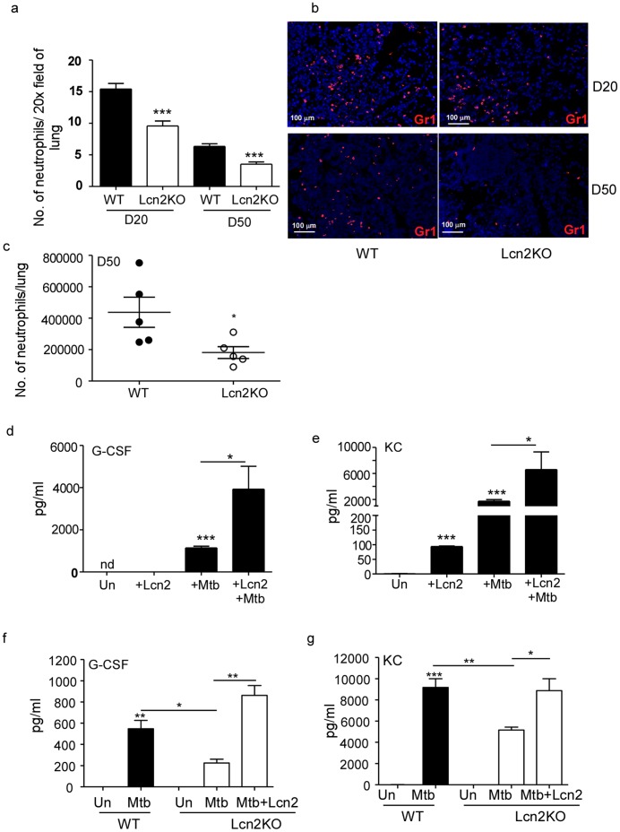
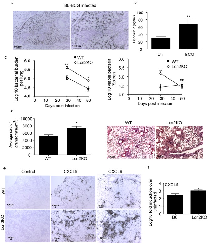
Pulmonary tuberculosis (TB), caused by the intracellular bacteria Mycobacterium tuberculosis, is a worldwide disease that continues to kill more than 1.5 million people every year worldwide. The accumulation of lymphocytes mediates the formation of the tubercle granuloma in the lung and is crucial for host protection against M.tuberculosis infection. However, paradoxically the tubercle granuloma is also the basis for the immunopathology associated with the disease and very little is known about the regulatory mechanisms that constrain the inflammation associated with the granulomas. Lipocalin 2 (Lcn2) is a member of the lipocalin family of proteins and binds to bacterial siderophores thereby sequestering iron required for bacterial growth. Thus far, it is not known whether Lcn2 plays a role in the inflammatory response to mycobacterial pulmonary infections. In the present study, using models of acute and chronic mycobacterial pulmonary infections, we reveal a novel role for Lcn2 in constraining T cell lymphocytic accumulation and inflammation by inhibiting inflammatory chemokines, such as CXCL9. In contrast, Lcn2 promotes neutrophil recruitment during mycobacterial pulmonary infection, by inducing G-CSF and KC in alveolar macrophages. Importantly, despite a common role for Lcn2 in regulating chemokines during mycobacterial pulmonary infections, Lcn2 deficient mice are more susceptible to acute M.bovis BCG, but not low dose M.tuberculosis pulmonary infection.


Conflict of interest statement

Competing Interests: The authors have declared that no competing interests exist.

Figures

Figure 1
Figure 1. Lcn2 is induced in murine lungs in response to low dose aerosolized M.tuberculosis infection.
Wild type (WT) mice were infected with ∼100 CFU M.tuberculosis via the aerosol route and at specific times after infection, lung tissue was harvested and processed to extract RNA and the expression of Lcn2 mRNA was determined by RT-PCR. Log 10 fold induction of Lcn2 gene in the lungs of M.tuberculosis-infected mice over lungs from uninfected controls is shown (a). WT M.tuberculosis-infected lungs (Mtb) on day 30 post infection or uninfected lungs (Un) were assayed for Lcn2 protein by ELISA (b). In situ hybridization with a Lcn2 cRNA probe was carried out in uninfected and M.tuberculosis-infected lung samples at various time points post infection (c). Original magnification. 100X-left panel; 200X-rightpanel). Lung fibroblasts, alveolar macrophages and tracheal epithelial cells were generated from WT lungs and cells were exposed to irradiated M.tuberculosis (100 µg/ml) for 48 hours. The induction of Lcn2 mRNA was measured by RT-PCR and the log 10 fold induction was measured in treated samples relative to untreated controls (d). The data points represent the mean and SD for four mice for each time point and one experiment representative of two shown. ***, p ≤0.0005.
Figure 2
Figure 2. Lcn2 is not required for protective immunity against low dose M.tuberculosis infection.
WT and Lcn2KO mice were infected with low dose aerosolized M.tuberculosis and the bacterial burden determined in the lungs at different time points post infection (a) and spleens and mediastinal lymph nodes (MLN) on day 50 post infection (b). Lymphocytes were isolated from the lung and the number of CD4+ IFNγ+ producing T cells were determined by intra cellular staining and flow cytometry following stimulation with PMA and Ionomycin (c). Lung from uninfected and infected WT and Lcn2KO mice were processed and the Log 10 induction of iNOS mRNA in infected samples relative to uninfected control samples was determined by RT-PCR (d). The data points represent the mean and SD for four mice for each time point and one experiment representative of two shown. ns-not significant.
Figure 3
Figure 3. Lcn2 regulates pulmonary inflammation during TB.
WT and Lcn2KO mice were infected with low dose aerosolized M.tuberculosis and formalin-fixed paraffin-embedded lungs were stained using H&E. Morphometric analysis of the average size of the granuloma in H&E stained sections of M.tuberculosis-infected WT and Lcn2KO lungs was determined and a representative section of the lung granuloma is shown for D20 and D50 post M.tuberculosis infection (a). Original magnification shown-100X. Lymphocytes were isolated from the lungs of WT and Lcn2KO M.tuberculosis-infected mice at day 20 and day 50 post infection and the number of CD3+ T cells determined by surface staining and flow cytometry (b). Lung sections from M.tuberculosis-infected WT and Lcn2KO mice at D50 post infection were stained for B220 (green) and CD3 (red) and shown at 200X magnification (c). Formalin-fixed, paraffin-embedded lung sections from WT and Lcn2KO from day 50 M.tuberculosis-infected mice were assayed for CXCL9 mRNA localization by ISH using a murine CXCL9 mRNA probe or control probe (d) and total lung CXCL9 mRNA levels induced quantitated using RT-PCR in day 20 (D20) or day 50 (D50) M.tuberculosis-infected mice (e). Original magnification-100X (d). The data points represent the mean and SD for four mice for each time point and one experiment representative of two shown. *, p ≤0.05. ***, p ≤0.0005.
Figure 4
Figure 4. Lcn2 is required for neutrophil recruitment to the lung during TB.
WT and Lcn2KO were infected with low dose aerosolized M.tuberculosis infection as described in Figure 2. Formalin-fixed paraffin-embedded lung sections from infected WT and Lcn2KO mice were stained for neutrophils (Gr1-red) by immunofluorescence. The number of neutrophils per 20X field was estimated at D20 and D50 (a) and a representative image is shown at 200X magnification (b). Lung cells were isolated from WT and Lcn2KO M.tuberculosis-infected lungs at D50 post infection and the total number of neutrophils (Gr1+CD11b+) determined by surface staining and flow cytometry (c). Alveolar macrophages isolated from WT mice were exposed to M.tuberculosis (100 µg/ml) alone, Lcn2 alone (10 µg/ml), or both Lcn2 (10 µg/ml) and M.tuberculosis (100 µg/ml) and levels of G-CSF (d), KC (e) were measured in culture supernatants after 48 hours. Alveolar macrophages were isolated from WT or Lcn2KO mice were exposed to M.tuberculosis (100 µg/ml) and exogenous Lcn2 (10 µg/ml) and the levels of G-CSF (f) and KC (g) determined in culture supernatant after 48 hours. The data points represent the mean and SD for 3–4 samples for each time point and one experiment representative of two shown. *, p ≤0.05. **, p ≤0.005. ***, p ≤0.0005.
Figure 5
Figure 5. Lcn2 regulates the M.bovis BCG induced inflammation and protection in murine lung.
Wild type (WT) mice were infected with 5×10⁶ CFU of M.bovis BCG by the intratracheal route and day 30 post-infection, formalin-fixed, paraffin-embedded lung sections were assayed for Lcn2 mRNA localization by ISH using a murine Lcn2 mRNA probe (a) and Lcn2 protein levels determined by ELISA (b). Bacterial burden was determined in the lungs and spleen of M.bovis BCG infected WT and Lcn2KO mice at the described time points (c). Formalin-fixed paraffin-embedded lungs were stained using H&E and morphometric analysis of the area occupied by the granuloma in H&E stained sections of M.bovis BCG infected WT and Lcn2KO lungs was determined (d). A representative section of the lung granuloma is shown at 100X. Formalin-fixed, paraffin-embedded lung sections from infected WT and Lcn2KO were assayed for CXCL9 mRNA localization by ISH using a murine CXCL9 mRNA probe or control probe (e) and total lung CXCL9 mRNA levels induced quantitated using RT-PCR in day 30 infected lungs (f). Original magnification 40X-left panel, middle panel; 100X-right panel (e).The data points represent the mean and SD for four mice for each time point and one experiment representative of two shown. *, p ≤0.05. **, p ≤0.005. ns-not significant.

Similar articles

Cited by

References

    1. Khader SA, Rangel-Moreno J, Fountain JJ, Martino CA, Reiley WW, et al. (2009) In a murine tuberculosis model, the absence of homeostatic chemokines delays granuloma formation and protective immunity. J Immunol 183: 8004–8014. - PMC - PubMed
    1. Seiler P, Aichele P, Bandermann S, Hauser AE, Lu B, et al. (2003) Early granuloma formation after aerosol Mycobacterium tuberculosis infection is regulated by neutrophils via CXCR3-signaling chemokines. Eur J Immunol 33: 2676–2686. - PubMed
    1. Kipnis A, Basaraba R, Orme I, Cooper A (2003) Role of chemokine CCL2 in the protective response to early murine pulmonary tuberculosis. Immunology 109: 547–551. - PMC - PubMed
    1. Peters W, Scott H, Chambers H, Flynn J, Charo I, et al. (2001) Chemokine receptor 2 serves an early and essential role in resistance to Mycobacterium tuberculosis . Proceedings of the National Academy of the Sciences USA 98: 7958–7963. - PMC - PubMed
    1. Scott H, Flynn J (2002) Mycobacterium tuberculosis in chemokine receptor 2-deficient mice: influence of dose on disease progression. Infection and Immunity 70: 5946–5954. - PMC - PubMed

Publication types

MeSH terms