Differential deactivation during mentalizing and classification of autism based on default mode network connectivity
- PMID: 23185536
- PMCID: PMC3501481
- DOI: 10.1371/journal.pone.0050064
Differential deactivation during mentalizing and classification of autism based on default mode network connectivity
Abstract
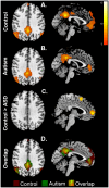
The default mode network (DMN) is a collection of brain areas found to be consistently deactivated during task performance. Previous neuroimaging studies of resting state have revealed reduced task-related deactivation of this network in autism. We investigated the DMN in 13 high-functioning adults with autism spectrum disorders (ASD) and 14 typically developing control participants during three fMRI studies (two language tasks and a Theory-of-Mind (ToM) task). Each study had separate blocks of fixation/resting baseline. The data from the task blocks and fixation blocks were collated to examine deactivation and functional connectivity. Deficits in the deactivation of the DMN in individuals with ASD were specific only to the ToM task, with no group differences in deactivation during the language tasks or a combined language and self-other discrimination task. During rest blocks following the ToM task, the ASD group showed less deactivation than the control group in a number of DMN regions, including medial prefrontal cortex (MPFC), anterior cingulate cortex, and posterior cingulate gyrus/precuneus. In addition, we found weaker functional connectivity of the MPFC in individuals with ASD compared to controls. Furthermore, we were able to reliably classify participants into ASD or typically developing control groups based on both the whole-brain and seed-based connectivity patterns with accuracy up to 96.3%. These findings indicate that deactivation and connectivity of the DMN were altered in individuals with ASD. In addition, these findings suggest that the deficits in DMN connectivity could be a neural signature that can be used for classifying an individual as belonging to the ASD group.
Conflict of interest statement
Figures


References
-
- Castelli F, Frith C, Happé F, Frith U (2002) Autism, asperger syndrome and brain mechanisms for the attribution of mental states to animated shapes. Brain 125: 1839–49. - PubMed
-
- Courchesne E, Pierce K, Schumann CM, Redcay E, Buckwalter JA, et al. (2007) Mapping early brain development in autism. Neuron 56: 399–413. - PubMed
-
- Cherkassky VL, Kana RK, Keller TA, Just MA (2006) Functional connectivity in a baseline resting-state network in autism. NeuroReport 17: 1687–90. - PubMed
-
- Kennedy DP, Courchesne E (2008) The intrinsic functional organization of the brain is altered in autism. NeuroImage 39: 1877–85. - PubMed
Publication types
MeSH terms
LinkOut - more resources
Full Text Sources
Other Literature Sources

