. 2012;7(11):e50142.
doi: 10.1371/journal.pone.0050142. Epub 2012 Nov 21.

Ribosomal binding site switching: an effective strategy for high-throughput cloning constructions

Affiliations

Ribosomal binding site switching: an effective strategy for high-throughput cloning constructions

Yangbo Hu et al. PLoS One. 2012.

Abstract

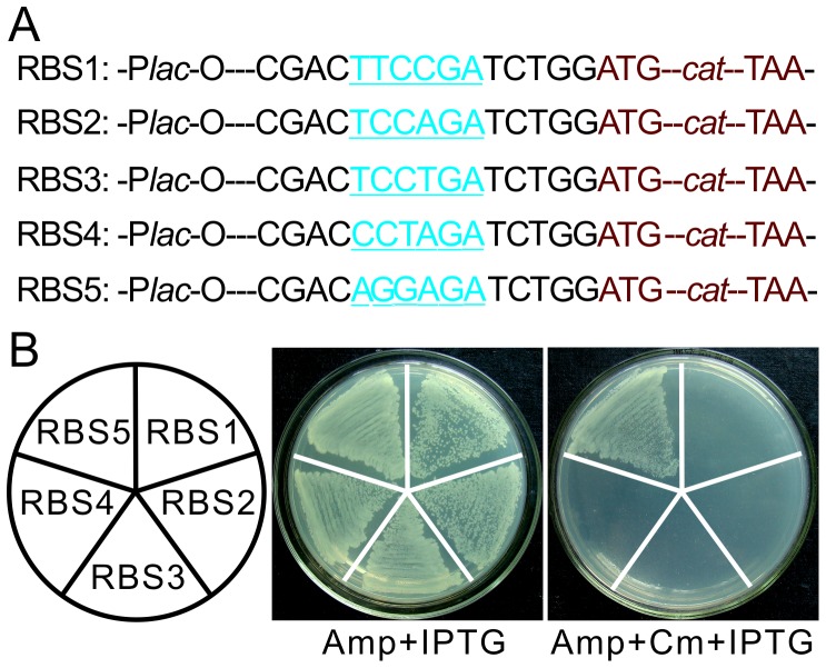
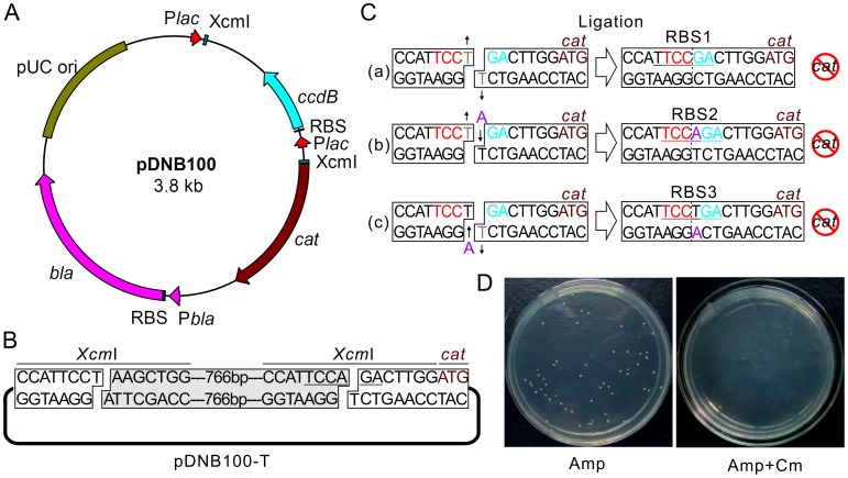
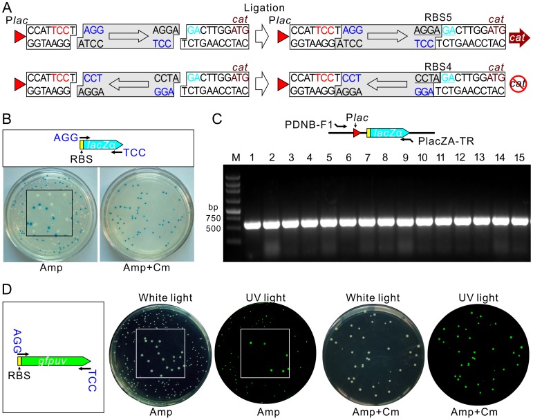
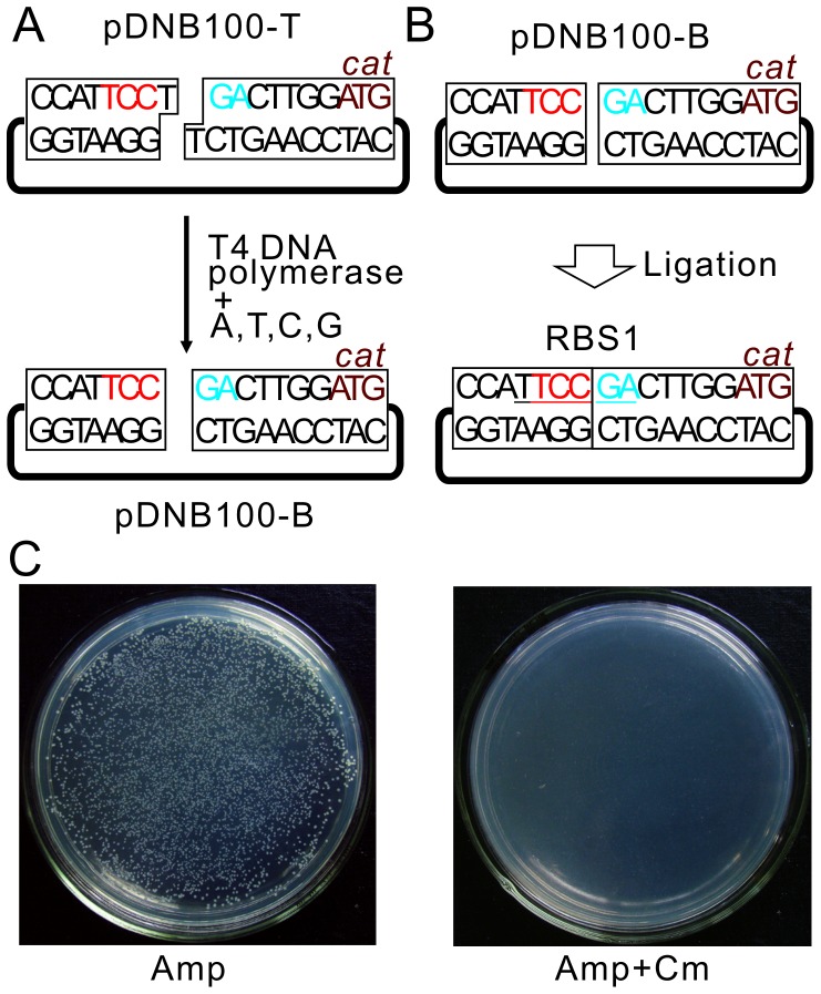
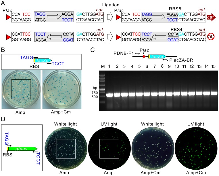
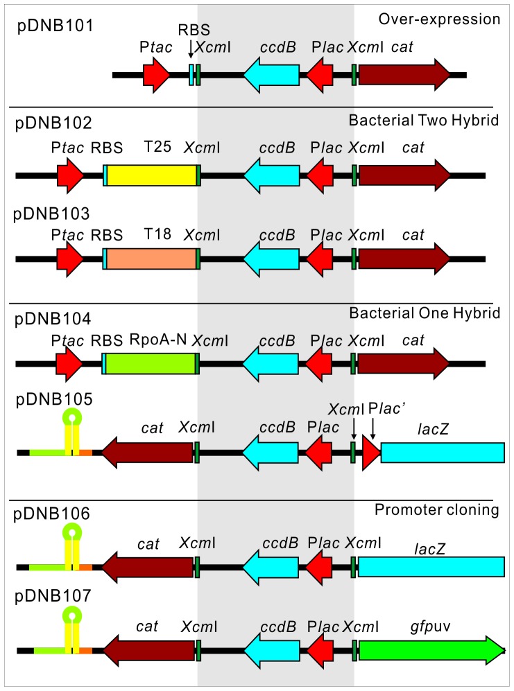
Direct cloning of PCR fragments by TA cloning or blunt end ligation are two simple methods which would greatly benefit high-throughput (HTP) cloning constructions if the efficiency can be improved. In this study, we have developed a ribosomal binding site (RBS) switching strategy for direct cloning of PCR fragments. RBS is an A/G rich region upstream of the translational start codon and is essential for gene expression. Change from A/G to T/C in the RBS blocks its activity and thereby abolishes gene expression. Based on this property, we introduced an inactive RBS upstream of a selectable marker gene, and designed a fragment insertion site within this inactive RBS. Forward and reverse insertions of specifically tailed fragments will respectively form an active and inactive RBS, thus all background from vector self-ligation and fragment reverse insertions will be eliminated due to the non-expression of the marker gene. The effectiveness of our strategy for TA cloning and blunt end ligation are confirmed. Application of this strategy to gene over-expression, a bacterial two-hybrid system, a bacterial one-hybrid system, and promoter bank construction are also verified. The advantages of this simple procedure, together with its low cost and high efficiency, makes our strategy extremely useful in HTP cloning constructions.


Conflict of interest statement

Competing Interests: Although the authors have filed a patent application (#CN20120076722.6) in China relating to the method developed in this article, this does not alter the authors' adherence to all the PLOS ONE policies on sharing data and materials. The authors declare there are no other relevant declarations relating to employment, consultancy, patents, products in development or marketed products, etc.

Figures

Figure 1
Figure 1. Influence of A/G content in the RBS sequence on gene expression.
(A) Design of five different RBS sequences (blue with underline) upstream of the cat gene (brown). All fragments were inserted downstream of the lac-O promoter (Plac-O) in pMD19-T vector and named RBS1 to RBS5. (B) Analysis of activities of these RBS sequences by Cm resistance. Five E. coli recombinant strains with plasmids containing RBS1 to RBS5 shown in left were respectively streaked on LB plates containing Amp+IPTG or Amp+Cm+IPTG.
Figure 2
Figure 2. Vector map of pDNB100 and its efficiency in removing vector self-ligation.
(A) Vector map of pDNB100. All key elements are labeled with different colors. (B) Frame of pDNB100-T. T tails are created by XcmI digestion of pDNB100 plasmid. The removed fragment is shown by the grey box. (C) Different situations for self-ligation of the pDNB100-T vector. The grey “T” represents the nucleotide that been removed during the ligation step or after been transformed into E. coli. The purple “A” represents nucleotide added to the plasmid by the bacterial DNA repair system. The start codon ATG for the cat gene is shown in brown. More details are given in the main text. (D) Transformants of pDNB100-T self-ligation product on LB plates with (right) or without (left) Cm.
Figure 3
Figure 3. Application of pDNB100-T in TA cloning construction.
(A) RBS sequences formed by forward and reverse insertion of PCR fragment. The boxed arrowhead indicates the orientation of the PCR fragment. The purple nucleotides are introduced by primers. (B) Cloning of a RBS containing a lacZα fragment. The upper panel shows the scheme for PCR amplification. Transformants on plate without (left) or with (right) Cm are shown. Expression of lacZα in E. coli DHα is detected by formation of blue/white colonies using X-gal as substrate and in the presence of IPTG. The square in the center of plate shows a magnified image. (C) PCR confirmation of blue colonies on plate containing Cm as shown in B. Paired positions for primers used in PCR are shown in the upper panel. (D) Cloning of a RBS containing a gfpuv fragment. The scheme for PCR amplification is also shown in the left panel. Transformants of ligated products on plates without or with Cm under white light and ultraviolet (UV) light are shown to illustrate the expression of GFPuv.
Figure 4
Figure 4. Overview of pDNB100-B preparation and its efficiency at eliminating self-ligation.
(A) Scheme for pDNB100-B vector construction. The T tails in pDNB100-T are treated with T4 DNA polymerase in the presence of four nucleotides. (B) Situation for pDNB100-B self-ligation. RBS sequence formed by vector self-ligation is underlined. (C) Transformants of pDNB100-B self-ligation product on LB plates in the absence (left) or presence of Cm (right).
Figure 5
Figure 5. Application of pDNB100-B in blunt end cloning construction.
(A) RBS sequences formed by forward and reverse insertion of PCR fragments. The boxed arrowhead indicates the orientation of PCR fragments. Initiation codon ATG for the cat gene is shown in brown. (B) Application of pDNB100-B in cloning of a RBS containing a lacZα fragment. The upper panel shows the scheme for PCR amplification. Transformants of ligated products on plate without (left) or with (right) Cm are shown. The expression of lacZα is detected by β-galactosidase activity. (C) PCR confirmation of blue colonies as shown in part B. Paired positions for primers used in PCR are shown in the upper panel. (D) Cloning of a RBS-containing gfpuv fragment using pDNB100-B vector. The scheme for PCR amplification is also shown in the left panel. Transformants of ligated products are spread onto plates with or without Cm to compare the efficiency of cat selection. Images under white or UV light are also compared to test the expression of GFPuv. The squares in the center of plate images show an enlarged section of the image.
Figure 6
Figure 6. Schematic of plasmids incorporating the RBS switching strategy in different cloning constructions.
Plasmids for gene over-expression (pDNB101), bacterial two-hybrid system (pDNB102 and pDNB103), bacterial one-hybrid system (pDNB104 and pDNB105) and promoter bank construction (pDNB106 and pDNB107) are presented. Each key element in the vector is labeled and a strong terminator upstream of the cat reporter gene in each of pDNB105, pDNB106 and pDNB107 is shown as a hairpin structure. The XcmI cassette in each plasmid is shown with grey background.

Similar articles

Cited by

References

    1. Scharf SJ, Horn GT, Erlich HA (1986) Direct cloning and sequence analysis of enzymatically amplified genomic sequences. Science 233: 1076–1078. - PubMed
    1. Aslanidis C, de Jong PJ (1990) Ligation-independent cloning of PCR products (LIC-PCR). Nucleic Acids Res 18: 6069–6074. - PMC - PubMed
    1. Hsiao K (1993) Exonuclease III induced ligase-free directional subcloning of PCR products. Nucleic Acids Res 21: 5528–5529. - PMC - PubMed
    1. Nour-Eldin HH, Hansen BG, Norholm MH, Jensen JK, Halkier BA (2006) Advancing uracil-excision based cloning towards an ideal technique for cloning PCR fragments. Nucleic Acids Res 34: e122. - PMC - PubMed
    1. Bubeck P, Winkler M, Bautsch W (1993) Rapid cloning by homologous recombination in vivo. Nucleic Acids Res 21: 3601–3602. - PMC - PubMed

Publication types