. 2012;7(11):e50264.
doi: 10.1371/journal.pone.0050264. Epub 2012 Nov 21.

Glucocorticoids regulation of FosB/ΔFosB expression induced by chronic opiate exposure in the brain stress system

Affiliations

Glucocorticoids regulation of FosB/ΔFosB expression induced by chronic opiate exposure in the brain stress system

Daniel García-Pérez et al. PLoS One. 2012.

Abstract

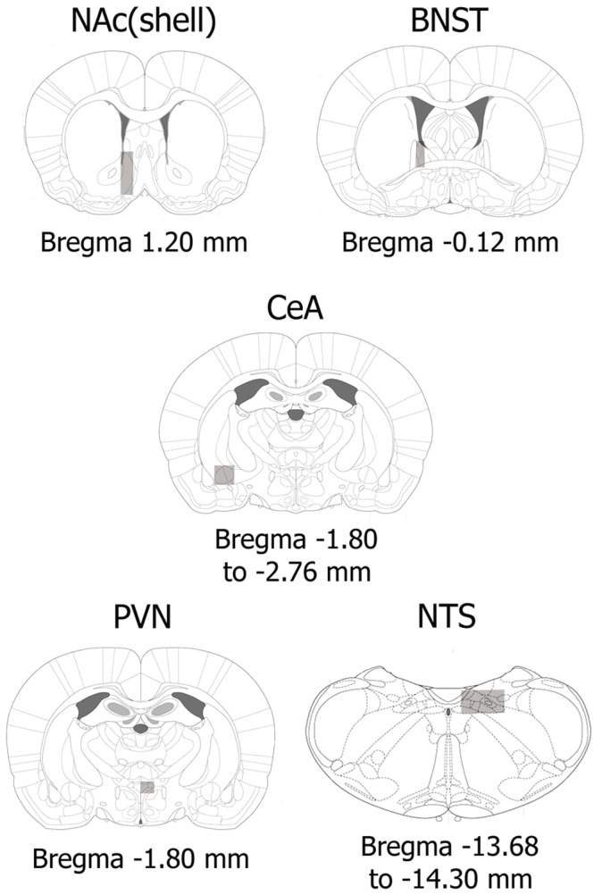
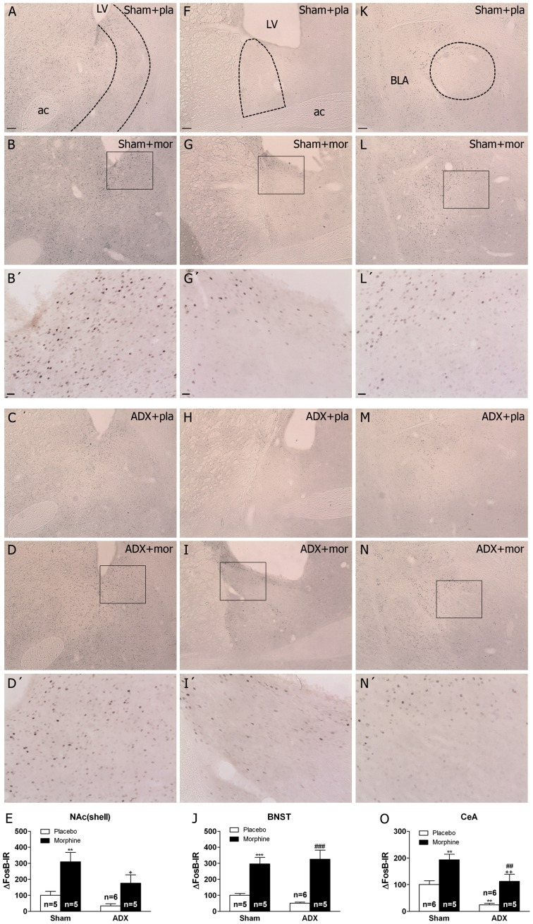
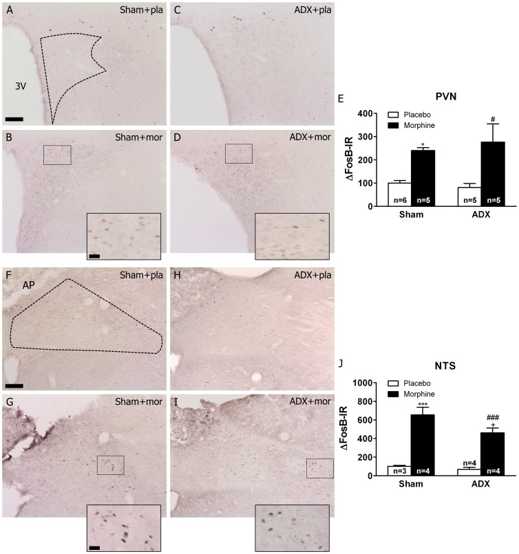
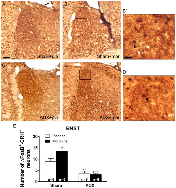
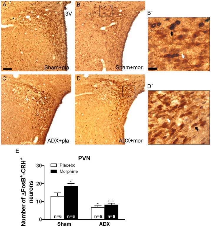
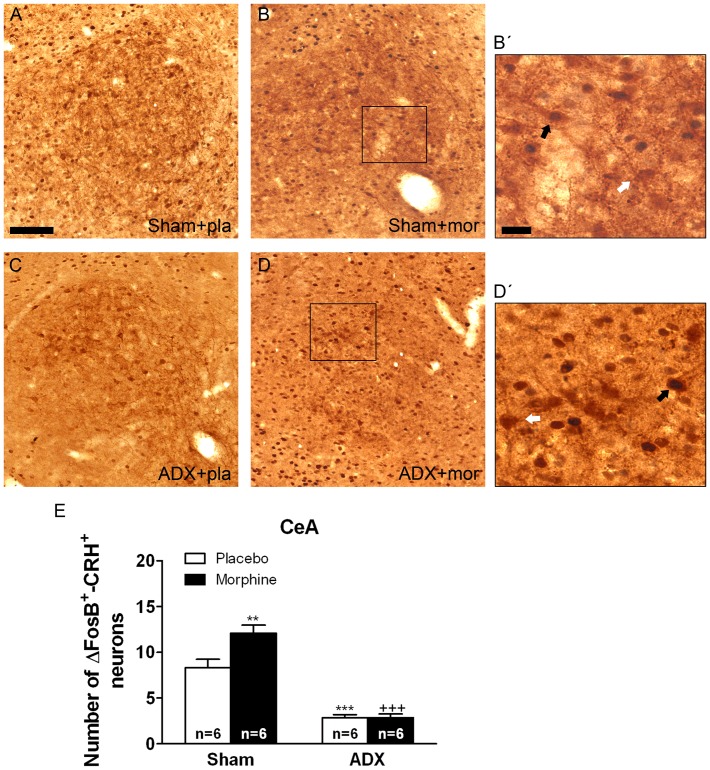
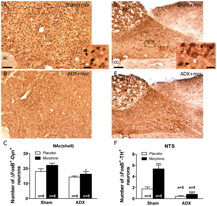
Chronic use of drugs of abuse profoundly alters stress-responsive system. Repeated exposure to morphine leads to accumulation of the transcription factor ΔFosB, particularly in brain areas associated with reward and stress. The persistent effects of ΔFosB on target genes may play an important role in the plasticity induced by drugs of abuse. Recent evidence suggests that stress-related hormones (e.g., glucocorticoids, GC) may induce adaptations in the brain stress system that is likely to involve alteration in gene expression and transcription factors. This study examined the role of GC in regulation of FosB/ΔFosB in both hypothalamic and extrahypothalamic brain stress systems during morphine dependence. For that, expression of FosB/ΔFosB was measured in control (sham-operated) and adrenalectomized (ADX) rats that were made opiate dependent after ten days of morphine treatment. In sham-operated rats, FosB/ΔFosB was induced after chronic morphine administration in all the brain stress areas investigated: nucleus accumbens(shell) (NAc), bed nucleus of the stria terminalis (BNST), central amygdala (CeA), hypothalamic paraventricular nucleus (PVN) and nucleus of the solitary tract noradrenergic cell group (NTS-A(2)). Adrenalectomy attenuated the increased production of FosB/ΔFosB observed after chronic morphine exposure in NAc, CeA, and NTS. Furthermore, ADX decreased expression of FosB/ΔFosB within CRH-positive neurons of the BNST, PVN and CeA. Similar results were obtained in NTS-A(2) TH-positive neurons and NAc pro-dynorphin-positive neurons. These data suggest that neuroadaptation (estimated as accumulation of FosB/ΔFosB) to opiates in brain areas associated with stress is modulated by GC, supporting the evidence of a link between brain stress hormones and addiction.


Conflict of interest statement

Competing Interests: The authors have declared that no competing interests exist.

Figures

Figure 1
Figure 1. Schematic drawings of coronal sections indicating brain areas (rectangles) in which FosB/ΔFosB positive nuclei were counted in the NAc(shell), BNST, CeA, PVN and NTS .
Figure 2
Figure 2. Adrenalectomy differentially regulates FosB/ΔFosB protein expression in the brain stress systems.
Photographs represent immunohistochemical detection of FosB/ΔFosB in the NAc(shell) (A-D), BNST (F-I) and CeA (K-N) from sham-operated and adrenalectomized (ADX) rats pretreated with placebo (pla) or morphine (mor) pellets for 10 days. B’, D’. G’, I’, L’ and N’ are high magnifications. Scale bar: 100 µm (30X, low magnification); 20 µm (100X, high magnification). LV, lateral ventricle; ac, anterior comisure; BLA, basolateral amygdala. E, J, O, quantitative analysis of FosB/ΔFosB immunoreactivity in the three nuclei from sham and ADX animal. Data correspond to the mean ± SEM (percent of control). Post hoc comparisons revealed a significant increase of FosB/ΔFosB protein expression in sham animals after chronic morphine exposure in the NAc(shell), BNST and CeA (**p<0.01; ***p<0.001 versus sham-placebo animals). In ADX-morphine dependent rats it was observed an attenuation of FosB/ΔFosB expression in the NAc(shell) and CeA compared with sham-dependent rats (+p<0.05; ++p<0.01 versus sham+morphine). In addition, ADX decreased basal FosB/ΔFosB expression in the CeA (**p<0.01 versus sham-placebo group. ##p<0.01; ###p<0.001 versus ADX+placebo).
Figure 3
Figure 3. Effects of adrenalectomy on FosB/ΔFosB protein expression in the PVN and NTS-A2 from morphine-dependent rats.
Photographs represent immunohistochemical detection of FosB/ΔFosB in the PVN (A-D), and NTS (F-I) neurons from sham-operated and ADX rats pretreated with placebo (pla) or morphine (mor) pellets for 10 days. Scale bar: 100 µm (70X, low magnification); 20 µm (200X, high magnification). 3 V, third ventricle; AP, area postrema; CC, canal central. Data represent the mean ± SEM (percent of control). E, J: quantitative analysis of FosB/ΔFosB-IR in the PVN and NTS, respectively. *p<0.05 ***p<0.001 versus sham+placebo; #p<0.05, ###p<0.001 versus ADX+placebo; +p<0.05 versus sham+morphine.
Figure 4
Figure 4. Adrenalectomy attenuated FosB/ΔFosB protein expression in the BNST CRH-positive neurons.
ADX and non-operated (sham) rats were made dependent on morphine (mor) for 10 days. Controls received placebo pellets (pla). Animals were perfused and the BNST was processed for double-labelled (FosB/ΔFosB and CRH) immunohistochemistry. Panels A-D show immunohistochemical detection of FosB/ΔFosB into CRH neurons after different treatments. Low and high magnifications (B’, D’) images show FosB/ΔFosB-positive (blue-black)/CRH-positive (brown) neurons (black arrow) and FosB/ΔFosB-negative/CRH-positive (white arrow) immunoreactivity. Scale bar: 100 µm (50X, low magnification); 20 µm (250X, high magnification). LV, lateral ventricle. E: quantitative analysis of FosB/ΔFosB-positive/CRH-positive neurons in the BNST. Data correspond to mean ± SEM. Post hoc test revealed a significant higher number of FosB/ΔFosB-positive nuclei into CRH immunoreactive neurons in morphine-dependent rats (**p<0.01 versus sham+placebo). ADX attenuated the increase of FosB/ΔFosB expression in CRH-positive neurons both in placebo- (**p<0.01 versus sham+placebo) and in morphine-treated animals (+++p<0.001 versus sham+morphine).
Figure 5
Figure 5. Adrenalectomy attenuated FosB/ΔFosB protein expression in the PVN CRH-positive neurons.
ADX and non-operated (sham) rats were made dependent on morphine (mor) for 10 days. Controls received placebo pellets (pla). Animals were perfused and the PVN was processed for double-labelled (FosB/ΔFosB and CRH) immunohistochemistry. Panels A-D show immunohistochemical detection of FosB/ΔFosB into CRH neurons after different treatments. Low and high magnifications (B’, D’) images show FosB/ΔFosB-positive (blue-black)/CRH-positive (brown) neurons (black arrow) and FosB/ΔFosB-negative/CRH-positive (white arrow) immunoreactivity. Scale bar: 100 µm (50X, low magnification); 20 µm (250X, high magnification). 3 V, third ventricle. E: quantitative analysis of FosB/ΔFosB-positive/CRH-positive neurons in the PVN. Data correspond to mean ± SEM. Post hoc test revealed a significant higher number of FosB/ΔFosB-positive nuclei into CRH immunoreactive neurons in morphine-dependent rats (*p<0.05 versus sham-placebo). ADX attenuated the increase of FosB/ΔFosB expression in CRH-positive neurons both in placebo- (*p<0.01 versus sham+placebo) and in morphine-treated animals (+++p<0.001 versus sham+morphine).
Figure 6
Figure 6. Adrenalectomy antagonized FosB/ΔFosB protein expression in the CeA CRH-positive neurons.
ADX and non-operated (sham) rats were made dependent on morphine (mor) for 10 days. Controls received placebo pellets (pla). Animals were perfused and the CeA was processed for double-labelled (FosB/ΔFosB and CRH) immunohistochemistry. Panels A-D show immunohistochemical detection of FosB/ΔFosB into CRH neurons after different treatments. Low and high magnifications (B’, D’) images show FosB/ΔFosB-positive (blue-black)/CRH-positive (brown) neurons (black arrow) and FosB/ΔFosB-negative/CRH-positive (white arrow) immunoreactivity. Scale bar: 100 µm (80X, low magnification); 20 µm (275X, high magnification). E: quantitative analysis of FosB/ΔFosB-positive/CRH-positive neurons in the CeA. Data correspond to mean ± SEM. Post hoc test revealed a significant higher number of FosB/ΔFosB-positive nuclei into CRH immunoreactive neurons in morphine-dependent rats (**p<0.01 versus sham+placebo). ADX attenuated the increase of FosB/ΔFosB expression in CRH-positive neurons both in placebo- (***p<0.001 versus sham+placebo) and in morphine-treated animals (+++p<0.001 versus sham+morphine).
Figure 7
Figure 7. Effects of adrenalectomy on FosB/ΔFosB protein expression in the NAc(shell) pro-DYN-positive neurons and in the NTS TH-positive neurons from morphine dependent rats.
ADX and non-operated (sham) rats were made dependent on morphine (mor) for 10 days. Controls received placebo pellets. Animals were perfused and the NAc and NTS were processed for double-labelled (FosB/ΔFosB and pro-DYN and FosB/ΔFosB and TH, respectively) immunohistochemistry. Panels A-B show immunohistochemical detection of FosB/ΔFosB into pro-DYN-positive neurons in sham- and ADX-morphine-dependent rats. Low and high magnifications images show FosB/ΔFosB-positive (blue-black)/pro-DYN-positive (brown) neurons (black arrow) and FosB/ΔFosB-negative/pro-DYN-positive (white arrow) immunoreactivity. Panels D-E show immunohistochemical detection of FosB/ΔFosB within TH-positive neurons in sham- and ADX-morphine-dependent rats. Low and high magnifications images show FosB/ΔFosB-positive (blue-black)/TH-positive (brown) neurons (black arrow) and FosB/ΔFosB-negative/TH-positive (white arrow) immunoreactivity. Scale bars: 100 µm (70X, low magnification); 20 µm (300X, high magnification). C: quantitative analysis of FosB/ΔFosB-positive/pro-DYN-positive neurons in the NAc(shell). F: quantitative analysis of FosB/ΔFosB-positive/TH-positive neurons in the NTS. Data correspond to mean ± SEM. +p<0.05 versus sham+morphine. ac, anterior comisure, AP, area postrema; cc, canal central. Data correspond to mean ± SEM. ADX significantly attenuated the increase of FosB/ΔFosB expression in pro-DYN-positive neuron in the NAc and in TH-positive neurons in the NTS from morphine-dependent animals (***p<0.001 versus sham+placebo; +p<0.05, +++p<0.001 versus sham+morphine).

Similar articles

Cited by

References

    1. Volkow ND, Skolnick P (2012) New Medications for Substance Use Disorders: Challenges and Opportunities. Neuropsychopharmacology 37: 290–292. - PMC - PubMed
    1. Robison AJ, Nestler EJ (2011) Transcriptional and epigenetic mechanisms of addiction. Nat Rev Neurosci 12: 623–637. - PMC - PubMed
    1. Hutchison ER, Okun E, Mattson MP (2009) The Therapeutic Potential of microRNAs in Nervous System Damage, Degeneration and Repair. Neuromolecular Med 11: 153–161. - PMC - PubMed
    1. Li MD, van der Vaart AD (2011) MicroRNAs in addiction: adaptation’s middlemen? Mol Psychiatry 16: 1159–1168. - PMC - PubMed
    1. Mumberg D, Lucibello FC, Schuermann M, Muller R (1991) Alternative splicing of fosB transcripts results in differentially expressed mRNAs encoding functionally antagonistic proteins. Genes Dev 5: 1212–1223. - PubMed

Publication types

MeSH terms