Separation of fNIRS signals into functional and systemic components based on differences in hemodynamic modalities
- PMID: 23185590
- PMCID: PMC3501470
- DOI: 10.1371/journal.pone.0050271
Separation of fNIRS signals into functional and systemic components based on differences in hemodynamic modalities
Abstract
In conventional functional near-infrared spectroscopy (fNIRS), systemic physiological fluctuations evoked by a body's motion and psychophysiological changes often contaminate fNIRS signals. We propose a novel method for separating functional and systemic signals based on their hemodynamic differences. Considering their physiological origins, we assumed a negative and positive linear relationship between oxy- and deoxyhemoglobin changes of functional and systemic signals, respectively. Their coefficients are determined by an empirical procedure. The proposed method was compared to conventional and multi-distance NIRS. The results were as follows: (1) Nonfunctional tasks evoked substantial oxyhemoglobin changes, and comparatively smaller deoxyhemoglobin changes, in the same direction by conventional NIRS. The systemic components estimated by the proposed method were similar to the above finding. The estimated functional components were very small. (2) During finger-tapping tasks, laterality in the functional component was more distinctive using our proposed method than that by conventional fNIRS. The systemic component indicated task-evoked changes, regardless of the finger used to perform the task. (3) For all tasks, the functional components were highly coincident with signals estimated by multi-distance NIRS. These results strongly suggest that the functional component obtained by the proposed method originates in the cerebral cortical layer. We believe that the proposed method could improve the reliability of fNIRS measurements without any modification in commercially available instruments.
Conflict of interest statement
Figures

 ) and four detectors (
 –
– ) were fixed directly above the left and right primary motor areas. The detector optodes on both sides were linearly aligned at distances of 10, 20, 30, and 40 mm from the source optode.
 dependency of the mutual information
 for participant 1. Upper two rows: body-tilting task. Middle two rows: breath-holding task. Bottom two rows: finger-tapping task. Left and Right indicate the measurement positions. 10 mm, 20 mm, 30 mm, and 40 mm indicate the source-detector distances.
 values from all experiments.


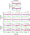
 conditions. Values of
  : −0.4 (left column), −0.6 (middle column), and −0.8 (right column) were used for the data at a source-detector distance of 30 mm during the finger-tapping task for participant 1. Upper two rows: hemodynamics estimated by the conventional method. Middle two rows: functional component by the proposed method. Bottom two rows: systemic component by the proposed method. Data were block averaged. Red and blue lines indicate oxy- and deoxyhemoglobin changes, respectively. Red and blue bands indicate SDs for oxy- and deoxyhemoglobin changes, respectively. Green line indicates the task period. “Left” and “Right” indicate the measurement positions. “L” (left) and “R” (right) indicate the side used during finger tapping.
Similar articles
-
Exploration of cerebral activation using hemodynamic modality separation method in high-density multichannel fNIRS.Annu Int Conf IEEE Eng Med Biol Soc. 2013;2013:1791-4. doi: 10.1109/EMBC.2013.6609869. Annu Int Conf IEEE Eng Med Biol Soc. 2013. PMID: 24110056
-
Event-related functional near-infrared spectroscopy (fNIRS) based on craniocerebral correlations: reproducibility of activation?Hum Brain Mapp. 2007 Aug;28(8):733-41. doi: 10.1002/hbm.20303. Hum Brain Mapp. 2007. PMID: 17080439 Free PMC article.
-
The physiological origin of task-evoked systemic artefacts in functional near infrared spectroscopy.Neuroimage. 2012 May 15;61(1):70-81. doi: 10.1016/j.neuroimage.2012.02.074. Epub 2012 Mar 9. Neuroimage. 2012. PMID: 22426347 Free PMC article.
-
Hemodynamic signals in fNIRS.Prog Brain Res. 2016;225:153-79. doi: 10.1016/bs.pbr.2016.03.004. Epub 2016 Apr 7. Prog Brain Res. 2016. PMID: 27130415 Review.
-
HomER: a review of time-series analysis methods for near-infrared spectroscopy of the brain.Appl Opt. 2009 Apr 1;48(10):D280-98. doi: 10.1364/ao.48.00d280. Appl Opt. 2009. PMID: 19340120 Free PMC article. Review.
Cited by
-
Cortical Speech Processing in Postlingually Deaf Adult Cochlear Implant Users, as Revealed by Functional Near-Infrared Spectroscopy.Trends Hear. 2018 Jan-Dec;22:2331216518786850. doi: 10.1177/2331216518786850. Trends Hear. 2018. PMID: 30022732 Free PMC article.
-
Inter-brain synchrony during mother-infant interactive parenting in 3-4-month-old infants with and without an elevated likelihood of autism spectrum disorder.Cereb Cortex. 2023 Dec 9;33(24):11609-11622. doi: 10.1093/cercor/bhad395. Cereb Cortex. 2023. PMID: 37885119 Free PMC article.
-
Separation of superficial and cerebral hemodynamics using a single distance time-domain NIRS measurement.Biomed Opt Express. 2014 Apr 10;5(5):1465-82. doi: 10.1364/BOE.5.001465. eCollection 2014 May 1. Biomed Opt Express. 2014. PMID: 24877009 Free PMC article.
-
Evaluating cortical responses to speech in children: A functional near-infrared spectroscopy (fNIRS) study.Hear Res. 2021 Mar 1;401:108155. doi: 10.1016/j.heares.2020.108155. Epub 2020 Dec 15. Hear Res. 2021. PMID: 33360183 Free PMC article.
-
Pre-operative Brain Imaging Using Functional Near-Infrared Spectroscopy Helps Predict Cochlear Implant Outcome in Deaf Adults.J Assoc Res Otolaryngol. 2019 Oct;20(5):511-528. doi: 10.1007/s10162-019-00729-z. Epub 2019 Jul 8. J Assoc Res Otolaryngol. 2019. PMID: 31286300 Free PMC article.
References
-
- Blasi A, Fox S, Everdell N, Volein A, Tucker L, et al. (2007) Investigation of depth dependent changes in cerebral haemodynamics during face perception in infants. Physics in medicine and biology 52: 6849–6864. - PubMed
-
- Yamada T, Umeyama S, Matsuda K (2009) Multidistance probe arrangement to eliminate artefacts in functional near-infrared spectroscopy. Journal of biomedical optics 14: 064034. - PubMed
Publication types
MeSH terms
Substances
LinkOut - more resources
Full Text Sources

