

Coating with a modular bone morphogenetic peptide promotes healing of a bone-implant gap in an ovine model
- PMID: 23185610
- PMCID: PMC3503930
- DOI: 10.1371/journal.pone.0050378
Coating with a modular bone morphogenetic peptide promotes healing of a bone-implant gap in an ovine model
Abstract
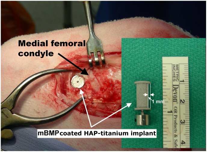
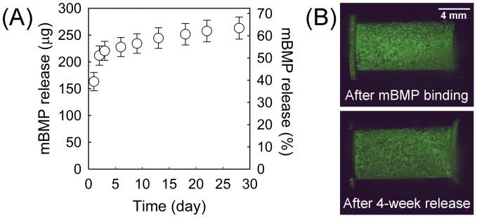
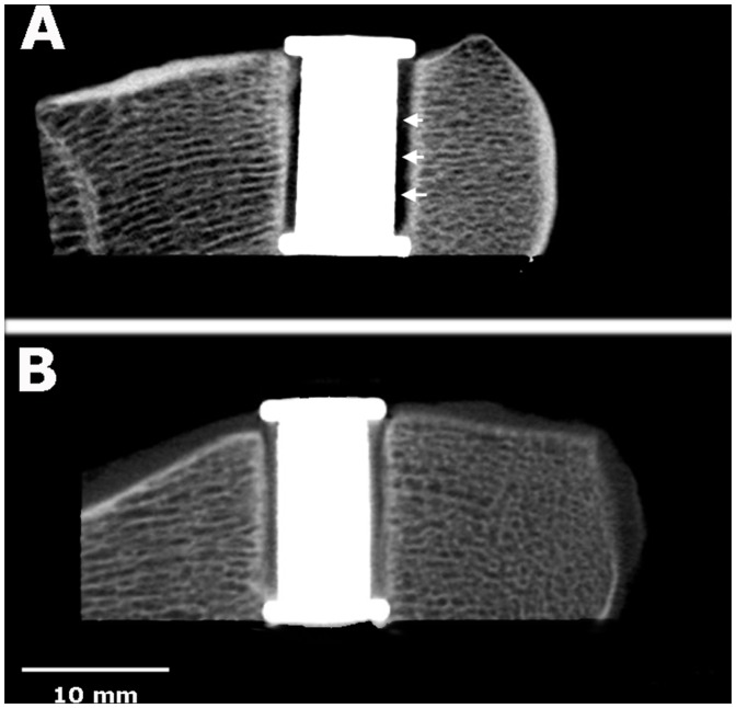
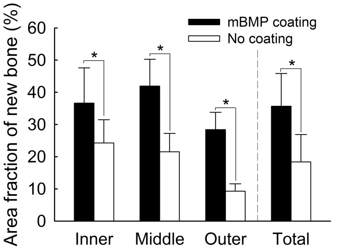
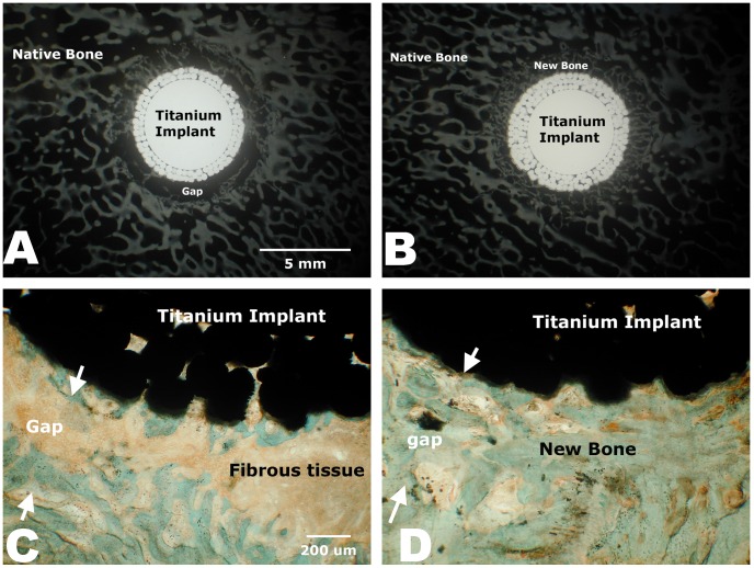
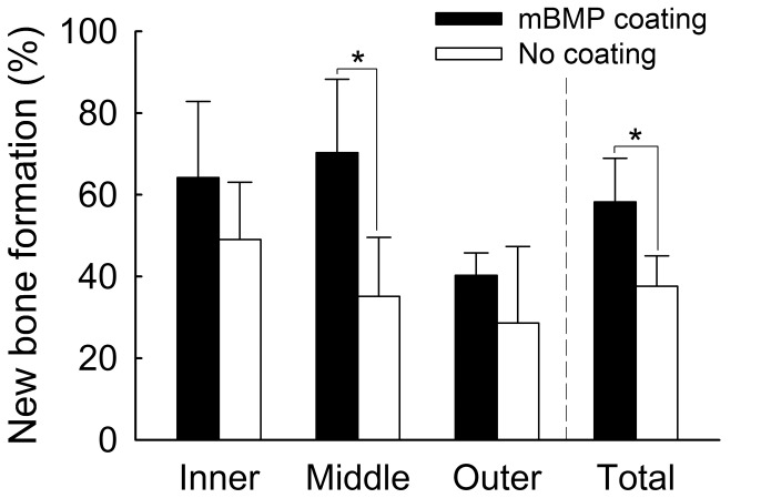
Despite the potential for growth factor delivery strategies to promote orthopedic implant healing, there is a need for growth factor delivery methods that are controllable and amenable to clinical translation. We have developed a modular bone growth factor, herein termed "modular bone morphogenetic peptide (mBMP)", which was designed to efficiently bind to the surface of orthopedic implants and also stimulate new bone formation. The purpose of this study was to coat a hydroxyapatite-titanium implant with mBMP and evaluate bone healing across a bone-implant gap in the sheep femoral condyle. The mBMP molecules efficiently bound to a hydroxyapatite-titanium implant and 64% of the initially bound mBMP molecules were released in a sustained manner over 28 days. The results demonstrated that the mBMP-coated implant group had significantly more mineralized bone filling in the implant-bone gap than the control group in C-arm computed tomography (DynaCT) scanning (25% more), histological (35% more) and microradiographic images (50% more). Push-out stiffness of the mBMP group was nearly 40% greater than that of control group whereas peak force did not show a significant difference. The results of this study demonstrated that mBMP coated on a hydroxyapatite-titanium implant stimulates new bone formation and may be useful to improve implant fixation in total joint arthroplasty applications.
Conflict of interest statement
Figures
Similar articles
-
[Improved osseointegration of titanium implants of different surface characteristics by the use of bone morphogenetic protein (BMP-3): an animal study performed at the metaphyseal bone bed in dogs].Z Orthop Ihre Grenzgeb. 2003 Nov-Dec;141(6):705-11. doi: 10.1055/s-2003-812411. Z Orthop Ihre Grenzgeb. 2003. PMID: 14679438 German.
-
Effects of a cell adhesion molecule coating on the blasted surface of titanium implants on bone healing in the rabbit femur.Int J Oral Maxillofac Implants. 2007 Jul-Aug;22(4):533-41. Int J Oral Maxillofac Implants. 2007. PMID: 17929513
-
Enhanced biocompatibility and osseointegration of calcium titanate coating on titanium screws in rabbit femur.J Huazhong Univ Sci Technolog Med Sci. 2017 Jun;37(3):362-370. doi: 10.1007/s11596-017-1741-9. Epub 2017 Jun 6. J Huazhong Univ Sci Technolog Med Sci. 2017. PMID: 28585129
-
Surface modification of implants in long bone.Biomatter. 2012 Jul-Sep;2(3):149-57. doi: 10.4161/biom.21563. Biomatter. 2012. PMID: 23507866 Free PMC article. Review.
-
Strontium in the bone-implant interface.Dan Med Bull. 2011 May;58(5):B4286. Dan Med Bull. 2011. PMID: 21535993 Review.
Cited by
-
Selection and identification of a novel bone-targeting peptide for biomedical imaging of bone.Sci Rep. 2020 Jun 29;10(1):10576. doi: 10.1038/s41598-020-67522-4. Sci Rep. 2020. PMID: 32601412 Free PMC article.
-
Biomaterial strategies for engineering implants for enhanced osseointegration and bone repair.Adv Drug Deliv Rev. 2015 Nov 1;94:53-62. doi: 10.1016/j.addr.2015.03.013. Epub 2015 Apr 8. Adv Drug Deliv Rev. 2015. PMID: 25861724 Free PMC article. Review.
-
Effects of BMP-12-releasing sutures on Achilles tendon healing.Tissue Eng Part A. 2015 Mar;21(5-6):916-27. doi: 10.1089/ten.TEA.2014.0001. Epub 2014 Dec 11. Tissue Eng Part A. 2015. PMID: 25354567 Free PMC article.
-
The use of recombinant human bone morphogenetic protein-2 (rhBMP-2) in maxillofacial trauma.Chin J Traumatol. 2017 Feb;20(1):1-3. doi: 10.1016/j.cjtee.2016.05.004. Epub 2017 Feb 9. Chin J Traumatol. 2017. PMID: 28236566 Free PMC article. Review.
-
Stable biofunctionalization of hydroxyapatite (HA) surfaces by HA-binding/osteogenic modular peptides for inducing osteogenic differentiation of mesenchymal stem cells.Biomater Sci. 2014;2:1779-1786. doi: 10.1039/C4BM00164H. Biomater Sci. 2014. PMID: 25642327 Free PMC article.
References
-
- Sachse A, Wagner A, Keller M, Wagner O, Wetzel WD, et al. (2005) Osteointegration of hydroxyapatite-titanium implants coated with nonglycosylated recombinant human bone morphogenetic protein-2 (BMP-2) in aged sheep. Bone 37: 699–710. - PubMed
-
- Lamberg A, Schmidmaier G, Soballe K, Elmengaard B (2006) Locally delivered TGF-beta1 and IGF-1 enhance the fixation of titanium implants: a study in dogs. Acta Orthop 77: 799–805. - PubMed
Publication types
MeSH terms
Substances
LinkOut - more resources
Full Text Sources
Miscellaneous
