. 2012;7(11):e50394.
doi: 10.1371/journal.pone.0050394. Epub 2012 Nov 21.

Peroxiredoxin 1 stimulates endothelial cell expression of VEGF via TLR4 dependent activation of HIF-1α

Affiliations

Peroxiredoxin 1 stimulates endothelial cell expression of VEGF via TLR4 dependent activation of HIF-1α

Jonah R Riddell et al. PLoS One. 2012.

Abstract

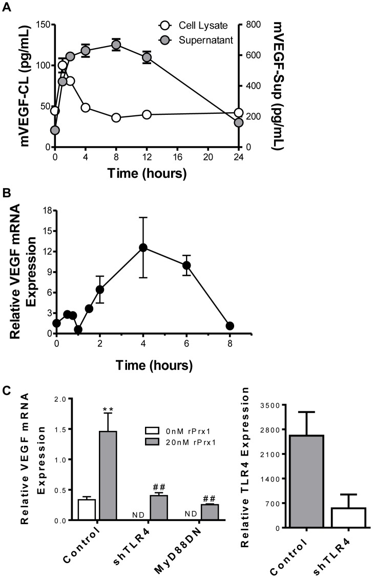
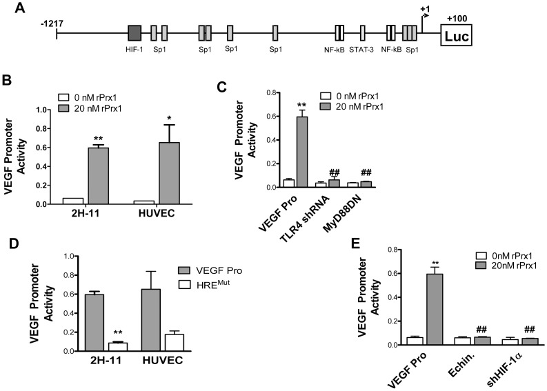
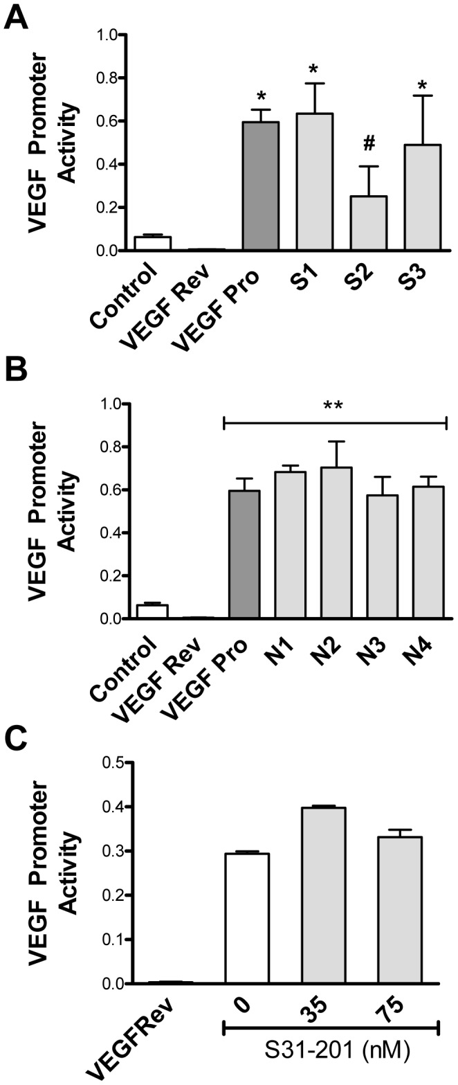
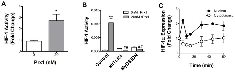
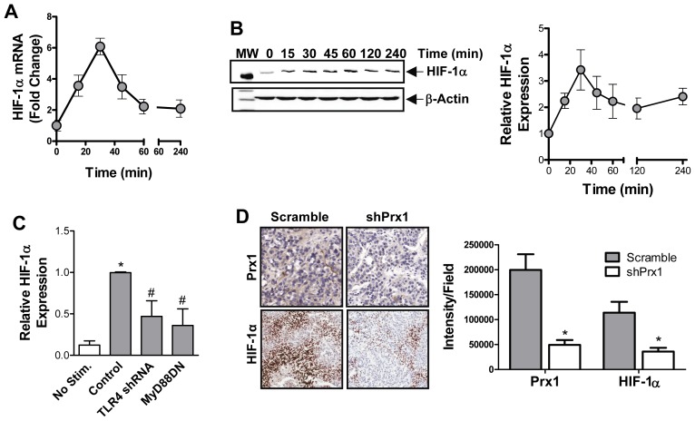
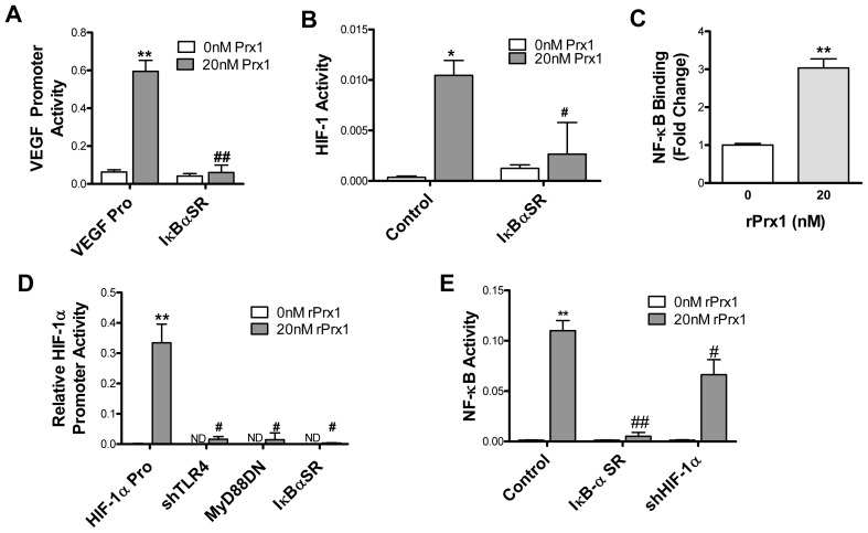
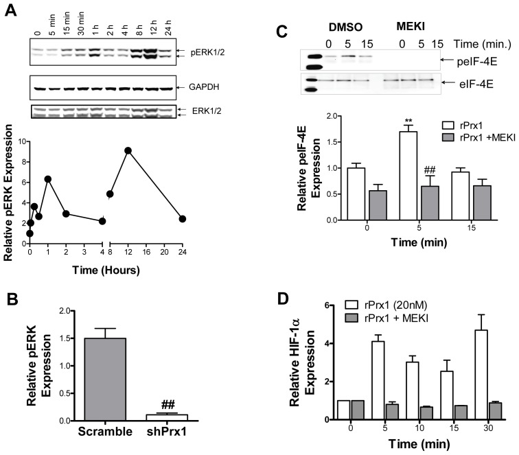
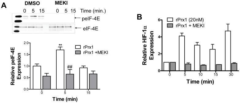
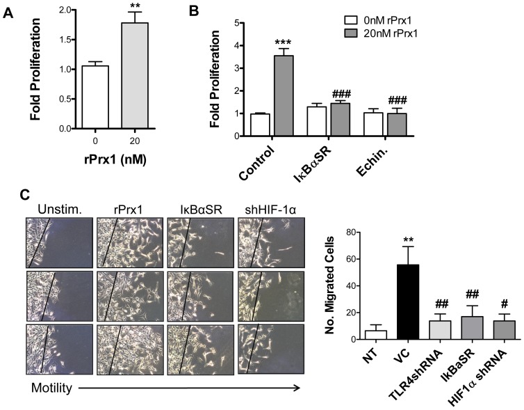
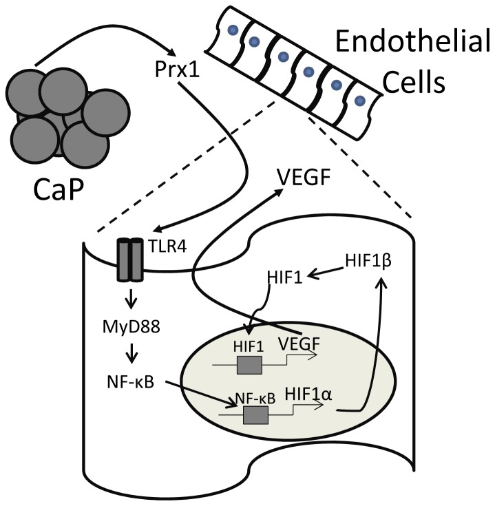
Chronic inflammation leads to the formation of a pro-tumorigenic microenvironment that can promote tumor development, growth and differentiation through augmentation of tumor angiogenesis. Prostate cancer (CaP) risk and prognosis are adversely correlated with a number of inflammatory and angiogenic mediators, including Toll-like receptors (TLRs), NF-κB and vascular endothelial growth factor (VEGF). Peroxiredoxin 1 (Prx1) was recently identified as an endogenous ligand for TLR4 that is secreted from CaP cells and promotes inflammation. Inhibition of Prx1 by CaP cells resulted in reduced expression of VEGF, diminished tumor vasculature and retarded tumor growth. The mechanism by which Prx1 regulates VEGF expression in normoxic conditions was investigated in the current study. Our results show that incubation of mouse vascular endothelial cells with recombinant Prx1 caused increases in VEGF expression that was dependent upon TLR4 and required hypoxia inducible factor-1 (HIF-1) interaction with the VEGF promoter. The induction of VEGF was also dependent upon NF-κB; however, NF-κB interaction with the VEGF promoter was not required for Prx1 induction of VEGF suggesting that NF-κB was acting indirectly to induce VEGF expression. The results presented here show that Prx1 stimulation increased NF-κB interaction with the HIF-1α promoter, leading to enhanced promoter activity and increases in HIF-1α mRNA levels, as well as augmented HIF-1 activity that resulted in VEGF expression. Prx1 induced HIF-1 also promoted NF-κB activity, suggesting the presence of a positive feedback loop that has the potential to perpetuate Prx1 induction of angiogenesis. Strikingly, inhibition of Prx1 expression in CaP was accompanied with reduced expression of HIF-1α. The combined findings of the current study and our previous study suggest that Prx1 interaction with TLR4 promotes CaP growth potentially through chronic activation of tumor angiogenesis.


Conflict of interest statement

Competing Interests: The authors have declared that no competing interests exist.

Figures

Figure 1
Figure 1. Prx1 stimulates endothelial cell expression of VEGF.
2H-11 cells were stimulated with 20 nM rPrx1 for various times. A. Culture media and cell lysates were harvested at 1, 2, 4, 8, 12 and 24 hours; VEGF protein levels were determined by ELISA. The mean values ± SD of 3 experiments are shown. The zero time point corresponds to VEGF levels present in untreated control cells. B. Total RNA was collected at 0.5, 1, 2, 4 and 6 hours; VEGF mRNA levels were determined by performing qPCR. A representative experiment of 3 repeats is shown. Results are presented as the average relative VEGR mRNA expression. The zero time point corresponds to VEGF levels present in untreated control cells. C. 2H-11 cells were transfected with control vectors (Control) or with vectors engineered to express shTLR4 or MyD88DN. The effect of shRNA on TLR4 expression is shown on the right. Cells were treated with rPrx118 hours after transfection; RNA was collected after 6 hours of stimulation and VEGF mRNA levels were measured by qPCR. Results are presented as the average relative VEGR mRNA expression. Error bars are ± SEM; n≥3. ** Represents P≤0.01 when compared to control untreated levels; ## represents P≤0.01 when compared to control stimulated values. ND = not done.
Figure 2
Figure 2. Prx1 induction of VEGF is dependent upon HIF-1.
A. The mouse VEGF promoter is depicted with selected promoter elements including sites for HIF-1, Sp1, NF-κB and STAT-3. B. 2H-11 and HUVEC cells were transfected with the VEGF promoter reporter plasmid (VEGF Pro) in the absence or presence of vectors engineered to express shTLR4 or a MyD88DN. Cells were stimulated with 20 nM Prx1 24 hours after transfection and VEGF promoter activity was measured 6 hours later. Results are presented as the average relative VEGF promoter activity. Error bars represent SEM; n≥3. B. 2H-11 cells were transfected with the VEGF promoter reporter plasmid (VEGF Pro) in the absence or presence of vectors engineered to express shTLR4 or a MyD88DN. Cells were stimulated with 20 nM Prx1 24 hours after transfection and VEGF promoter activity was measured 6 hours later. Results are presented as the average relative VEGF promoter activity. Error bars represent SEM; n≥3. D. 2H-11 cells were transfected with the VEGF promoter reporter plasmid (VEGF Pro) or a VEGF promoter reporter plasmid in which the HRE was mutated (HREMUT). Cells were stimulated with 20 nM Prx1 24 hours after transfection and VEGF promoter activity was measured 6 hours later. Results are presented as the average relative VEGF promoter activity; error bars represent SEM; n≥3. ND = not done. E. 2H-11 cells were transfected with the VEGF Pro in the absence or presence of a vector engineered to express shHIF-1α. Cells were stimulated with 20 nM Prx1 24 hours after transfection and VEGF promoter activity was measured 6 hours later. In some cases cells were treated with echinomycin (Echin) for 16 h prior to addition of rPrx1. Results are presented as the average relative VEGF promoter activity. Error bars represent SEM; n≥3. ** Represents P≤0.01 when compared to control untreated levels; ## represents P≤0.01 when compared to control stimulated values.
Figure 3
Figure 3. Direct stimulation of VEGF promoter activity by Prx1 depends on Sp1 but not NF-κB or STAT-3.
2H-11 cells were transfected with the VEGF promoter luciferase reporter construct (VEGF Pro), a control in which the VEGF promoter was reversed (VEGF Rev) or with VEGF promoter constructs mutated at either the (A) SP1 sites (S1: −75 to −61; S2: −65 to −51; S3 −52 to −38) or (B) NF-κB sites (N1/N2: −227 to −213; N3/4: −120 to −106). Cells were either untreated (Control) or stimulated with 20 nM Prx1 24 hours after transfection and promoter activity was measured 6 hours later. Results are presented as the average relative VEGF promoter activity; error bars represent SEM. C. 2H-11 cells were transfected with a control in which the VEGF promoter was reversed (VEGF Rev) or with the VEGF promoter luciferase reporter construct (VEGF Pro). Transfected cells were treated with S31–201 for 3 hours followed by treatment with 20 nM rPrx1 for 6 hours. Promoter activity was measured. Results are presented as the average relative VEGF promoter activity; error bars represent SEM. * Represents P≤0.05 when compared to untreated control levels. ** Represents P≤0.01 when compared to untreated control levels; # represents P≤0.05 when compared to VEGF Pro stimulated values.
Figure 4
Figure 4. Prx1 regulates HIF-1 activity.
A. 2H-11 endothelial cells were stimulated with rPrx1; cells were harvested at 1 hour. ChIP assays were performed by immunoprecipitation with antibodies to HIF-1α and qPCR amplification of isolated DNA fragments with VEGF promoter specific primers. Results are normalized to qPCR levels of the VEGF promoter prior to enrichment through IP. Results are reported as a fold change to untreated cells. Error bars represent SEM; n≥3. B. 2H-11 cells were transfected with a HIF-1 responsive firefly luciferase promoter construct containing three HRE sites (Control) in the presence or absence of plasmids encoding shTLR4 or MyD88. Transfected cells were treated with rPrx1 (20 nM); luciferase assays were performed at 6 h. Results are presented as the average relative HIF-1 activity; error bars represent SEM; n≥3. * represents P≤0.05 when compared to control untreated levels;** represents P≤0.01 when compared to control untreated levels; ## represents P≤0.01 when compared to control stimulated values. C. 2H-11 cells were treated with 20 nM rPrx1; nuclear and cytoplasmic fractions were isolated from cells at the indicated time points. Cell fractions were separated by electrophoresis and probed for expression of HIF-1α, laminin β1 and α-Tubulin. Fold increase in HIF-1α protein expression relative to the untreated control is shown. Error bars represent SEM.
Figure 5
Figure 5. Prx1 regulates HIF-1α expression.
A. 2H-11 cells were stimulated with 20 nM rPrx1. Total RNA was collected at various times after stimulation and qPCR was performed with HIF-1α specific primers. The mean fold change in HIF-1α mRNA expression relative to levels present in untreated cells is shown. Error bars represent SEM; n≥3. B. Endothelial cells were stimulated with rPrx1 (20 nM) for the indicated time periods; cell lysates were generated and a Western blot was performed with antibodies specific to HIF-1α and β-Actin. Quantization of HIF-1α expression from 3 experiments is shown (right); error bars represent SEM. C. 2H-11 cells were transfected with control vector or vectors engineered to express shTLR4 and MyD88DN. Cell lysates were generated from untreated (0 nM rPrx1) and treated (20 nM rPrx1) transfected cells 45 minutes after stimulation. HIF-1α levels were determined by Western analysis. Results are depicted as the mean HIF-1α levels from three experiments; error bars represent SEM. D. PC-3M scramble and shPrx1 tumors were harvested from mice when they reached 150 mm3. Prx1 and HIF-1α expression was determined by IHC. Quantization of the IHC is shown on the right and represented as HIF-1α expression (Intensity) per field. * Represents P≤0.05 when compared to control untreated levels; # represents P≤0.05 when compared to control stimulated values or levels in scramble PC-3M tumors.
Figure 6
Figure 6. Prx1 stimulates HIF-1α expression and HIF-1 activity through NF-κB.
A. 2H-11 cells were transfected with the VEGF promoter luciferase reporter construct alone (VEGF Pro) or with a vector encoding for IκB-αSR. Cells were stimulated with 20 nM Prx1 24 hours after transfection and VEGF promoter activity was measured 6 hours later. Results are presented as the average relative VEGF promoter activity; error bars represent SEM. B. HIF-1 activity was assessed using the HIF-1 responsive luciferase reporter. 2H-11 endothelial cells were transfected with the HIF-1 reporter alone (Control) or with an expression vector for an IκB-α SR. Cells were stimulated with 20 nM Prx1 24 hours after transfection and VEGF promoter activity was measured 6 hours later. Results are presented as the average relative VEGF promoter activity. Error bars represent SEM; n≥3. C. 2H-11 endothelial cells were stimulated with rPrx1; cells were harvested at 1 hour. ChIP assays were performed by immunoprecipitation with antibodies to NF-κB and qPCR amplification of isolated DNA fragments with HIF-1α promoter specific primers. Results are normalized to qPCR levels of the HIF-1α promoter prior to enrichment through IP. Results are reported as a fold change to untreated cells. Error bars represent SEM; n≥3. D. 2H-11 endothelial cells were transfected with the HIF-1α promoter (HIF-1α Pro) alone or with an expression vectors encoding for shRNA specific for TLR4 (shTLR4), MyD88DN or IκB-αSR. Cells were stimulated with 20 nM Prx1 24 hours after transfection and HIF-1α promoter activity was measured 6 hours later. Results are presented as the average relative HIF-1α promoter activity; error bars represent SEM; n≥3. ND = not done. D. 2H-11 cells were transfected with NF-κB responsive reporter construct alone (Control) or with vectors encoding for IκB-αSR or shHIF-1α. Cells were stimulated with 20 nM Prx1 24 hours after transfection and NF-κB responsive reporter activity was measured 6 hours later. Results are presented as the average relative NF-κB activity; error bars represent SEM. ** Represents P≤0.01 when compared to control untreated levels; # represents P≤0.05 and ## represents P0.01 when compared to Control stimulated values. N≥3.
Figure 7
Figure 7. Prx1 induces phosphorylation of ERK in vitro and in vivo.
A. 2H-11 endothelial cells were stimulated with rPrx1 (20 nM) for the indicated time periods; cell lysates were generated and a Western blot was performed with antibodies specific to pERK1/2, total ERK, and GAPDH. A representative blot is shown; quantization of relative pERK expression from 3 experiments is shown on the right. B. PC-3M scramble and shPrx1 tumors were harvested at 150 mm3. Tumor lysates were separated by electrophoresis and stained with antibodies specific to phosphorylated ERK1/2, total ERK1/2, β-Actin and Prx1. A representative blot is shown; quantization of relative pERK expression is shown on the right. Error bars represent SEM; ## represents P≤0.01.
Figure 8
Figure 8. Prx1 Activation of ERK and eIF-4E is Required for HIF-1α Induction.
A/B. 2H-11 cells were treated with DMSO or U0126 (MEKI) prior to Prx1 stimulation. C. Cell lysates were generated and total eIF-4E was isolated by immunoprecipitation. Fractions were separated by electrophoresis and stained for expression of phosphorylated or total eIF-4E. A representative blot is shown; quantization of relative peIF-4E expression from 3 experiments is shown below; error bars represent SEM, n = 3. D. Cell lysates were generated and a Western blot was performed with antibodies specific to HIF-1α and GAPDH. Quantization of relative HIF-1α expression from 3 experiments is shown; error bars represent SEM. ** Represents P≤0.01 when compared to control untreated levels; ## represents P≤0.01 when compared to control stimulated values.
Figure 9
Figure 9. Prx1 stimulation of endothelial cell proliferation and motility depends on NF-κB, and HIF-1.
A. HUVEC were stimulated with 20 nM Prx1 and proliferation was measured 24 hours later by SRB assay. Results are presented as the average fold proliferation over untreated control cells; error bars represent SEM. n≥3. ** Represents P≤0.001 when compared to control untreated levels. B. 2H-11 cells were transfected with control vectors or vectors encoding for IκB-αSR. Cells were stimulated with 20 nM Prx1 24 hours after transfection and proliferation was measured 24 hours later by MTT assay. In some cases cells were incubated with echinomycin 16 h prior to addition of Prx1. Results are presented as the average fold proliferation over untreated control cells; error bars represent SEM. n≥3. *** Represents P≤0.0001 when compared to control untreated levels; ### represents P≤0.0001 when compared to control stimulated values. C. 2H-11 cells were transfected with control vectors or vectors encoding for IκB-αSR or shHIF-1α. Cell migration was tested by mechanically removing cells from a section of the culture dish 24 hours after transfection and culturing the remaining cells in the absence (Unstim.) or presence of 20 nM rPrx1 for 24 hours. Images from 3 independent experiments are shown. Quantization of the number of migrated cells is shown to the right. Results are presented as the average number of cells; error bars represent SEM. n≥3. ** Represents P≤0.01 when compared to control untreated levels; ## represents P≤0.01 when compared to control stimulated values; # represents P≤0.05 when compared to control stimulated values.
Figure 10
Figure 10. Prx1 secreted from prostate tumor cells stimulates VEGF release from endothelial cells.
A schematic model is shown in which Prx1 secreted from prostate cancer cells (CaP) interact with TLR4 expressed on the surface of endothelial cells. Interaction of Prx1 with TLR4 results in activation of NF-κB and stimulation of HIF-1α gene expression. HIF-1α interacts with HIF-1β to form HIF-1. HIF-1 migrates into the nucleus and stimulates expression of VEGF, which in turn stimulates proliferation and migration of endothelial cells.

Similar articles

Cited by

References

    1. Coussens LM, Werb Z (2002) Inflammation and cancer. Nature 420: 860–867. - PMC - PubMed
    1. Grivennikov SI, Greten FR, Karin M (2010) Immunity, inflammation, and cancer. Cell 140: 883–899. - PMC - PubMed
    1. Grivennikov SI, Karin M (2010) Inflammation and oncogenesis: a vicious connection. Curr Opin Genet Dev 20: 65–71. - PMC - PubMed
    1. Mantovani A, Allavena P, Sica A, Balkwill F (2008) Cancer-related inflammation. Nature 454: 436–444. - PubMed
    1. Shukla S, Maclennan GT, Marengo SR, Resnick MI, Gupta S (2005) Constitutive activation of P I3 K-Akt and NF-kappaB during prostate cancer progression in autochthonous transgenic mouse model. Prostate 64: 224–239. - PubMed

Publication types

MeSH terms