

Titanium dioxide (TiO2) nanoparticles preferentially induce cell death in transformed cells in a Bak/Bax-independent fashion
- PMID: 23185639
- PMCID: PMC3503962
- DOI: 10.1371/journal.pone.0050607
Titanium dioxide (TiO2) nanoparticles preferentially induce cell death in transformed cells in a Bak/Bax-independent fashion
Abstract
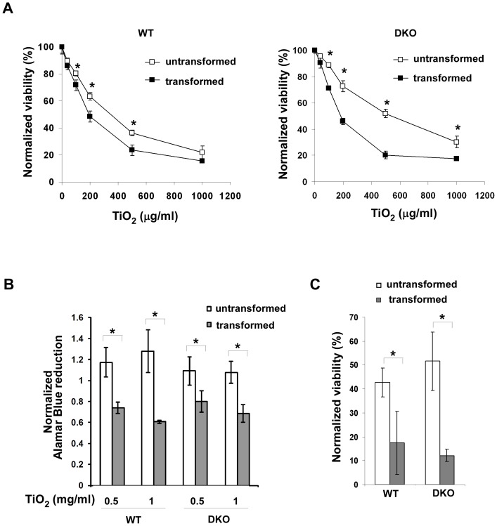
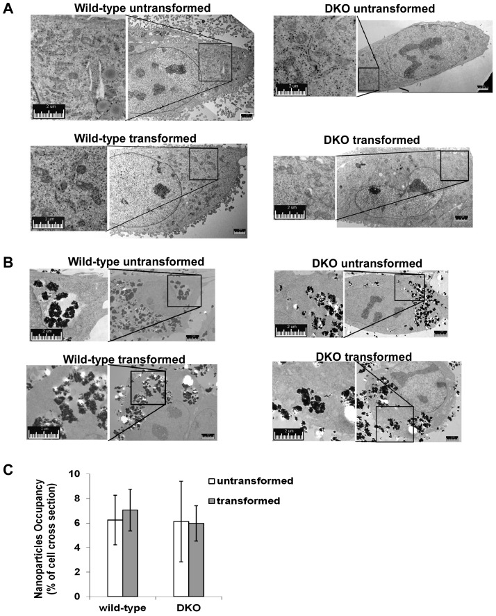
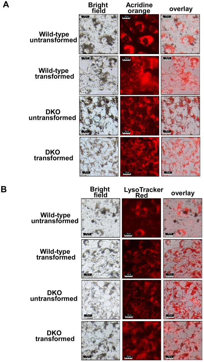
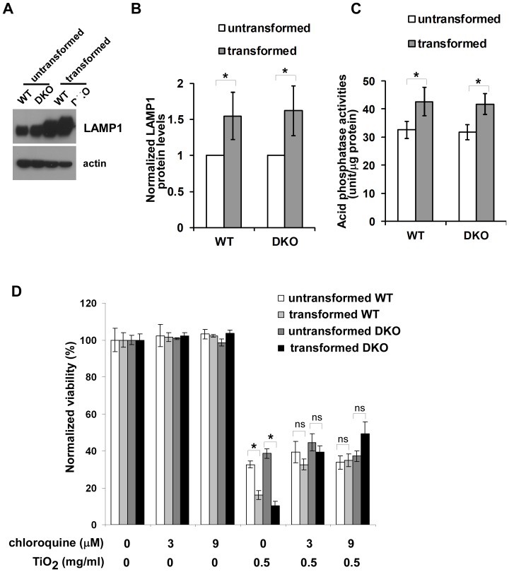
While the cytotoxic effects of titanium dioxide (TiO(2)) nanoparticles have been under intense investigation, the molecular mechanisms of this cytotoxicity remain unknown. Here we investigated the influence of oncogenic transformation and a major apoptotic signaling pathway on cellular responses to TiO(2) nanoparticles. Isogenic wild-type (WT) and apoptosis-resistant (Bak(-/-)Bax(-/-)) cell lines with and without tumorigenic transformation were examined. TiO(2) nanoparticles preferentially reduced viability of tumorigenic cells in a dose-dependent fashion compared with their untransformed counterparts. Importantly, the elevated cytotoxicity of TiO(2) nanoparticles was independent of a major Bak/Bax-dependent apoptosis pathway. Because transformation does not affect cellular fluid-phase endocytosis or nanoparticle uptake, it is likely that the increased cytotoxicity in tumor cells is due to the interaction between TiO(2) nanoparticles and the lysosomal compartment. Overall, our data indicate that TiO(2) nanoparticles induce cytotoxicity preferentially in transformed cells independent of a major apoptotic signaling pathway.
Conflict of interest statement
Figures


Similar articles
-
Titanium dioxide induces apoptotic cell death through reactive oxygen species-mediated Fas upregulation and Bax activation.Int J Nanomedicine. 2012;7:1203-14. doi: 10.2147/IJN.S28647. Epub 2012 Mar 5. Int J Nanomedicine. 2012. PMID: 22419868 Free PMC article.
-
Titanium dioxide (TiO2) nanoparticles induce JB6 cell apoptosis through activation of the caspase-8/Bid and mitochondrial pathways.J Toxicol Environ Health A. 2009;72(19):1141-9. doi: 10.1080/15287390903091764. J Toxicol Environ Health A. 2009. PMID: 20077182
-
Inhibition of the ER Ca2+ pump forces multidrug-resistant cells deficient in Bak and Bax into necrosis.J Cell Sci. 2009 Dec 15;122(Pt 24):4481-91. doi: 10.1242/jcs.055772. Epub 2009 Nov 17. J Cell Sci. 2009. PMID: 19920074
-
Pro-apoptotic Bax is the major and Bak an auxiliary effector in cytokine deprivation-induced mast cell apoptosis.Cell Death Dis. 2010 May 13;1(5):e43. doi: 10.1038/cddis.2010.20. Cell Death Dis. 2010. PMID: 21364649 Free PMC article.
-
BCL-2 proteins and apoptosis: Recent insights and unknowns.Biochem Biophys Res Commun. 2018 May 27;500(1):26-34. doi: 10.1016/j.bbrc.2017.06.190. Epub 2017 Jul 1. Biochem Biophys Res Commun. 2018. PMID: 28676391 Review.
Cited by
-
Involvement of Bcl-2 Activation and G1 Cell Cycle Arrest in Colon Cancer Cells Induced by Titanium Dioxide Nanoparticles Synthesized by Microwave-Assisted Hybrid Approach.Front Bioeng Biotechnol. 2020 Jul 15;8:606. doi: 10.3389/fbioe.2020.00606. eCollection 2020. Front Bioeng Biotechnol. 2020. PMID: 32760701 Free PMC article.
-
Assessment of TiO2 Nanoparticle Impact on Surface Morphology of Chinese Hamster Ovary Cells.Materials (Basel). 2022 Jun 29;15(13):4570. doi: 10.3390/ma15134570. Materials (Basel). 2022. PMID: 35806697 Free PMC article.
-
NLRP3 inflammasome, oxidative stress, and apoptosis induced in the intestine and liver of rats treated with titanium dioxide nanoparticles: in vivo and in vitro study.Int J Nanomedicine. 2019 Mar 15;14:1919-1936. doi: 10.2147/IJN.S192382. eCollection 2019. Int J Nanomedicine. 2019. PMID: 30936694 Free PMC article.
-
Nanoparticles Biosynthesized by Fungi and Yeast: A Review of Their Preparation, Properties, and Medical Applications.Molecules. 2015 Sep 11;20(9):16540-65. doi: 10.3390/molecules200916540. Molecules. 2015. PMID: 26378513 Free PMC article. Review.
-
Photodynamic N-TiO2 Nanoparticle Treatment Induces Controlled ROS-mediated Autophagy and Terminal Differentiation of Leukemia Cells.Sci Rep. 2016 Oct 4;6:34413. doi: 10.1038/srep34413. Sci Rep. 2016. PMID: 27698385 Free PMC article.
References
-
- Newman MD, Stotland M, Ellis JI (2009) The safety of nanosized particles in titanium dioxide- and zinc oxide-based sunscreens. J Am Acad Dermatol 61: 685–692. - PubMed
-
- Marquis BJ, Love SA, Braun KL, Haynes CL (2009) Analytical methods to assess nanoparticle toxicity. Analyst 134: 425–439. - PubMed
-
- Nel A, Xia T, Madler L, Li N (2006) Toxic potential of materials at the nanolevel. Science 311: 622–627. - PubMed
Publication types
MeSH terms
Substances
Grants and funding
LinkOut - more resources
Full Text Sources
Research Materials
