. 2012 Nov 27:9:259.
doi: 10.1186/1742-2094-9-259.

The gamma chain subunit of Fc receptors is required for alpha-synuclein-induced pro-inflammatory signaling in microglia

Affiliations

The gamma chain subunit of Fc receptors is required for alpha-synuclein-induced pro-inflammatory signaling in microglia

Shuwen Cao et al. J Neuroinflammation. .

Abstract

Background: The protein alpha-synuclein (α-SYN), which is found in the Lewy bodies of dopamine-producing (DA) neurons in the substantia nigra (SN), has an important role in the pathogenesis of Parkinson's disease (PD). Previous studies have shown that neuroinflammation plays a key role in PD pathogenesis. In an AAV-synuclein mouse model of PD, we have found that over-abundance of α-SYN triggers the expression of NF-κB p65, and leads to microglial activation and DA neurodegeneration. We also have observed that Fcγ receptors (FcγR), proteins present on the surface of microglia that bind immunoglobulin G (IgG) and other ligands, are key modulators of α-SYN-induced neurodegeneration.

Methods: In order to study the role of FcγRs in the interactions of α-SYN and microglia, we treated the primary microglial cultures from wild-type (WT) and FcγR-/- mice with aggregated human α-SYN in vitro.

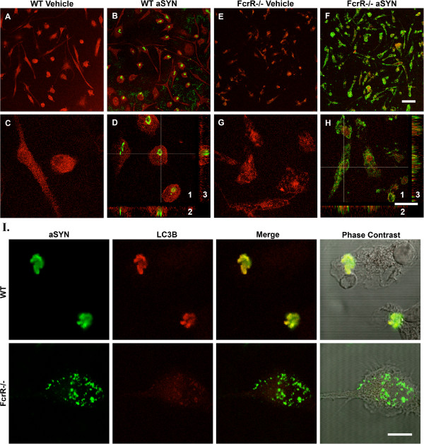
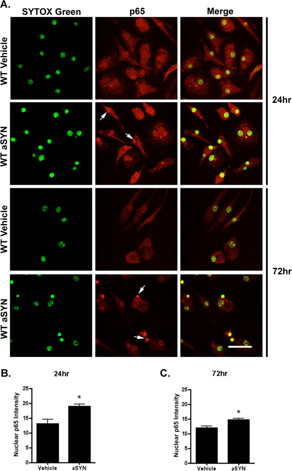
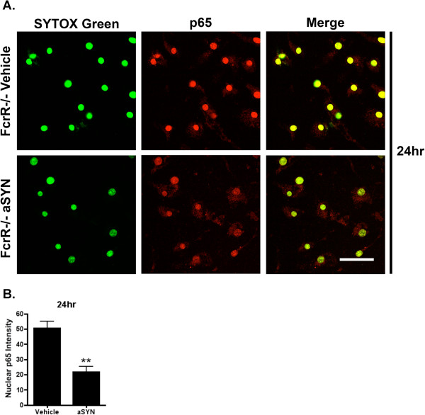
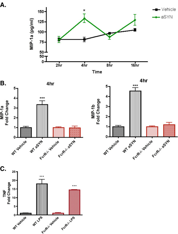
Results: Using immunocytochemistry, we found that α-SYN was taken up by both WT and FcγR-/- microglia, however, their patterns of internalization were different, with aggregation in autophagosomes in WT cells and more diffuse localization in FcγR-/- microglia. In WT microglia, α-SYN induced the nuclear accumulation of NF-κB p65 protein and downstream chemokine expression while in FcγR-/- mouse microglia, α-SYN failed to trigger the enhancement of nuclear NF-κB p65, and the pro-inflammatory signaling was reduced.

Conclusions: Our results suggest that α-SYN can interact directly with microglia and can be internalized and trafficked to autophagosomes. FcγRs mediate this interaction, and in the absence of the gamma chain, there is altered intracellular trafficking and attenuation of pro-inflammatory NF-κB signaling. Therefore, blocking either FcγR signaling or downstream NF-κB activation may be viable therapeutic strategies in PD.


Figures

Figure 1
Figure 1
α-SYN internalization in WT and FcγR−/−mouse primary microglia. (A-H) 72 h after the treatment of aggregated human α-SYN, WT and FcγR−/− mouse primary microglia were immunostained for human α-SYN (Green) and the microglial marker CD45 (Red). Condensed α-SYN was observed in the WT microglia. In FcγR−/− microglia, there was still uptake of aggregated human α-SYN but the pattern of intracellular α-SYN was quite different, with diffuse labeling of small puncta throughout the cytoplasm. Scale bars: panel A, B, E, F bar=50 μm; panel C, D, G, H bar=20 μm. Image 1 in Panels D and H are confocal images of WT and FcγR−/− mouse primary microglia treated with aggregated human α-SYN, respectively; 2 and 3 in Panel D and H are transverse and lengthwise images of z-stack series of the indicated cells. (I) 24 h after the treatment of aggregated human α-SYN, WT and FcγR−/− mouse primary microglia were immunostained for human α-SYN (Green) and autophagosomal marker LC3B (Red). α-SYN co-localized with LC3B in WT but not FcγR−/− microglia. Phase contrast images show the morphology of the cell. Scale bar=10 μm.
Figure 2
Figure 2
α-SYN-induced NF-κB activation in WT mouse primary microglia. (A) 24 h and 72 h after the treatment of either vehicle or 500 nM aggregated human α-SYN, cells were stained for NF-κB p65 protein (Red) and SYTOX Green to show the nucleus. At both time points, α-SYN-treated microglia exhibited increased immunoreactivity for NF-κB p65, and the nuclear accumulation of NF-κB p65 was quite distinct compared with the vehicle treated ones. Scale bar=40 μm. Arrows indicate the enrichment of nuclear NF-κB p65. (B, C) Quantification of 24 h and 72 h nuclear NF-κB p65 intensity. At least four images from each group were analyzed using ImageJ software. At both time points α-SYN-treated WT microglia had markedly enhanced nuclear NF-κB p65 staining compared with the vehicle-treated controls. *P<0.05, α-SYN vs. vehicle, t-test.
Figure 3
Figure 3
α-SYN-induced NF-κB activation was blocked in FcγR−/−mouse primary microglia. (A) 24 h after treatment with either vehicle or 500 nM aggregated human α-SYN, FcγR−/− microglia were stained for NF-κB p65 protein (Red) and SYTOX Green. α-SYN-treated microglia exhibited attenuated immunoreactivity for NF-κB p65 compared with the vehicle-treated ones. Scale bar=40 μm. (B) Quantification of 24 h nuclear NF-κB p65 intensity. After the treatment of α-SYN, FcγR−/− microglia had a significant decrease in nuclear p65. **P<0.01, α-SYN vs. vehicle, t-test.
Figure 4
Figure 4
FcγR−/−attenuated α-SYN-induced NF-κB signaling and downstream expression of pro-inflammatory molecules. (A) MIP-1α ELISA on the conditioned media collected 2, 4, 8, and 16 h from the vehicle and α-SYN-treated WT primary microglia. At 4 h, but not the other time points, the MIP-1α level was significantly increased with α-SYN treatment compared with the vehicle controls. *P<0.05, α-SYN vs. vehicle, t-test. (B) Conditioned media were collected 4 h and 24 h after the treatment of vehicle or aggregated human α-SYN on WT and FcγR−/− microglia for a 25-plex mouse cytokine/chemokine assay. At 4 h, MIP-1α and MIP-1β were significantly increased with α-SYN treatment in WT microglia but not FcγR−/− microglia compared with vehicle-treated ones. All chemokine expression levels were normalized to the level of vehicle-treated WT or FcγR−/− microglia, respectively. ***P <0.001 WT α-SYN vs. WT vehicle. One-way ANOVA with Tukey’s multiple comparison test. (C) Conditioned media collected from primary WT and FcγR−/− microglia treated overnight with 100 ng/mL LPS. Both WT and FcγR−/− microglia respond normally to TLR4 stimulation. TNF expression levels were normalized to the level of vehicle-treated WT or FcγR−/− microglia, respectively. ***P <0.001 WT LPS vs. WT vehicle, FcγR−/− LPS vs. FcγR−/− vehicle. One-way ANOVA with Tukey’s multiple comparison test.

References

    1. Spillantini MG, Schmidt ML, Lee VM, Trojanowski JQ, Jakes R, Goedert M. Alpha-synuclein in Lewy bodies. Nature. 1997;388:839–840. doi: 10.1038/42166. - DOI - PubMed
    1. Hardy J, Cai H, Cookson MR, Gwinn-Hardy K, Singleton A. Genetics of Parkinson’s disease and parkinsonism. Ann Neurol. 2006;60:389–398. doi: 10.1002/ana.21022. - DOI - PubMed
    1. Polymeropoulos MH, Lavedan C, Leroy E, Ide SE, Dehejia A, Dutra A, Pike B, Root H, Rubenstein J, Boyer R, Stenroos ES, Chandrasekharappa S, Athanassiadou A, Papapetropoulos T, Johnson WG, Lazzarini AM, Duvoisin RC, Di Iorio G, Golbe LI, Nussbaum RL. Mutation in the alpha-synuclein gene identified in families with Parkinson’s disease. Science. 1997;276:2045–2047. doi: 10.1126/science.276.5321.2045. - DOI - PubMed
    1. McGeer PL, Itagaki S, Boyes BE, McGeer EG. Reactive microglia are positive for HLA-DR in the substantia nigra of Parkinson’s and Alzheimer’s disease brains. Neurology. 1988;38:1285–1291. doi: 10.1212/WNL.38.8.1285. - DOI - PubMed
    1. Brochard V, Combadiere B, Prigent A, Laouar Y, Perrin A, Beray-Berthat V, Bonduelle O, Alvarez-Fischer D, Callebert J, Launay JM, Duyckaerts C, Flavell RA, Hirsch EC, Hunot S. Infiltration of CD4+ lymphocytes into the brain contributes to neurodegeneration in a mouse model of Parkinson disease. J Clin Invest. 2009;119:182–192. - PMC - PubMed

Publication types

MeSH terms

LinkOut - more resources