. 2012 Dec;122(12):4569-79.
doi: 10.1172/JCI62422. Epub 2012 Nov 26.

The mitochondrial heme exporter FLVCR1b mediates erythroid differentiation

Affiliations

The mitochondrial heme exporter FLVCR1b mediates erythroid differentiation

Deborah Chiabrando et al. J Clin Invest. 2012 Dec.

Abstract

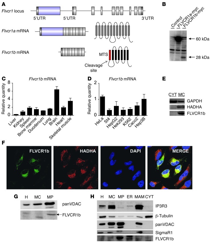
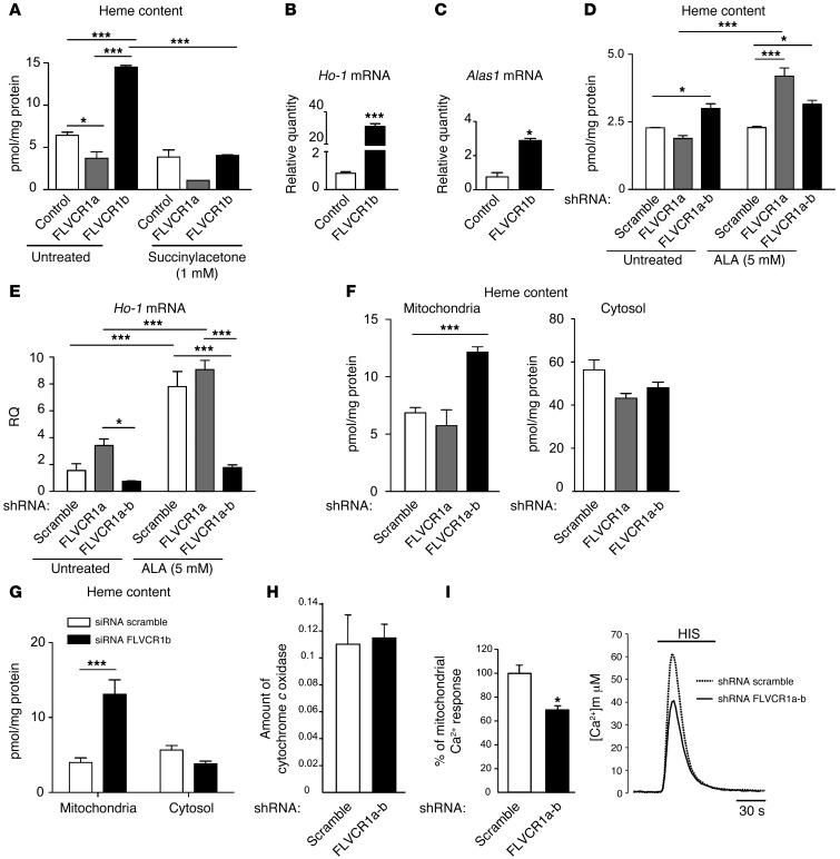
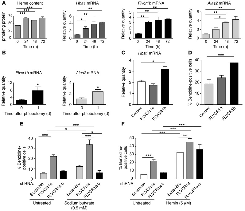
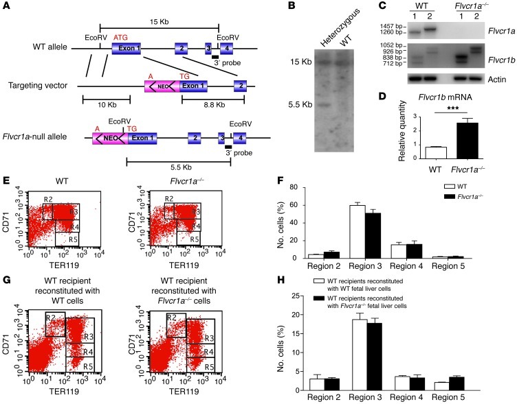
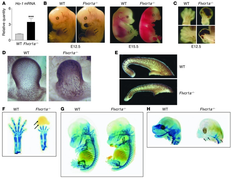
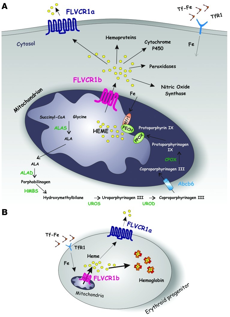
Feline leukemia virus subgroup C receptor 1 (FLVCR1) is a cell membrane heme exporter that maintains the balance between heme levels and globin synthesis in erythroid precursors. It was previously shown that Flvcr1-null mice died in utero due to a failure of erythropoiesis. Here, we identify Flvcr1b, a mitochondrial Flvcr1 isoform that promotes heme efflux into the cytoplasm. Flvcr1b overexpression promoted heme synthesis and in vitro erythroid differentiation, whereas silencing of Flvcr1b caused mitochondrial heme accumulation and termination of erythroid differentiation. Furthermore, mice lacking the plasma membrane isoform (Flvcr1a) but expressing Flvcr1b had normal erythropoiesis, but exhibited hemorrhages, edema, and skeletal abnormalities. Thus, FLVCR1b regulates erythropoiesis by controlling mitochondrial heme efflux, whereas FLVCR1a expression is required to prevent hemorrhages and edema. The aberrant expression of Flvcr1 isoforms may play a role in the pathogenesis of disorders characterized by an imbalance between heme and globin synthesis.


Figures

Figure 1
Figure 1. Identification of a mitochondrial isoform of Flvcr1.
(A) Schematic representation of Flvcr1 genetic locus, Flvcr1a and Flvcr1b transcripts, and the predicted protein structures. The MTS in FLVCR1b is indicated in red; cleavage at position 38 results in a protein with 6 hydrophobic transmembrane domains. See also Supplemental Figure 1. (B) Western blot analysis of total protein extracts of HeLa cells overexpressing FLVCR1a-myc, FLVCR1b-myc, or the control vector. Antibody against myc was used. (C) qRT-PCR analysis of Flvcr1b mRNA levels in mouse tissues and (D) human cell lines. Values represent mean ± SEM. n = 6. (E) Western blot analysis of fractionated protein extracts of HeLa cells overexpressing FLVCR1b-myc showing that FLVCR1b is localized in the mitochondrial enriched fraction. Antibodies against GAPDH (cytosolic marker), HADHA (mitochondrial marker), and myc (to detect FLVCR1b) were used. (F) Immunofluorescence analysis of HeLa cells overexpressing FLVCR1b-myc showing the colocalization between FLVCR1b (detected with an antibody against myc) and HADHA. See also Supplemental Figures 2 and 3. Original magnification, ×63. (G) Endogenous FLVCR1b mitochondrial localization revealed by immunoblot analysis. Homogenate, crude, and pure mitochondrial fraction, respectively, before and after Percoll gradient separation prepared from HeLa cells. 20 μg of proteins were loaded on 15% SDS-polyacrylamide gels. (H) Detection of endogenous FLVCR1b by immunoblotting in HEK293 fractionation. IP3R3 and SigmaR1 were used as markers of MAM, tubulin as a marker of cytosol, and VDAC as a marker of mitochondria. 50 μg of proteins were loaded on 15% SDS-polyacrylamide gels. H, homogenate; MC, crude mitochondria; MP,pure mitochondria; CYT, cytosol.
Figure 2
Figure 2. FLVCR1b overexpression increases intracellular heme content, while its silencing results in heme accumulation in mitochondria.
(A) Heme levels in HeLa cells overexpressing FLVCR1a-myc, FLVCR1b-myc, or a control vector. Succinylacetone was used as a competitive inhibitor of heme biosynthesis. n = 6. Two-way ANOVA. (B and C) qRT-PCR analysis of Ho-1 mRNA and Alas1 mRNA in FLVCR1b-overexpressing HeLa cells compared with control. n = 6. t test. (D and E) Heme content and qRT-PCR analysis of Ho-1 transcript levels in HeLa cells in which the expression of both FLVCR1a and FLVCR1b, or FLVCR1a alone, was downregulated using specific shRNA. Heme biosynthesis was stimulated with ALA. n = 6. Two-way ANOVA. (F) Heme content in mitochondria and cytosol from HeLa cells, in which the expression of both FLVCR1a and FLVCR1b, or FLVCR1a alone, was downregulated using specific shRNA compared with control cells. n = 6. One-way ANOVA. (G) Heme content in mitochondria and cytosol isolated from HeLa cells in which FLVCR1b expression was downregulated using a specific siRNA against the 5′ UTR of Flvcr1b with respect to controls. n = 6. Two-way ANOVA. (H) Quantity of cytochrome c oxidase in control versus silenced FLVCR1a-b HeLa cells. n = 4. (I) Mitochondrial Ca2+ responses to agonist stimulation in control and silenced FVLCR1a-b HeLa cells. Data were normalized to mean of the control group. Traces are representative of 12 experiments from 3 preparations. t test. Values represent mean ± SEM. *P < 0.05; ***P < 0.001.
Figure 3
Figure 3. FLVCR1b controls erythroid differentiation in vitro.
(A) Heme content and qRT-PCR analysis of Hba1, Flvcr1b, and Alas2 mRNAs in K562 cells treated with 5 mM ALA to induce in vitro erythroid differentiation. n = 6. One-way ANOVA. (B) qRT-PCR analysis of spleen Flvcr1b and Alas2 mRNA levels following the stimulation of erythropoiesis by phlebotomy. n = 6. t test. (C) qRT-PCR analysis of Hba1 mRNA and (D) benzidine staining of K562 cells overexpressing FLVCR1a-myc, FLVCR1b-myc, or a control vector. n = 6. One-way ANOVA. (E) Benzidine staining of K562 cells in which the expression of both FLVCR1a and FLVCR1b, or FLVCR1a alone, was downregulated using specific shRNA, compared with control cells. Erythroid differentiation was induced with sodium butyrate (0.5 mM, 72 hours). n = 6. Two-way ANOVA. (F) Benzidine staining of silenced K562 cells stimulated with hemin (5 μM; 72 hours). The percentage of benzidine-positive cells is indicated. Values represent mean ± SEM. n = 6. Two-way ANOVA. *P < 0.05; **P < 0.01; ***P < 0.001.
Figure 4
Figure 4. FLVCR1b controls erythroid differentiation in vivo.
(A) Schematic representation of the targeting strategy. The start codon in the first exon of the Flvcr1 gene was disrupted by the insertion of the neomycin resistance cassette by homologous recombination in mouse ES cells. (B) Southern blot analysis on genomic DNA extracted from a recombinant and a wild-type clone, digested with EcoRV, and hybridized with the probe shown in A. (C) RT-PCR analysis of Flvcr1 isoforms levels in wild-type and Flvcr1a–/– embryos at E13.5. Two different pairs of primers were used in lanes 1 and 2. (D) qRT-PCR analysis of Flvcr1b mRNA on E13.5 wild-type and Flvcr1a–/– embryos. n = 6. t test. ***P < 0.001. (E) Erythroid differentiation is completely normal in Flvcr1a–/– fetal liver. Representative flow cytometric analyses of E14.5 liver cells from wild-type and Flvcr1a–/– embryos immunostained with antibodies to CD71 and Ter119. Regions R2–R5 corresponding to different maturational stages are indicated. The percentage of cells in each population is reported (F). n = 6. (G) Representative flow cytometric analyses of bone marrow cells, isolated from mice transplanted with wild-type or Flvcr1a–/– fetal liver cells, immunostained with antibodies to CD71 and Ter119. Regions R2–R5 corresponding to different maturational stages are indicated. The percentage of cells in each population is reported (H). n = 6. Values represent mean ± SEM.
Figure 5
Figure 5. Flvcr1a–/– embryos show hemorrhages, edema, and skeletal malformations.
(A) qRT-PCR analysis showing the upregulation of Ho-1 mRNA in Flvcr1a–/– embryos. Values represent mean ± SEM. n = 6. t test. ***P < 0.001. (B) Multifocal and extended hemorrhages and edema in E12.5 and E15.5 Flvcr1a–/– embryos. (C) Enlarged view of E12.5 wild-type and Flvcr1a–/– limbs. (D and E) Whole-mount immunohistochemical analysis of E11.5 wild-type and Flvcr1a–/– embryos using anti-PECAM antibody showing abnormal vasculogenesis in Flvcr1a knockout animal. An enlarged view of the anterior limb (D) and tail (E) is shown. (FH) Skeletal malformations in E14.5 Flvcr1a–/– embryos were analyzed by alcian blue/alizarin red staining. (F) An enlarged view of the anterior limb of E14.5 embryos: digits do not form properly in the Flvcr1a–/– embryo. Different grades of skeletal malformations are shown: Meckel’s cartilage (arrows) is reduced in the Flvcr1a–/– embryo (G); head cartilages are severely compromised and the lower jaw (arrows) completely absent in the Flvcr1a–/– embryo (H).
Figure 6
Figure 6. A model for FLVCR1 isoforms function.
(A) A schematic representation of heme biosynthesis is shown (see text for details). Transferrin-bound iron (Tf-Fe) is taken up by cells through transferrin receptor 1 (TfR1), and iron is transferred to the mitochondrion for heme biosynthesis. It was reported that the mitochondrial iron importer MITOFERRIN1 (MFRN1) and FECH are part of the same complex in the inner mitochondrial membrane. FLVCR1b could work in close association with this complex to allow heme export out of the mitochondrion for incorporation into new hemoproteins. Heme not used for hemoprotein synthesis is exported out of the cell through the cell-surface isoform FLVCR1a. (B) During erythroid differentiation, the expression of FLVCR1b in the mitochondrion regulates heme export into the cytosol, allowing hemoglobinization of erythroid precursors. At the cell membrane, FLVCR1a regulates the export of heme in excess. The data reported here suggest that decreased expression of the membrane heme exporter FLVCR1a and increased expression of FLVCR1b are fundamental for proper differentiation of erythroid progenitors.

Comment in

References

    1. Chen JJ. Regulation of protein synthesis by the heme-regulated eIF2alpha kinase: relevance to anemias. Blood. 2007;109(7):2693–2699. - PMC - PubMed
    1. Furuyama K, Kaneko K, Vargas PD. Heme as a magnificent molecule with multiple missions: heme determines its own fate and governs cellular homeostasis. Tohoku J Exp Med. 2007;213(1):1–16. doi: 10.1620/tjem.213.1. - DOI - PubMed
    1. Quigley JG, et al. Identification of a human heme exporter that is essential for erythropoiesis. Cell. 2004;118(6):757–766. doi: 10.1016/j.cell.2004.08.014. - DOI - PubMed
    1. Keel SB, et al. A heme export protein is required for red blood cell differentiation and iron homeostasis. Science. 2008;319(5864):825–828. doi: 10.1126/science.1151133. - DOI - PubMed
    1. Ponka P. Cell biology of heme. Am J Med Sci. 1999;318(4):241–256. doi: 10.1097/00000441-199910000-00004. - DOI - PubMed

Publication types

MeSH terms