. 2012 Dec 15;11(24):4597-612.
doi: 10.4161/cc.22871. Epub 2012 Nov 27.

Alternatively spliced protein arginine methyltransferase 1 isoform PRMT1v2 promotes the survival and invasiveness of breast cancer cells

Affiliations

Alternatively spliced protein arginine methyltransferase 1 isoform PRMT1v2 promotes the survival and invasiveness of breast cancer cells

R Mitchell Baldwin et al. Cell Cycle. .

Abstract

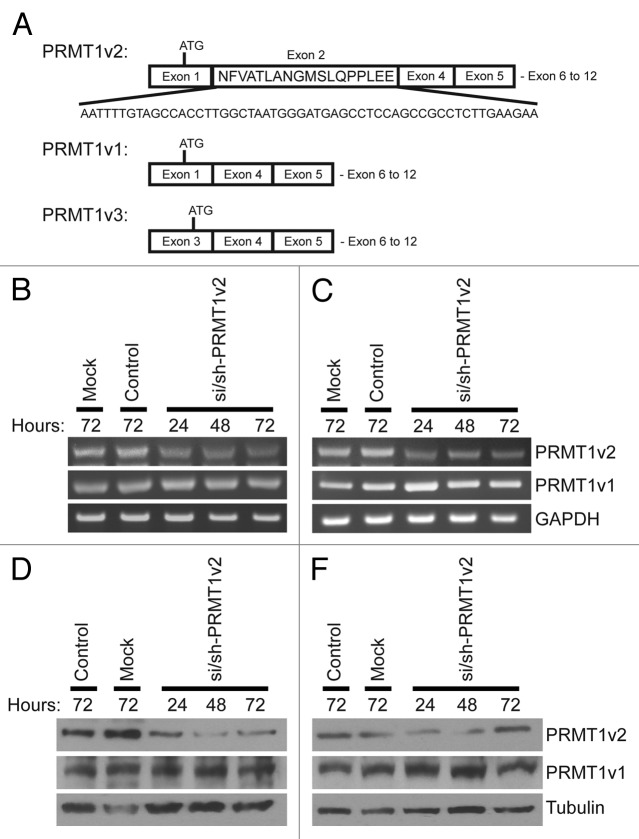
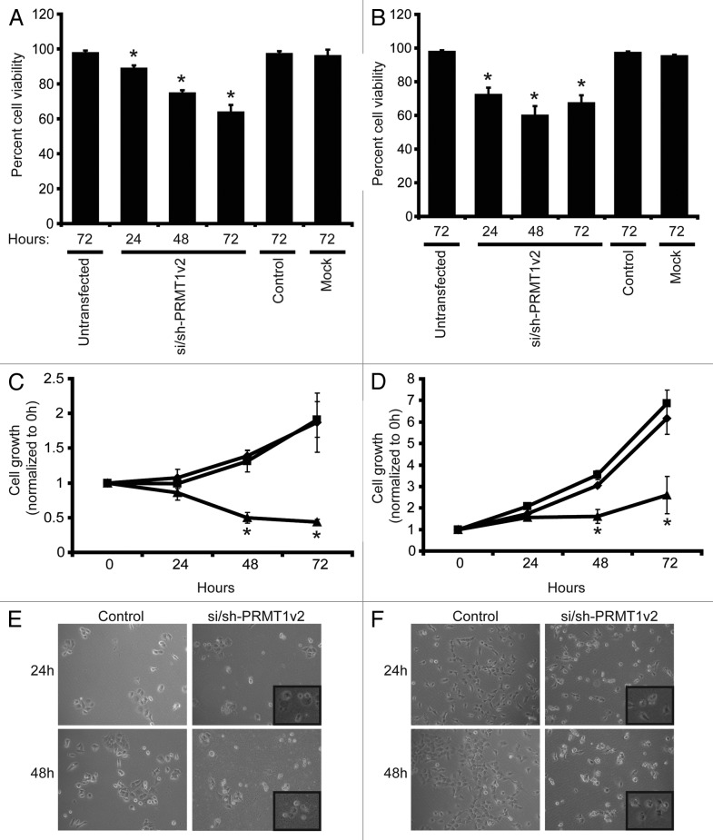
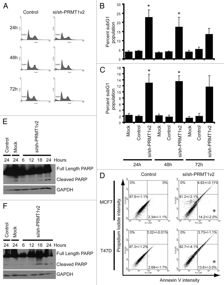
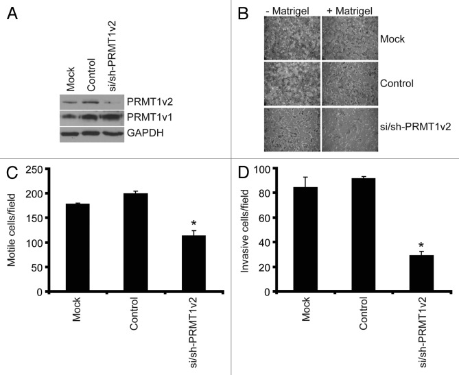
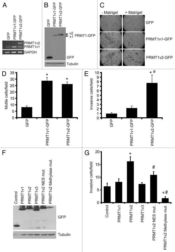
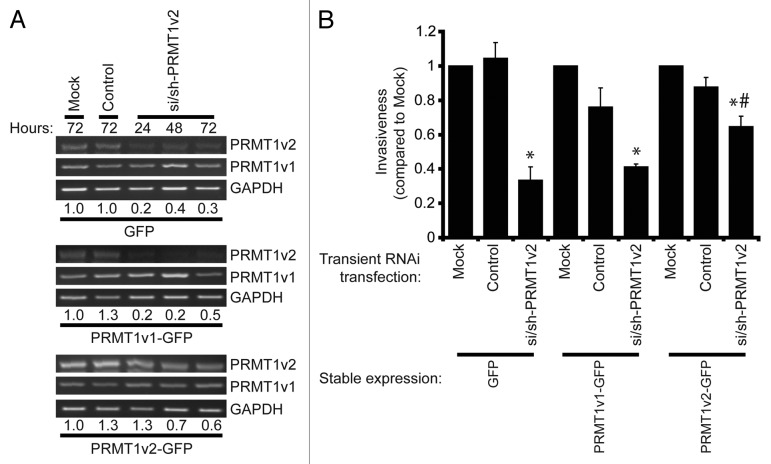
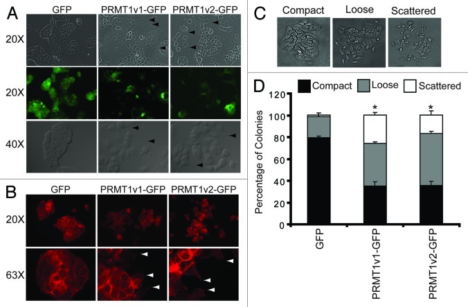
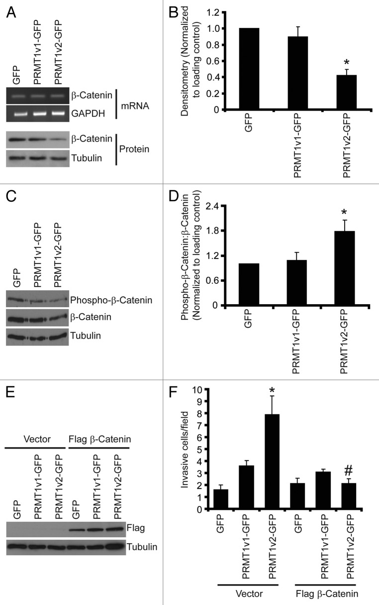
Protein arginine methylation is catalyzed by protein arginine methyltransferases (PRMTs) and plays an important role in many cellular processes. Aberrant PRMT expression has been observed in several common cancer types; however, their precise contribution to the cell transformation process is not well understood. We previously reported that the PRMT1 gene generates several alternatively spliced isoforms, and our initial biochemical characterization of these isoforms revealed that they exhibit distinct substrate specificity and subcellular localization. We focus here on the PRMT1v2 isoform, which is the only predominantly cytoplasmic isoform, and we have found that its relative expression is increased in breast cancer cell lines and tumors. Specific depletion of PRMT1v2 using RNA interference caused a significant decrease in cancer cell survival due to an induction of apoptosis. Furthermore, depletion of PRMT1v2 in an aggressive cancer cell line significantly decreased cell invasion. We also demonstrate that PRMT1v2 overexpression in a non-aggressive cancer cell line was sufficient to render them more invasive. Importantly, this novel activity is specific to PRMT1v2, as overexpression of other isoforms did not enhance invasion. Moreover, this activity requires both proper subcellular localization and methylase activity. Lastly, PRMT1v2 overexpression altered cell morphology and reduced cell-cell adhesion, a phenomenon that we convincingly linked with reduced β-catenin protein expression. Overall, we demonstrate a specific role for PRMT1v2 in breast cancer cell survival and invasion, underscoring the importance of identifying and characterizing the distinct functional differences between PRMT1 isoforms.


Figures

None
Figure 1. Specific depletion of PRMT1v2. Schematic of PRMT1v2, PRMT1v1 and PRMT1v3 coding exon structure (A). Alternative splicing causes inclusion of exon 2 in the PRMT1v2 coding sequence. Total RNA was collected from MCF7 and T47D cells transfected with si/sh-PRMT1v2 at 24, 48 and 72 h post-transfection and mock and control siRNA (control) transfected cells at 72 h. PCR analysis of cDNA generated from total RNA using PRMT1 primers shows depletion of PRMT1v2 mRNA with no effect on PRMT1v1 in MCF7 (B) and T47D (C) cells. GAPDH serves as a loading control. Total protein lysates were collected from MCF7 and T47D cells transfected with si/sh-PRMT1v2 at 24, 48 and 72 h post-transfection and from mock and control transfected cells at 72 h. Western blot analysis using a PRMT1v2-specific antibody shows effective depletion of protein expression in MCF7 (D) and T47D (E) cells. No effect on PRMT1v1 protein expression was observed. Tubulin serves as a loading control.
None
Figure 2. Depletion of PRMT1v2 affects breast cancer cell viability and growth. Viable cell numbers were determined by viable cell counts using trypan blue exclusion 24, 48 and 72 h following mock, control or si/sh-PRMT1v2 transfection in MCF7 (A) and T47D (B) cells. Data are expressed as a percentage of viable cell counts to total cell counts and are the mean ± standard error of three independent experiments performed in triplicate (*p < 0.05). MCF7 (C) and T47D (D) cells were plated at equal numbers and then mock (♦), control (■) or si/sh-PRMT1v2 transfected (▲) and assessed by MTT assay at 0, 24, 48 and 72 h post-transfection. Data are the mean ± standard error of three independent experiments with six replicates per experiment (*p < 0.05). Morphology of control and si/sh-PRMT1v2 transfected MCF7 (E) and T47D (F) cells 24 and 48 h post-transfection. Images were taken at 20X magnification with insets focused on an area in the field of view.
None
Figure 3. Depletion of PRMT1v2 induces apoptosis. Representative flow cytometric analyses of propidium iodide (PI)-stained MCF7 cells following control or si/sh-PRMT1v2 transfection at 24, 48 and 72 h (A). Percentage of subG1 population for MCF7 (B) and T47D (C) cells. Data are the mean ± standard error of five (for MCF7) and four (for T47D) independent experiments (*p < 0.05). Representative flow cytometric analyses of Annexin V and PI co-staining of MCF7 and T47D cells following control or si/sh-PRMT1v2 transfection for 24 h (D). Percentages in each quadrant represent the mean ± standard error of three independent experiments (*p < 0.05). Total protein lysates were collected from MCF7 and T47D cells that were mock and control transfected for 24 h or transfected with si/sh-PRMT1v2 for 6, 12, 18 and 24 h. Western blot analysis for the expression of PARP shows the appearance of its cleavage product in MCF7 (E) and T47D (F) cells transfected with si/sh-PRMT1v2. GAPDH was used as a loading control.
None
Figure 4. Depletion of PRMT1v2 inhibits breast cancer cell motility and invasion. MDA-MB-231 cells depleted of PRMT1v2 were assessed for effects on motility and invasion. Western analysis for PRMT1v2 and PRMT1v1 following 24 h depletion (A). GAPDH serves as a loading control. Following 24 h of PRMT1v2 depletion, cells replated at equal numbers into Transwell chambers and incubated for an additional 24 h. Motility was analyzed using Transwell chambers without a Matrigel (- Matrigel). Invasion was analyzed using Transwell chambers containing a Matrigel layer (+ Matrigel). Representative images of cells that have passed through the Transwell chamber -/+ Matrigel at 20X magnification (B). Cell numbers that passed through the Transwell chambers in the absence (C: motile cells/field) or presence (D: invasive cells/field) of Matrigel were determined. Data represents the mean ± standard error of three independent experiments (*p < 0.05 comparing to control).
None
Figure 5. Overexpression of PRMT1v2 in non-invasive breast cancer cells enhances invasion. Total RNA was collected from MCF7 cells stably expressing GFP, GFP-tagged PRMT1v1 (PRMT1v1-GFP) and GFP-tagged PRMT1v2 (PRMT1v2-GFP). PCR analysis of cDNA generated from total RNA using PRMT1 primers (A). GAPDH serves as a loading control. Total protein lysates from MCF7 cells stably expressing GFP, PRMT1v1-GFP or PRMT1v2-GFP were analyzed by western blotting using an anti-GFP antibody (B). Tubulin serves as a loading control. MCF7 stably expressing GFP, PRMT1v1-GFP or PRMT1v2-GFP were analyzed for motility and invasion using Transwell chambers without (- Matrigel) or with a Matrigel layer (+ Matrigel). Cells were plated into Transwell chambers at the same density and the numbers of cells that crossed the chamber membrane was counted after 72 h. Representative images of cells that have passed through the Transwell chamber -/+ Matrigel at 20X magnification (C). Cell numbers that passed through the chamber mebranes without a Matrigel layer (D: motile cells/field) or containing a Matrigel layer (E: invasive cells/field) were counted. Data represents the mean ± standard error of four independent experiments (*p < 0.05 comparing to GFP and #p < 0.05 comparing to PRMT1v1-GFP). MCF7 cells were transiently transfected with plasmid constructs containing a C-terminal EGFP-tagged PRMT1v1, PRMT1v2, PRMT1v3, PRMT1v2 NES mutant or a PRMT1v2 catalytically inactive methylase mutant for 24 h. Western analysis for GFP shows expression of each PRMT1 isoform and PRMT1v2 mutants in MCF7 cells 24 h post transfection (F). To examine invasion, following 24 h transfection, cells were seeded into Matrigel containing Transwell chambers and incubated for 72 h. Cell numbers that passed through the chamber membranes were counted (G). Data represents the mean ± standard error of three independent experiments (*p < 0.05 comparing to control, #p < 0.05 comparing to PRMT1v2).
None
Figure 6. Overexpression of PRMT1v2 partially rescues the inhibition of invasion resulting from PRMT1v2 depletion. MCF7 cells stably expressing GFP, PRMT1v1-GFP or PRMT1v2-GFP were transfected with si/sh-PRMT1v2. Total RNA was collected 24, 48 and 72 h post-si/sh-PRMT1v2 transfection and 72 h post-transfection for mock and control samples. PCR analysis of cDNA generated from total RNA using PRMT1 primers shows levels of PRMT1v2 and PRMT1v1 mRNA (A). Endogenous and exogenous PRMT1v2 levels are detected as a single band in this PCR analysis as the primers do not differentiate the two species. GAPDH serves as a loading control. The densitometry values (indicated below the respective lanes) for the samples in the representative PCRs were normalized to mock transfected cells. MCF7 cells stably expressing GFP, PRMT1v1-GFP or PRMT1v2-GFP were mock, control or si/sh-PRMT1v2 transfected for 24 h. Twenty-four hours post-transfection, cells were replated at equal numbers into Transwell chambers containing a Matrigel layer and incubated for 72 h. Cell numbers that passed through the chambers were counted (B). Data represents the mean ± standard error of three independent experiments (*p < 0.05 compared with matched control transfection, #p < 0.05 compared with MCF7 GFP transfected with si/sh-PRMT1v2).
None
Figure 7. Overexpression of PRMT1v2 in MCF7 cells alters their morphology. Representative phase contrast and GFP fluorescence images of MCF7 cell stably expressing GFP, PRMT1v1-GFP or PRMT1v2-GFP proteins (A, arrowsheads indicate the presence of filopodia and lamellipodia, magnifications are indicated to the left). Fluorescence images of actin filament staining using FITC-conjugated phalloidin in MCF7 cells stably expressing GFP, PRMT1v1-GFP and PRMT1v2-GFP (B, arrowsheads indicate the presence of filopodia and lamellipodia, magnifications are indicated to the left). A colony dispersion assay was used to quantify the effects on colony formation. Representative images of compact, loose and scattered colonies (C). Distribution of compact, loose and scattered colonies in MCF7 cells stably expressing GFP, PRMT1v1-GFP or PRMT1v2-GFP (D). Ten thousand cells were plated and grown as described in ref. . Approximately 70 colonies were counted for each cell line per experiment. Colonies were scored by three independent investigators. Data are the mean ± standard error from four independent experiments (*p < 0.05, comparing compact colony numbers).
None
Figure 8. PRMT1v2 expression causes a decrease in β-catenin expression. Total RNA was collected from MCF7 cells stably expressing GFP, PRMT1v1-GFP or PRMT1v2-GFP. PCR analysis of cDNA generated from total RNA using β-catenin primers (A, mRNA). GAPDH serves as a loading control. Total protein lysates collected from MCF7 cells stably expressing GFP, PRMT1v1-GFP or PRMT1v2-GFP and were analyzed by western blotting for the expression of β-catenin protein (A, protein). Tubulin serves as a loading control. Densitometry of β-catenin protein expression normalized to loading control. Values are expressed relative to MCF7 GFP expressing cells (B). Data represents the mean ± standard error of seven independent experiments (*p < 0.01 comparing to GFP or PRMT1v1-GFP). Western blotting for levels of phosphorylated β-catenin using a phospho-specific antibody that recognizes phosphorylation at Ser33, Ser37 and Thr41 as well as total β-catenin and tubulin (C). Densitometry was used to determine the ratio of phosphorylated β-catenin to total β-catenin protein expression normalized to loading control (D). Values are expressed relative to MCF7 GFP-expressing cells. Data represents the mean ± standard error of three independent experiments (*p < 0.05 compared with GFP and PRMT1v1-GFP). MCF7 cells stably expressing GFP, PRMT1v1-GFP or PRMT1v2-GFP were transiently transfected with an expression plasmid containing flag-tagged β-catenin. Western blot analysis for flag 24 h post-transfection (E). Tubulin serves as a loading control. Following 24 h, transfection with flag-tagged β-catenin containing plasmid cells were replated at equal numbers in Transwell chambers containing a Matrigel layer and incubated for an additional 72 h. Cell numbers that passed through the chambers were counted (F). Data represents the mean ± standard error of three independent experiments (*p < 0.05 comparing to control, #p < 0.05 comparing to PRMT1v2 vector control).

References

    1. Bedford MT, Clarke SG. Protein arginine methylation in mammals: who, what, and why. Mol Cell. 2009;33:1–13. doi: 10.1016/j.molcel.2008.12.013. - DOI - PMC - PubMed
    1. Hart M, Concordet JP, Lassot I, Albert I, del los Santos R, Durand H, et al. The F-box protein beta-TrCP associates with phosphorylated beta-catenin and regulates its activity in the cell. Curr Biol. 1999;9:207–10. doi: 10.1016/S0960-9822(99)80091-8. - DOI - PubMed
    1. Zurita-Lopez CI, Sandberg T, Kelly R, Clarke SG. Human Protein Arginine Methyltransferase 7 (PRMT7) is a Type III Enzyme Forming omega-NG-Monomethylated Arginine Residues. J Biol Chem. 2012;287:29801–14. doi: 10.1074/jbc.M111.336271. - DOI - PMC - PubMed
    1. Boisvert FM, Chénard CA, Richard S. Protein interfaces in signaling regulated by arginine methylation. Sci STKE. 2005;2005:re2. doi: 10.1126/stke.2712005re2. - DOI - PubMed
    1. Ong SE, Mittler G, Mann M. Identifying and quantifying in vivo methylation sites by heavy methyl SILAC. Nat Methods. 2004;1:119–26. doi: 10.1038/nmeth715. - DOI - PubMed

Publication types

MeSH terms

Substances