. 2012 Dec 15;11(24):4589-96.
doi: 10.4161/cc.22853. Epub 2012 Nov 27.

Identification of NCF2/p67phox as a novel p53 target gene

Affiliations

Identification of NCF2/p67phox as a novel p53 target gene

Dafne Italiano et al. Cell Cycle. .

Abstract

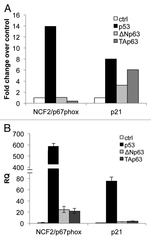
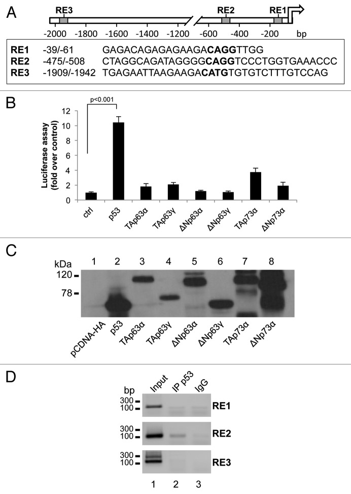
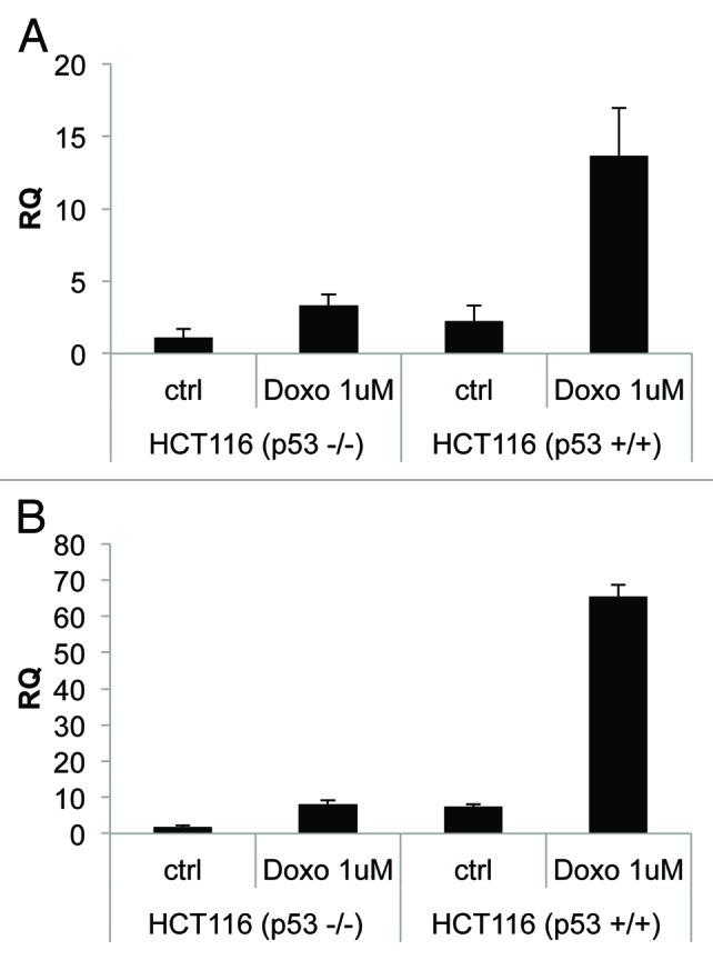
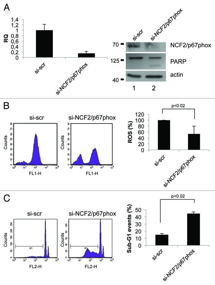
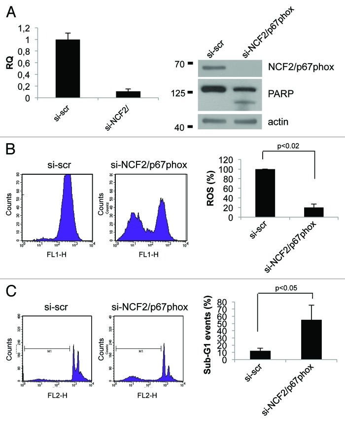
Analysis of microarrays performed in p53-, TAp63α- and ΔNp63α-inducible SaOs-2 cell lines allowed the identification of NCF2 mRNA upregulation in response to p53 induction. NCF2 gene encodes for p67phox, the cytosolic subunit of the NADPH oxidase enzyme complex. The recruitment of p67phox to the cell membrane causes the activation of the NADPH oxidase complex followed by the generation of NADP+ and superoxide from molecular oxygen. The presence of three putative p53 binding sites on the NCF2 promoter was predicted, and the subsequent luciferase and chromatin immunoprecipitation assays showed the activation of NCF2 promoter by p53 and its direct binding in vivo to at least one of the sites, thus confirming the hypothesis. NCF2 upregulation was also confirmed by real-time PCR in several cell lines after p53 activation. NCF2 knockdown by siRNA results in a significant reduction of ROS production and stimulates cell death, suggesting a protective function of Nox2-generated ROS in cells against apoptosis. These results provide insight into the redox-sensitive signaling mechanism that mediates cell survival involving p53 and its novel target NCF2/p67phox.


Figures

None
Figure 1. mRNA expression of NCF2/p67phox in SaOs-2 and in H1299 by p53 family members. (A) Microarray in SaOs-2 inducible clones for each transcription factor. Expression of NCF2/p67phox was measured 24 h after induction with doxycycline. p21 is used as a positive control. (B) Real-time PCR analysis of NCF2/p67phox expression following transfection of p53, ΔN or Tap63 in non-small cell lung carcinoma H1299 cells. p21 is used as a positive control.
None
Figure 2. p53 transcriptionally activates NCF2/p67phox. (A) Map of the human NCF2/p67phox promoter. The promoter possesses three putative p53-responsive elements as indicated by the boxes. (B) p53 induces NCF2/p67phox promoter activity. HEK293 cells were transfected with pGL3-p67phox and the transactivators cloned in pcDNA vectors to evaluate the promoter activity by luciferase assay. Results are shown as the mean of three independent experiments. (C) Following transfection, HEK293 cells were lysed, and a western blot was performed using an anti-HA antibody to verify the transcription factors expression. The figure shows a representative experiment. (D) p53 binds to NCF2/p67phox promoter on RE2. Chromatin immunoprecipitation was performed in p53-inducible SaOs-2 cells using an anti-p53 antibody (IP p53) or a non-specific serum IgG. Non-immunoprecipitated chromatin was loaded as a positive control (input).The figure shows one representative experiment of three.
None
Figure 3. Endogenous-activated p53 induces NCF2/p67phox expression. (A) Colorectal carcinoma HCT116 cells, available in both p53 negative or positive clones, were treated with doxorubicin 1 μM to induce activation of p53. Cells were collected 24 h after treatment and real-time PCR was performed. (B) p21 is used as a positive control. Results are shown as the mean of three independent experiments.
None
Figure 4. Inhibition of NCF2/p67phox in HCT116 cells decreases ROS and induces apoptosis. (A) HCT116 cells were transfected with either a control siRNA (si-scr) or specific NCF2/p67phox-targeting siRNA (si-NCF2/p67phox) and collected after 48 h. NCF2/p67phox protein and transcript levels were examined by western blot and real-time PCR, respectively. The western blot shown is one representative experiment. Real-time PCR shows the mean of three independent experiments. Actin is shown as a loading control. (B) ROS levels were assayed using a DCFDA staining and FACS analysis. Left panel shows one representative experiment of four independent ones. Right panel shows the mean of four independent experiments as a percentage respect to the control (presented as 100%). (C) Apoptosis levels were assayed by propidium iodide staining and FACS analysis. Percentage of sub-G1 events (M1) is shown. Left panel shows one representative experiment of three. Right panel shows the mean of three independent experiments.
None
Figure 5. Inhibition of NCF2/p67phox in HaCat cells decreases ROS and induces apoptosis. (A) HaCat cells were transfected with either a control siRNA (si-scr) or specific NCF2/p67phox-targeting siRNA (si-NCF2/p67phox) and collected after 48 h. NCF2/p67phox protein and transcript levels were examined by western blot and real-time PCR, respectively. Western blot for both whole and cleaved PARP was performed to assay apoptosis. The western blot is one representative experiment. Real-time PCR shows the mean of three independent experiments. Actin is shown as a loading control. (B) ROS levels were assayed using a DCFDA staining and FACS analysis. The mean of three independent experiments as a percentage respect to the control (presented as 100%) is shown. (C) Apoptosis levels were assayed by PI staining and FACS analysis. Percentage of sub-G1 events (M1) is shown for one of three experiments. The mean of three independent experiments is shown on the right panel.

Comment in

Similar articles

Cited by

References

    1. Craige SE, Keaney JF., Jr. Polyploidy and dysregulated ROS signaling: the school of hard Nox? Cell Cycle. 2009;8:797. doi: 10.4161/cc.8.6.8336. - DOI - PubMed
    1. Brown DI, Griendling KK. Nox proteins in signal transduction. Free Radic Biol Med. 2009;47:1239–53. doi: 10.1016/j.freeradbiomed.2009.07.023. - DOI - PMC - PubMed
    1. Geiszt M, Leto TL. The Nox family of NAD(P)H oxidases: host defense and beyond. J Biol Chem. 2004;279:51715–8. doi: 10.1074/jbc.R400024200. - DOI - PubMed
    1. Nauseef WM. Biological roles for the NOX family NADPH oxidases. J Biol Chem. 2008;283:16961–5. doi: 10.1074/jbc.R700045200. - DOI - PMC - PubMed
    1. Geering B, Simon HU. A novel signaling pathway in TNFα-induced neutrophil apoptosis. Cell Cycle. 2011;10:2821–2. doi: 10.4161/cc.10.17.16747. a. - DOI - PubMed

Publication types