Review
. 2012 Nov-Dec;18(6):584-90.
doi: 10.1097/PPO.0b013e3182745ad4.

Endoscopic palliation of pancreatic cancer

Affiliations
Review

Endoscopic palliation of pancreatic cancer

Gregory A Coté et al. Cancer J. 2012 Nov-Dec.

Abstract

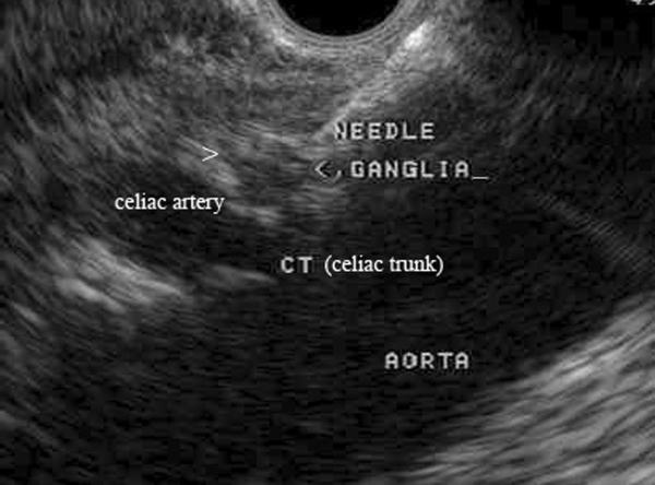
Endoscopy has an increasingly important role in the palliation of patients with pancreatic ductal adenocarcinoma. Endoscopic biliary drainage is still requested in the majority of patients who present with obstructive jaundice, and the increased use of self-expandable metallic stents has reduced the incidence of premature stent occlusion. First-line use of metallic stents is expected to be utilized more frequently as neoadjuvant protocols are improved. The efficacy of endoscopy for palliating gastroduodenal obstruction has advanced with the development of through-the-scope, self-expandable gastroduodenal stents. There have been advances in pain management, with endoscopic ultrasound-guided celiac plexus neurolysis reducing opiate requirements and pain for patients with unresectable malignancy. Future applications of endoscopy in pancreatic cancer may include fine-needle injection of chemotherapeutic and other agents into the lesion itself. This review will summarize the evidence of endoscopy in the management of patients with pancreatic cancer.


Figures

Figure 1
Figure 1. Occlusion of a plastic bile duct stent
Due to their limited diameter, plastic stents may occlude after 2–3 months due to the development of a bacterial biofilm and precipitation of bile/sludge along the internal margin of the stent. For this reason, use of a 10Fr stent is preferred in the setting of malignant biliary obstruction when surgery is anticipated in the next three months.
Figure 2A–B
Figure 2A–B. Distal bile duct obstruction: Deployment of a self-expandable metallic stent (SEMS)
Cholangiogram confirms a distal bile duct stricture (A). A self-expandable metallic stent (SEMS) is deployed (note contrast flow immediately following deployment), with the proximal margin of the stent > 2cm below the hepatic bifurcation (B). This permits safe creation of a choledochojejunostomy if surgery is performed at a later date.
Figure 3
Figure 3. Recommended algorithm for biliary drainage in patients with PDAC
PDAC = pancreatic ductal adenocarcinoma; SEMS = self-expandable metallic stents; PDAC SEMS are currently approved by the FDA for use in patients who have an inoperable, malignant bile duct stricture. ¥Surgical resection without preoperative biliary drainage is reasonable if the procedure can be arranged in a timely fashion and the patient has no significant symptoms related to biliary obstruction (e.g., cholangitis, pruritis refractory to medical therapy).
Figure 4A–E
Figure 4A–E. Endoscopic deployment of a gastroduodenal stent
An obstructing malignant stricture is visualized in the duodenal sweep (A) and demarcated using fluoroscopy (B). A previous biliary metallic stent is only seen on fluoroscopy. A balloon catheter is used to advance a 0.035” stiff guidewire across the stricture, aided by a combination of endoscopy and fluoroscopy (C). The endoscope is withdrawn to the antrum, where the stent catheter is advanced over the guidewire and centered across the stricture (D). The stent is deployed by slowly withdrawing its sheath, allowing its proximal margin to flare in the antrum (E).Reproduced, permission pending, from Cote GA and Edmundowicz SA
Figure 5
Figure 5. EUS-guided celiac neurolysis
A 22 gauge needle is inserted into a celiac ganglion identified by endoscopic ultrasound. Factors associated with a better response include direct injection of celiac ganglia (when visualized) and absence of tumor invasion of the celiac plexus.

Similar articles

Cited by

References

    1. Ross WA, Wasan SM, Evans DB, et al. Combined EUS with FNA and ERCP for the evaluation of patients with obstructive jaundice from presumed pancreatic malignancy. Gastrointest Endosc. 2008;68:461–6. - PubMed
    1. Moss AC, Morris E, Leyden J, et al. Malignant distal biliary obstruction: a systematic review and meta-analysis of endoscopic and surgical bypass results. Cancer Treat Rev. 2007;33:213–21. - PubMed
    1. Ballinger AB, McHugh M, Catnach SM, et al. Symptom relief and quality of life after stenting for malignant bile duct obstruction. Gut. 1994;35:467–70. - PMC - PubMed
    1. Pereira-Lima JC, Jakobs R, Maier M, et al. Endoscopic stenting in obstructive jaundice due to liver metastases: does it have a benefit for the patient? Hepatogastroenterology. 1996;43:944–8. - PubMed
    1. Moss AC, Morris E, Mac Mathuna P. Palliative biliary stents for obstructing pancreatic carcinoma. Cochrane Database Syst Rev. 2006:CD004200. - PubMed

Publication types

MeSH terms