

Mansonella ozzardi corneal lesions in the Amazon: a cross-sectional study
- PMID: 23187969
- PMCID: PMC3533123
- DOI: 10.1136/bmjopen-2012-001266
Mansonella ozzardi corneal lesions in the Amazon: a cross-sectional study
Abstract
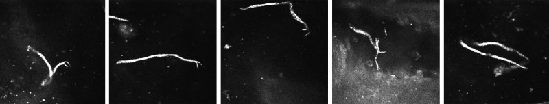
Objectives: To characterise and confirm the presence of Mansonella ozzardi microfilariae in the cornea by biomicroscopy and corneal confocal microscopy.
Design: Cross-sectional study.
Settings: Clinical practice study in patients from rural communities in Coari city on the Solimões river, Amazonas state, Brazil.
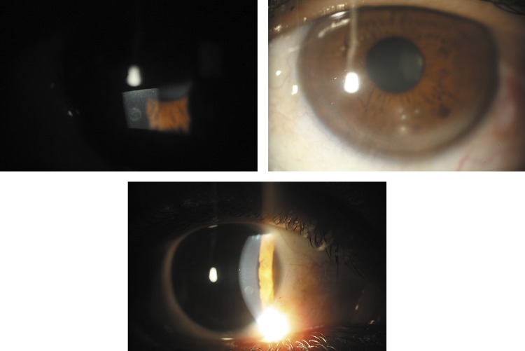
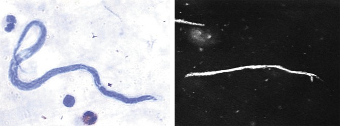
Participants: The eyes of 212 consecutive volunteer patients were examined using a flash light and their blood checked for the presence of microfilariae by an expert microscopist. Patients with suspicious corneal lesions (characterised as nummular keratitis) were submitted to biomicroscopy, fundoscopy and corneal confocal microscopy evaluation (CCME). In two patients, a biopsy of the limbal conjunctiva adjacent to the nummular keratitis was carried out and blood collected from the surgical wound for microfilariae investigation by thick blood film examination.
Primary and secondary outcome measures: Positive correlation between corneal biomicroscopic and confocal lesions and M ozzardi microfilaremia.
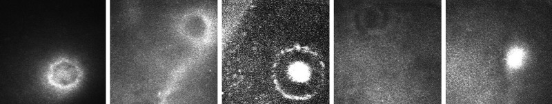
Results: Of the 212 patients, 56 (26.4%) were positive for microfilaremia. 22 patients with nummular keratitis identified under flash light examination underwent biomicroscopy and CCME. Corneal lesions were positively correlated to microfilaremia (p=0.0001). At biomicroscopy, lesions were classified as quiescent or active. At CCME, lesions were categorised as circular or filiform. The associations between corneal lesions, CCME findings and microfilaremia are shown.
Conclusions: We describe M ozzardi microfilariae in the cornea and the associated eye pathology. Further studies using ocular tissue PCR and other imaging techniques would be helpful.
Figures
References
-
- Branco BC, Chamon W, Belfort R, et al. Ocular findings in Pauiní (Southwest of the Brazilian Amazon) and possible corneal lesions caused by Mansonella. Arq Bras Oftalmol 1998;61:647–82
-
- Martins M, Pessoa FA, de Medeiros MB, et al. Mansonella ozzardi in Amazonas, Brazil: prevalence and distribution in the municipality of Coari, in the middle Solimões River. Mem Inst Oswaldo Cruz 2010;105:246–53 - PubMed
-
- Deane MP. Sobre a incidência de filárias humanas em Manaus, estado do Amazonas. Rev Serv Esp Saude Publ 1949;2:849–58
-
- Oliveira wr. (Filarial infestation in inhabitants of Vila Pereira, territory of Roraima (Brazil)). Rev Inst Med Trop Sao Paulo 1963;5:287–8 - PubMed
-
- D'Andretta, Pio da Silva CM, Kameyana F. Ocorrência da mansonelose entre índios do alto Xingu. Rev Soc Bras Med Trop 1969;3:11
LinkOut - more resources
Full Text Sources