. 2013 Jan 9;32(1):100-11.
doi: 10.1038/emboj.2012.309. Epub 2012 Nov 27.

The alarmin Mrp8/14 as regulator of the adaptive immune response during allergic contact dermatitis

Affiliations

The alarmin Mrp8/14 as regulator of the adaptive immune response during allergic contact dermatitis

Beatrix Petersen et al. EMBO J. .

Abstract

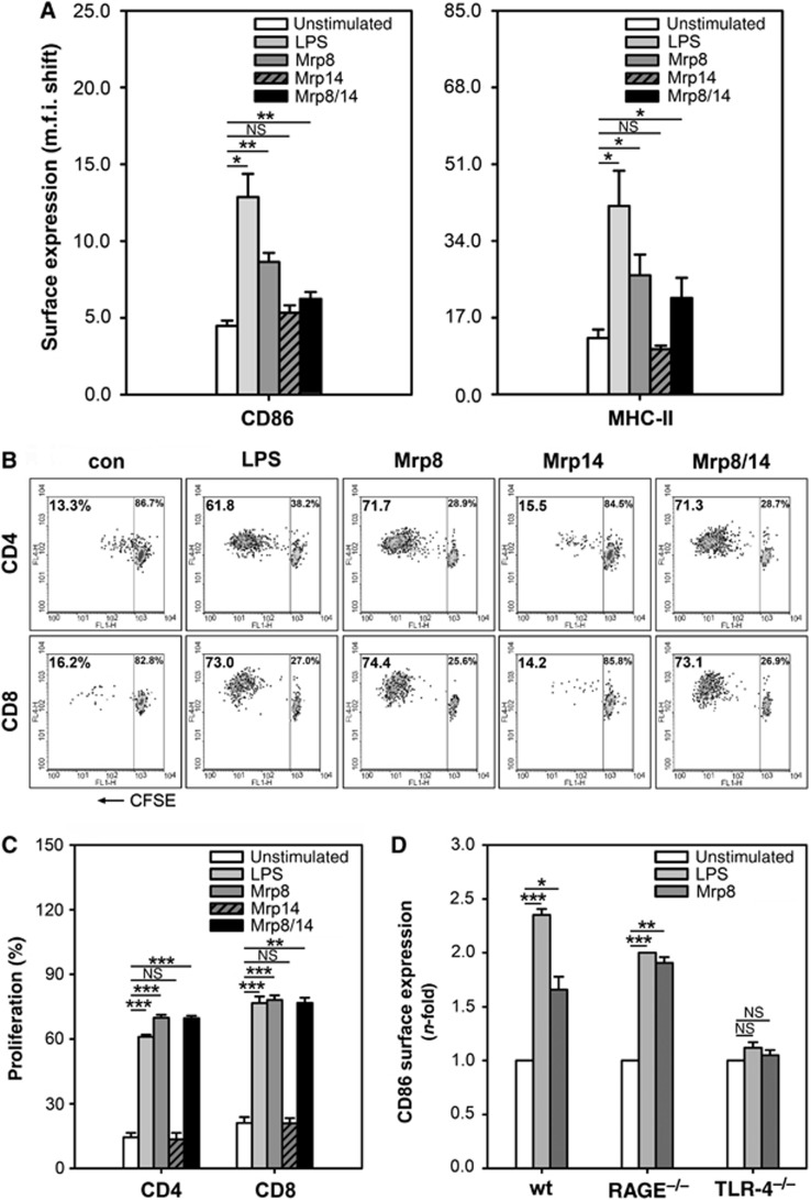
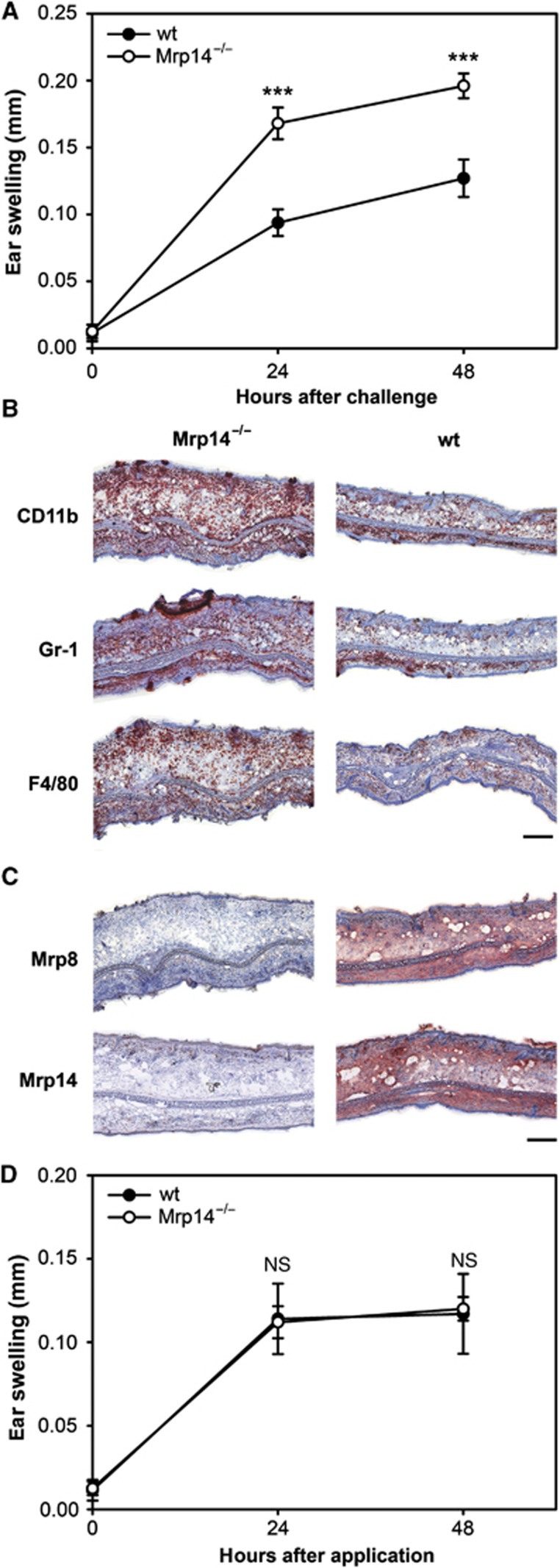
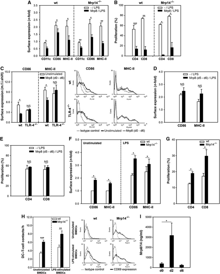
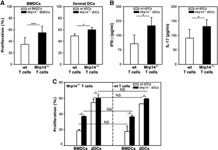
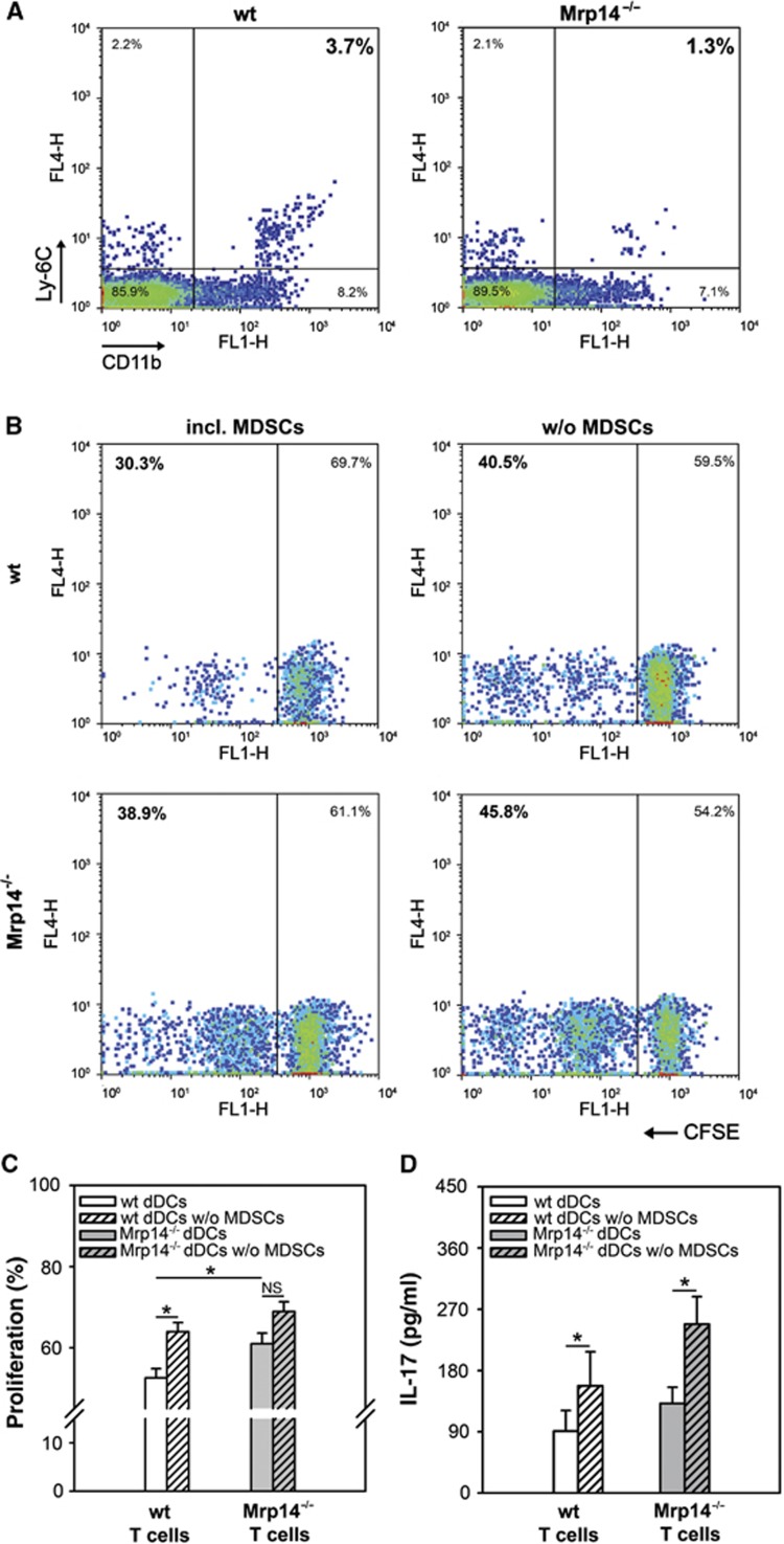
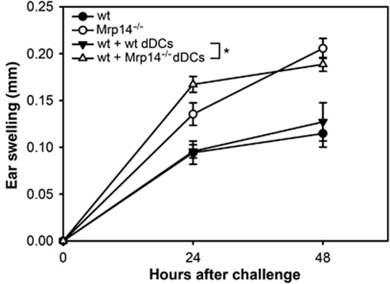
Mrp8 and Mrp14 are endogenous alarmins amplifying inflammation via Toll-like receptor-4 (TLR-4) activation. Due to their pro-inflammatory properties, alarmins are supposed to enhance adaptive immunity via activation of dendritic cells (DCs). In contrast, analysing a model of allergic contact dermatitis (ACD) we observed a more severe disease outcome in Mrp8/14-deficient compared to wild-type mice. This unexpected phenotype was associated with an enhanced T-cell response due to an accelerated maturation of DCs in Mrp8/14-deficient mice. Accordingly, Mrp8, the active component of the heterocomplex, inhibits early DC maturation and antigen presentation in a TLR-4-dependent manner. Transfer of DCs purified from the local lymph nodes of sensitized Mrp8/14-deficient to wild-type mice determined the outcome of ACD. Our results link a pro-inflammatory role of the endogenous TLR-4 ligand Mrp8/14 to a regulatory function in adaptive immunity, which shows some similarities with the 'hygiene hypothesis' regarding continuous TLR-4 stimulation and decreased risk of allergy.


Conflict of interest statement

The authors declare that they have no conflict of interest.

Figures

Figure 1
Figure 1
Mrp8/14-induced TLR-4-mediated activation of iBMDCs. (A) Expression of activation markers CD86 and MHC-II on in vitro generated iBMDCs from C57BL/6 mice. iBMDCs were activated for 48 h in the presence of Mrp8 or Mrp14 (8 μg/ml), Mrp8/14 (20 μg/ml), or LPS (300 ng/ml). Unstimulated iBMDCs served as control. M.f.i. shifts were determined by flow cytometry (three independent experiments; mean±s.e.m.). (B, C) Proliferation of CFSE-labelled CD4+ and CD8+ allogenic spleen T cells from BALB/c mice after 5 days of culture with unstimulated, Mrp8-, Mrp14-, Mrp8/14-, or LPS-stimulated C57BL/6 BMDCs (DC:T-cell ratio was 1:5). Panel (B) shows results from one of three independent experiments which are summarized in (C) (mean±s.e.m.). (D) Comparison of CD86 expression of unstimulated, LPS- (300 ng/ml) and Mrp8- (8 μg/ml) stimulated BMDCs from C57BL/6 wt, RAGE−/−, and TLR-4−/− mice. Following 48 h of stimulation, m.f.i. shifts were measured by flow cytometry and normalized to unstimulated control cells (three independent experiments; mean±s.e.m.). *P<0.05, **P<0.01, ***P<0.001, and NS=not significant.
Figure 2
Figure 2
(AC) DNFB-induced ACD response in ears of C57BL/6 wt and Mrp14−/− mice. (A) Ear swelling of wt and Mrp14−/− mice after elicitation of ACD. Mice were sensitized by painting 0.4% DNFB in acetone/olive oil on the shaved abdomen. After 6 days, the right ears were challenged with 0.4% DNFB in acetone/olive oil. The left ears were treated with acetone/olive oil only and served as controls. Ear thickness was measured 24 and 48 h after challenge. Swelling was determined by calculating the difference between the control ear and the challenged ear (four independent experiments with each 8–15 mice per group; mean±s.e.m.) (***P<0.001). (B, C) Histological analyses of ear sections from DNFB-treated wt and Mrp14−/− mice 48 h after challenge. Sections were stained for (B) immigrated immune cell (CD11b+ myeloid cells, Gr-1+ granulocytes, F4/80+ macrophages) and (C) expression of Mrp8 and Mrp14. (B, C) scale bar, 250 μm. (D) Croton oil-induced ICD response in ears of C57BL/6 wt and Mrp14−/− mice. Mice were painted with 10 μl 3% croton oil in acetone/olive oil (4:1) onto the right ears. The left ears served as controls and were treated with acetone/olive oil only. Ear thickness was measured 24 and 48 h after the treatment. Ear swelling was determined by calculating the difference between the untreated ear and the treated ear (three independent experiments with five mice per group each; mean±s.e.m.).
Figure 3
Figure 3
Extracellular Mrp8 impairs DC differentiation. (A) C57BL/6 wt and Mrp14−/−BMDCs generated in the presence of 8 μg/ml Mrp8 [Mrp8/LPS] were stimulated with 300 ng/ml LPS for 48 h on day 6. CD11c, CD86, and MHC-II surface expression was compared to conventionally LPS-stimulated BMDCs [-/LPS] (four independent experiments; mean±s.e.m.). (B) Proliferation of allogenic T cells (BALB/c) induced by C57BL/6 BMDCs generated as described in (A) (DC:T cells, 1:5) (four independent experiments; mean±s.e.m.). (C) CD86 and MHC-II expression on C57BL/6 wt and TLR-4−/− BMDCs generated in the absence or presence of 8 μg/ml Mrp8 [Mrp8 (d0–d6)]. The right part shows one of three independent experiments that are summarized in the left part (mean±s.e.m.). (D) Phenotype of C57BL/6 wt BMDCs stimulated with 8 μg/ml Mrp8 on day 5 for 24 h [Mrp8 (d5–d6/LPS)]. On day 6, 300 ng/ml LPS was added for further 48 h. CD86 and MHC-II expression was compared to conventionally LPS-stimulated BMDCs [-/LPS] (three independent experiments; mean±s.e.m.). (E) Proliferation of allogenic T cells (BALB/c) induced by C57BL/6 wt BMDCs generated as described in (D) (DC:T cells, 1:5) (three independent experiments; mean±s.e.m.). (F) Accelerated BMDC differentiation due to the lack of Mrp8/14. CD86 and MHC-II expression on wt and Mrp14−/− BMDCs stimulated for 48 h with LPS (300 ng/ml) (right) or left unstimulated (left). M.f.i. shifts were normalized to unstimulated wt cells (four independent experiments; mean±s.e.m.). (G) Proliferation of allogenic spleen T cells (BALB/c) induced by C57BL/6 wt or Mrp14−/− BMDCs (DC:T cells, 1:5) (four independent experiments; mean±s.e.m.). (H) Improved ability of Mrp14−/− BMDCs to contact T cells. Allogenic T cells (BALB/c) were embedded with unstimulated or LPS-stimulated wt and Mrp14−/− BMDCs (C57BL/6) in 3-D collagen gels. Interactions were monitored by time-lapse microscopy. Left part: Number of DC–T-cell contacts per hour (two independent experiments; 40 DCs evaluated per condition). Right part: Embedded T cells were analysed for CD69 surface expression. (I) Mrp8/14 serum levels of DNFB-sensitized wt mice. Sera of 10 mice were analysed (two independent experiments, five mice each, mean±s.e.m.) *P<0.05, **P<0.01, ***P<0.001, and NS, not significant.
Figure 4
Figure 4
Increased DC-mediated T-cell response in Mrp14−/− mice during ACD. (A) Proliferation of wt and Mrp14−/− T cells during ACD. Wt and Mrp14−/− spleen T cells isolated from DNFB-challenged C57BL/6 mice were cultured with DNBS-loaded wt or Mrp14−/− BMDCs of naive C57BL/6 mice (left) or in vivo-primed EpCAM-negative dDCs from ear-draining LNs of the allergic wt or Mrp14−/− C57BL/6 mice (right) (DC:T-cell ratio was 1:5). Proliferation was measured after 5 days of co-culture (four independent experiments; mean±s.e.m.). (B) Expression of pro-inflammatory cytokines IFN-γ and IL-17 by spleen T cells of DNFB-challenged wt and Mrp14−/− C57BL/6 mice in response to stimulation with wt or Mrp14−/− DNFB-primed EpCAM-negative dDCs from the same ACD mice, respectively (three independent experiments; mean±s.e.m.). (C) Proliferation of Mrp14−/− T cells (left) or wt T cells (right) from spleens of DNFB-challenged C57BL/6 mice co-cultured with either DNBS-charged BMDCs of naive C57BL/6 mice or in vivo-primed EpCAM-negative dDCs from ear-draining LNs of the allergic wt or Mrp14−/− C57BL/6 mice. T-cell proliferation was measured after 5 days of co-culture (three independent experiments; mean±s.e.m.). *P<0.05, ***P<0.001, and NS, not significant.
Figure 5
Figure 5
Increased T-cell response during ACD is partly dependent on the number of MDSCs. (A) Quantitative analysis of CD11b+Ly-6C+ MDSCs in LN suspensions from wt and Mrp14−/− mice 48 h after ACD elicitation with DNFB (*P=0.04). Results from one of four independent experiments are shown. (B, C) Proliferation of T cells from allergic wt or Mrp14−/− mice (C57BL/6) induced by MDSC-depleted EpCAM-negative dDC fractions (B, right and C white or grey hatched bars) in comparison to untreated EpCAM-negative dDCs (B, left and C white or grey bars) from the same DNFB-challenged wt and Mrp14−/− mice. Proliferation was determined by CFSE dilution (four independent experiments). *P<0.05 and NS, not significant. (D) Release of the pro-inflammatory cytokine IL-17 by spleen T cells from DNFB-treated wt and Mrp14−/− mice (C57BL/6) in response to MDSC-depleted EpCAM-negative dDCs or untreated EpCAM-negative dDCs of the same mice (three independent experiments; mean±s.e.m.). *P<0.05.
Figure 6
Figure 6
Wt mice develop an increased ear swelling after adoptive transfer of DNFB-primed Mrp14−/− dDCs. EpCAM-negative dDCs were isolated from wt and Mrp14−/− mice 48 h after challenge with DNFB. Subsequently, two identical groups of sensitized wt mice were injected intravenously with these freshly isolated wt or Mrp14−/− dDCs (1.6 × 10⁶ dDCs per mouse) immediately before challenge with 0.4% DNFB. Ear swelling of both groups was evaluated 24 and 48 h after challenge. Data were compared to ear swelling of DNFB-challenged wt and Mrp14−/− mice. Data are from five mice per group (mean±s.e.m.). *P<0.05.

Similar articles

Cited by

References

    1. Banchereau J, Steinman RM (1998) Dendritic cells and the control of immunity. Nature 392: 245–252 - PubMed
    1. Björk P, Björk A, Vogl T, Stenström M, Liberg D, Olsson A, Roth J, Ivars F, Leanderson T (2009) Identification of human S100A9 as a novel target for treatment of autoimmune disease via binding to quinoline-3-carboxamides. PLoS Biol 7: e97d. - PMC - PubMed
    1. Bour H, Peyron E, Gaucherand M, Garrigue J, Desvignes C, Kaiserlian D, Revillard J, Nicolas J (2005) Major histocompatibility complex class I-restricted CD8+ T cells and class II-restricted CD4+ T cells, respectively, mediate and regulate contact sensitivity to dinitrofluorobenzene. Eur J Immunol 25: 3006–3010 - PubMed
    1. Cheng P, Corzo CA, Luetteke N, Yu B, Nagaraj S, Bui MM, Ortiz M, Nacken W, Sorg C, Vogl T, Roth J, Gabrilovich DI (2008) Inhibition of dendritic cell differentiation and accumulation of myeloid-derived suppressor cells in cancer is regulated by S100A9 protein. J Exp Med 205: 2235–2249 - PMC - PubMed
    1. Ege MJ, Mayer M, Normand A, Genuneit J, Cookson W, Braun-Fahrländer C, Heederik D, Piarroux R, von Mutius E (2011) Exposure to environmental microorganisms and childhood asthma. N Engl J Med 364: 701–709 - PubMed

Publication types

MeSH terms