. 2013 Feb;83(2):429-38.
doi: 10.1124/mol.112.082271. Epub 2012 Nov 27.

Canonical transient receptor channel 5 (TRPC5) and TRPC1/4 contribute to seizure and excitotoxicity by distinct cellular mechanisms

Affiliations

Canonical transient receptor channel 5 (TRPC5) and TRPC1/4 contribute to seizure and excitotoxicity by distinct cellular mechanisms

Kevin D Phelan et al. Mol Pharmacol. 2013 Feb.

Abstract

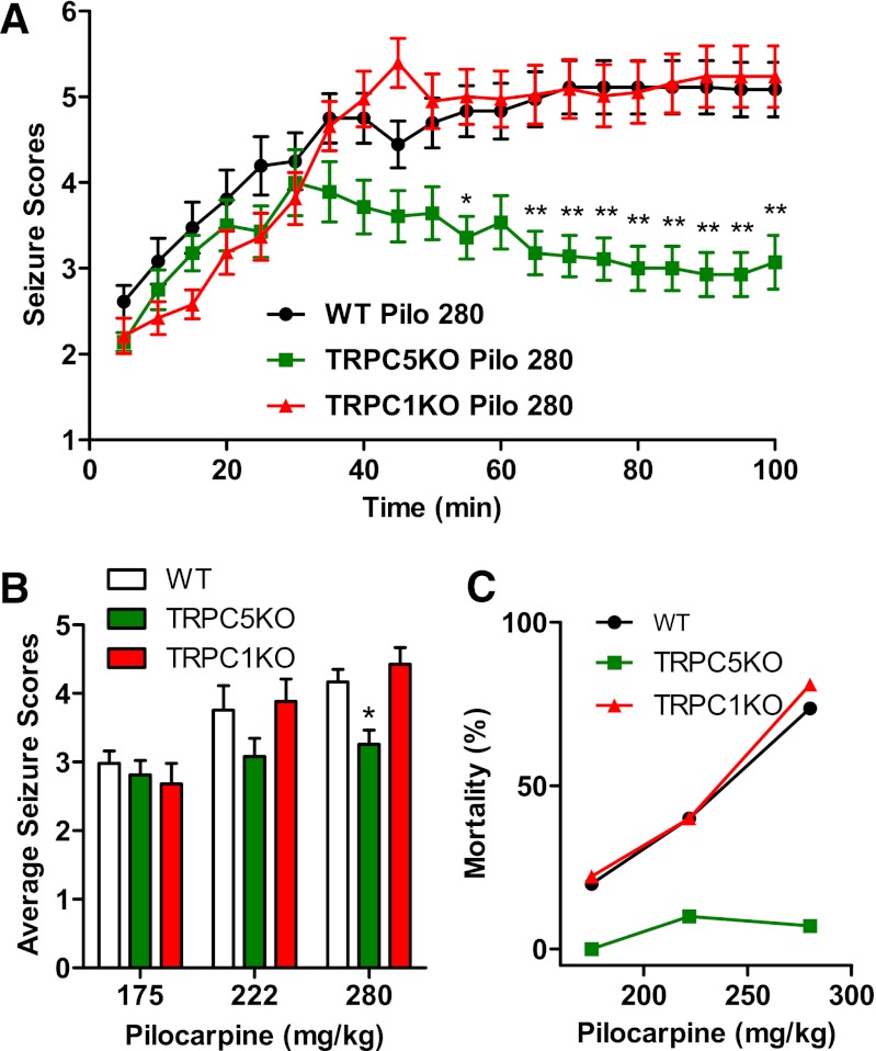
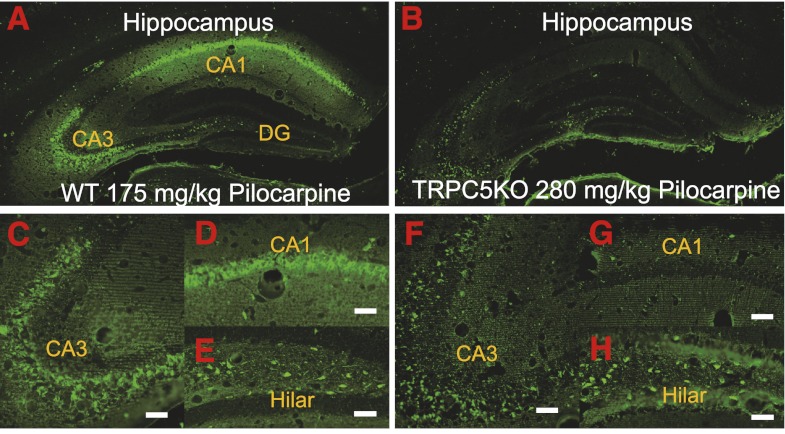
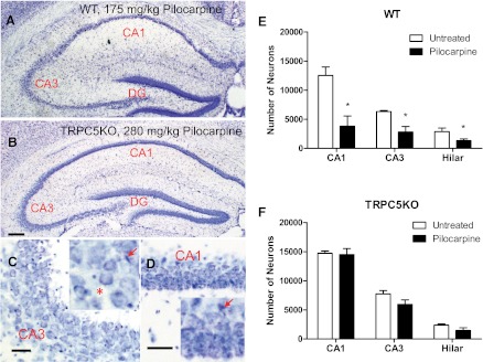
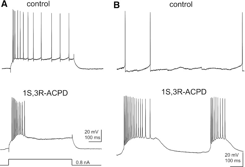
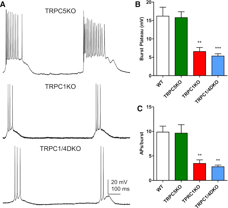
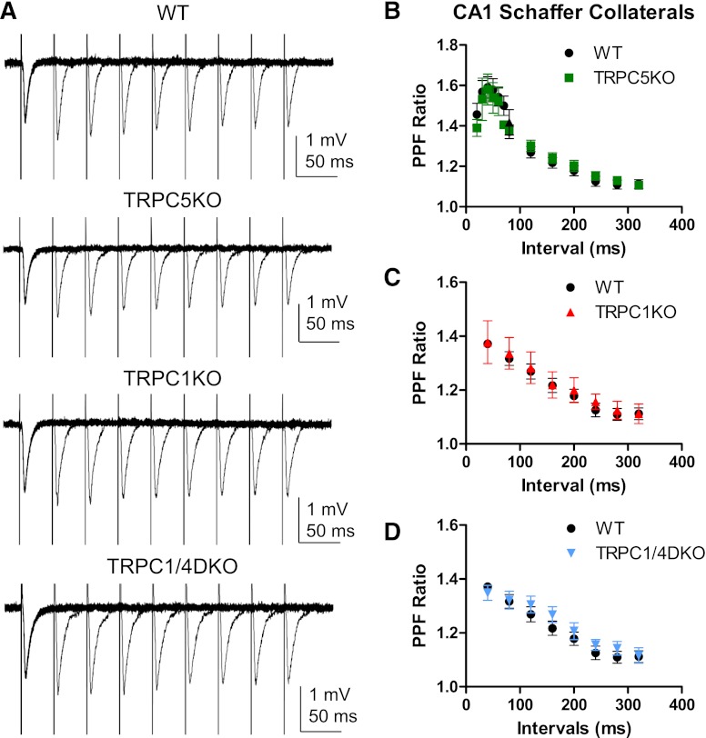
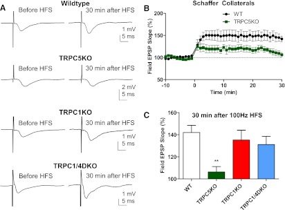
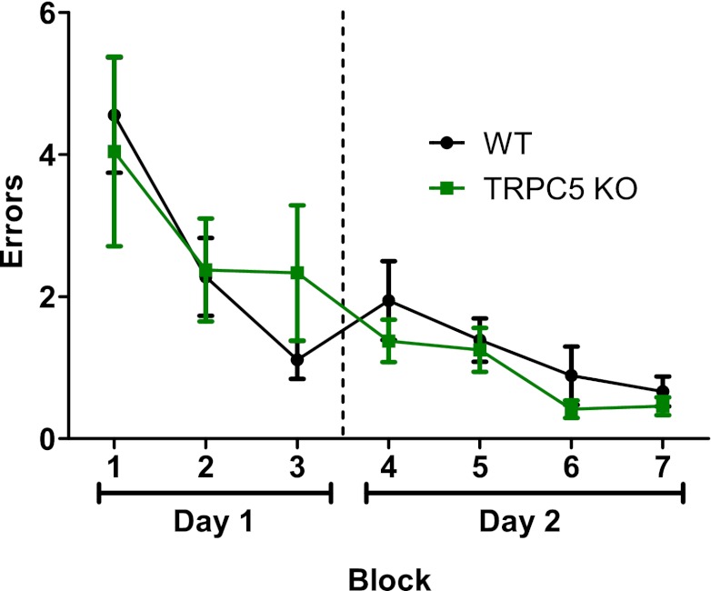
Seizures are the manifestation of highly synchronized burst firing of a large population of cortical neurons. Epileptiform bursts with an underlying plateau potential in neurons are a cellular correlate of seizures. Emerging evidence suggests that the plateau potential is mediated by neuronal canonical transient receptor potential (TRPC) channels composed of members of the TRPC1/4/5 subgroup. We previously showed that TRPC1/4 double-knockout (DKO) mice lack epileptiform bursting in lateral septal neurons and exhibit reduced seizure-induced neuronal cell death, but surprisingly have unaltered pilocarpine-induced seizures. Here, we report that TRPC5 knockout (KO) mice exhibit both significantly reduced seizures and minimal seizure-induced neuronal cell death in the hippocampus. Interestingly, epileptiform bursting induced by agonists for metabotropic glutamate receptors in the hippocampal CA1 area is unaltered in TRPC5 KO mice, but is abolished in TRPC1 KO and TRPC1/4 DKO mice. In contrast, long-term potentiation is greatly reduced in TRPC5 KO mice, but is normal in TRPC1 KO and TRPC1/4 DKO mice. The distinct changes from these knockouts suggest that TRPC5 and TRPC1/4 contribute to seizure and excitotoxicity by distinct cellular mechanisms. Furthermore, the reduced seizure and excitotoxicity and normal spatial learning exhibited in TRPC5 KO mice suggest that TRPC5 is a promising novel molecular target for new therapy.


Figures

Fig. 1.
Fig. 1.
Confirmation of TRPC5 knockout. (A) Intron-exon organization of the Mus musculus TRPC5 gene. Open boxes, untranslated exonic sequence. Closed boxes, translated open reading frame. *Stop codon. Closed triangles between exons 4 and 5, and between exons 5 and 6, loxP sites (ATAAC T TCGT ATAGC ATACA TTATA CGAAG TTAT). 3F, 4R, 6F, and 7R, PCR primers (sequences available upon request). The lengths of the amplicons, including primers, are depicted. (B) Diagram of the expected disruption after excision of exon 5 by the action of the cre recombinase expressed as a transgene under control of the Sox2 promoter (Sox2-cre; Jackson Laboratories). (C) RT-PCR analysis of brain mRNA from a TRPC5 control mouse (WT) and a TRPC5 knockout (KO) mouse. The depicted image is of the electrophoretic migration in a 1.5% agarose gel of the amplicons obtained using the primers depicted in A and B. The results confirmed that in TRPC5 KO mice a TRPC5 mRNA is made that is predicted to lack the coding sequence corresponding to exon 5. This was confirmed by sequencing the amplicon. The mRNA is predicted to encode a 414-amino-acid protein that corresponds to TRPC5[1-413] plus a non-natural valine. (D–E) Confocal images showing immunohistochemical staining of TRPC5 in the hippocampal CA1 and CA3 region. Note the cytoplasmic TRPC5 immunostaining (green) in the soma surrounding the nucleus (blue, stained with DAPI) and the lack of TRPC5 immunostaining in the TRPC5 KO mice. Scale bar: 10 μm.
Fig. 2.
Fig. 2.
Pilocarpine-induced seizures were significantly reduced in TRPC5 KO mice. (A) The time course of pilocarpine-induced seizures in WT, TRPC5KO, and TRPC1KO mice after a single injection of pilocarpine (280 mg/kg, i.p.). Pooled data (mean ± S.E.M.) was plotted (n = 18, 14, and 19 for WT, TRPC5KO, and TRPC1 KO, respectively). See Phelan et al. (2012) for description of seizure scale. Note statistically significantly reduced seizure scores in TRPC5 KO mice at the late phase after pilocarpine injection [P < 0.001, two-way analysis of variance (ANOVA)]. *P < 0.05; **P < 0.01, Bonferroni post hoc tests against WT. (B) Average seizure scores were lower in TRPC5 KO compared with WT mice (P < 0.01, two-way ANOVA). *P < 0.05, Bonferroni post hoc tests against WT. Pooled data (mean ± S.E.M.) was plotted (n = 24, 10, and 18, for WT at 175, 222, and 280 mg/kg pilocarpine, respectively; n = 8, 10, and 14 for TRPC5 KO at 175, 222, and 280 mg/kg pilocarpine, respectively; n = 9, 15, and 19 for TRPC1 KO at 175, 222, and 280 mg/kg pilocarpine, respectively). (C) Mortality in the first 24 hours after pilocarpine injections was reduced in TRPC5 KO mice (n = 8, 10, and 14) compared with WT mice (n = 24, 10, and 18) and TRPC1KO mice (n = 9, 15, and 19).
Fig. 3.
Fig. 3.
TRPC5 KO mice exhibited greatly reduced FJC-positive neurons in the hippocampus after pilocarpine-induced severe seizures. Neuronal cell death induced by pilocarpine-induced seizures was assessed in selected groups of mice with comparable average seizure scores above 3 for WT (3.79 ± 0.14, n = 6) and TRPC5 KO mice (3.48 ± 0.09, n = 6). Mice with average seizure scores ≤3 did not show any FJC-positive neurons. Representative images of FJC-stained neurons in the hippocampus of WT (A, C–E) and TRPC5 KO mice (B, F–H) (2-day survival: WT, 175 mg/kg; TRPC5 KO, 280 mg/kg). Scale bars: 0.25 mm (A, B); 0.05 mm (C, D, F, G); 0.04 mm (E, H).
Fig. 4.
Fig. 4.
Reduced neuronal cell death after pilocarpine-induced seizures in the hippocampus of TRPC5 KO mice. (A–B) Nissl staining in a representative transverse section through the hippocampus in WT and TRPC5 KO mice after severe seizures induced by pilocarpine. Note the normal appearance of the pyramidal cell layer in (B) TRPC5 KO in contrast to the severe gliosis and a loss of normal pyramidal neurons in (A) WT. (C–D) High-power photomicrographs illustrating the lack of gliosis and sparing of CA1 and CA3 neurons in TRPC5 KO mice. Scale bars: 0.5 mm (A–B), 25 μm (C–D). (E–F) Serial coronal sections (50 μm) from mice with similar pilocarpine-induced seizures were stained with Nissl and surviving neurons (with stained cytoplasm and round nuclei) were counted using Stereologer with a 100× oil-immersion objective. The manifestation of seizure-induced degeneration typically is a loss of normal pyramidal cells (*) and occasionally the condensed pyramidal cell nucleus indicated by arrows in the insets of C and D. Pooled data (mean ± S.E.M.) were plotted (n = 4, 5 for untreated and pilocarpine-treated WT mice; n = 3, 5 for untreated and pilocarpine-treated TRPC5 KO mice). In WT mice, the number of neurons was statistically significantly reduced in the CA1, CA3, and hilar regions after severe seizures induced by pilocarpine (P < 0.05, unpaired t tests). On the other hand, in TRPC5 KO mice, the number of neurons was not statistically significantly reduced after pilocarpine-induced severe seizures in any of the three areas (unpaired t tests).
Fig. 5.
Fig. 5.
The effect of 1S,3R-ACPD on the firing pattern of CA1 pyramidal neurons. (A) Representative traces showing the firing patterns evoked by a depolarizing current step (500 ms) in a CA1 pyramidal neuron recorded from adult WT mice (Vh= −70 mV). Note the spike adaptation under control conditions and the burst in the presence of 30 μM 1S,3R-ACPD, a mGluR agonist. (B) Spontaneous firing in the same CA1 pyramidal neuron under the control conditions and in the presence of 30 μM 1S,3R-ACPD.
Fig. 6.
Fig. 6.
Spontaneous burst firing induced by mGluR activity is normal in TRPC5 KO mice, but reduced in TRPC1 KO and TRPC1/4 DKO mice. (A) Representative current-clamp recordings showing the spontaneous burst firing induced by 30 μM 1S,3R-ACPD in CA1 pyramidal neurons in adult TRPC5 KO, TRPC1 KO, and TRPC1/4 DKO mice. (B) The amplitude of the plateau underlying the burst is comparable in the WT and TRPC5 KO mice, but significantly reduced in TRPC1 KO and TRPC1/4 DKO mice. Amplitudes were measured for three randomly selected bursts in each neuron and then averaged. Pooled data (mean ± S.E.M.) were plotted (n = 4, 4, 5, 5 for WT, TRPC5 KO, TRPC1 KO, and TRPC1/4 DKO mice, respectively). (C) The duration of each burst was quantified by the number of action potentials within each burst, and three random bursts from each CA1 pyramidal neuron were analyzed to obtain the average number of spikes per burst. Pooled data (mean ± S.E.M.) were plotted (n = 4, 4, 5, 5 for WT, TRPC5 KO, TRPC1 KO, and TRPC1/4 DKO mice, respectively). **P < 0.01, ANOVA and Tukey’s post hoc test.
Fig. 7.
Fig. 7.
Normal paired-pulse facilitation in TRPC5 KO, TRPC1 KO, and TRPC1/4 DKO mice. (A) Representative traces of paired-pulse facilitation (PPF) of Schaffer collateral field excitatory postsynaptic potential (fEPSP) in WT, TRPC5 KO, TRPC1KO, and TRPC1/4 DKO mice. A pair of electric stimuli with increasing intervals (40, 80, 120, 160, 200, 240, 280, and 320 ms) was delivered at 10-second intervals, and the resulting pair of fEPSPs was recorded. (B–D) The averaged PPF ratios (the peak of the second EPSP over the peak of the first EPSP in each pair) and standard errors were plotted (n = 5-15, 5-13, 6, 6 for WT, TRPC5 KO, TRPC1 KO and TRPC1/4DKO, respectively). Note the peak of PPF occurs around 40–50 ms intervals and the subsequent exponential decay at greater intervals. There was no statistically significant difference between WT and TRPC5 KO mice, between WT and TRPC1 KO mice, or between WT and TRPC1/4DKO mice (two-way ANOVA).
Fig. 8.
Fig. 8.
Reduced high-frequency stimuli-induced long-term potentiation (LTP) in the CA1 region in TRPC5 KO, but not in TRPC1 KO and TRPC1/4 DKO mice. (A) Representative traces of Schaffer collateral field excitatory postsynaptic potential (fEPSP) recorded before and 30 minutes after high-frequency stimuli (HFS; 100 Hz, 1 second; repeated three times with a 20-second interval) in WT, TRPC5 KO, TRPC1 KO, and TRPC1/4 DKO mice. Traces shown were the average of 12 consecutive recordings collected at 0.2 Hz. (B) Field EPSP slopes for each minute were determined by averaging 12 consecutive fEPSP recordings in each mouse, and the normalized mean and standard error were plotted (P < 0.01 for genotype effects, two-way ANOVA; n = 14, 9 for WT and TRPC5KO mice, respectively). (C) The averaged fEPSP slope 30 minutes after 100 Hz HFS in WT (n = 14), TRPC5 KO (n = 9), TRPC1 KO (n = 12), and TRPC1/4DKO mice (n = 9). Note the statistically significantly reduced LTP in TRPC5 KO mice; the LTP was normal in TRPC1 KO and TRPC1/4 DKO mice. **P < 0.01, ANOVA and Tukey’s post hoc tests.
Fig. 9.
Fig. 9.
TRPC5 KO mice exhibited normal spatial learning in radial arm water maze. Adult male WT (n = 6) and TRPC5 KO mice (n = 8) were tested in a radial arm water maze using the method described previously by Alamed et al. (2006). There was no statistically significant difference between WT and TRPC5 KO mice (two-way ANOVA).

References

    1. Alamed J, Wilcock DM, Diamond DM, Gordon MN, Morgan D. (2006) Two-day radial-arm water maze learning and memory task; robust resolution of amyloid-related memory deficits in transgenic mice. Nat Protoc 1:1671–1679 - PubMed
    1. Azouz R, Jensen MS, Yaari Y. (1996) Ionic basis of spike after-depolarization and burst generation in adult rat hippocampal CA1 pyramidal cells. J Physiol 492:211–223 - PMC - PubMed
    1. Barton ME, Shannon HE. (2005) Behavioral and convulsant effects of the (S) enantiomer of the group I metabotropic glutamate receptor agonist 3,5-DHPG in mice. Neuropharmacology 48:779–787 - PubMed
    1. Birnbaumer L, Yildirim E, Abramowitz J. (2003) A comparison of the genes coding for canonical TRP channels and their M, V and P relatives. Cell Calcium 33:419–432 - PubMed
    1. Borges K, Gearing M, McDermott DL, Smith AB, Almonte AG, Wainer BH, Dingledine R. (2003) Neuronal and glial pathological changes during epileptogenesis in the mouse pilocarpine model. Exp Neurol 182:21–34 - PubMed

Publication types

MeSH terms

Substances