. 2013 Jan 18;288(3):1856-70.
doi: 10.1074/jbc.M112.394528. Epub 2012 Nov 27.

Small misfolded Tau species are internalized via bulk endocytosis and anterogradely and retrogradely transported in neurons

Affiliations

Small misfolded Tau species are internalized via bulk endocytosis and anterogradely and retrogradely transported in neurons

Jessica W Wu et al. J Biol Chem. .

Abstract

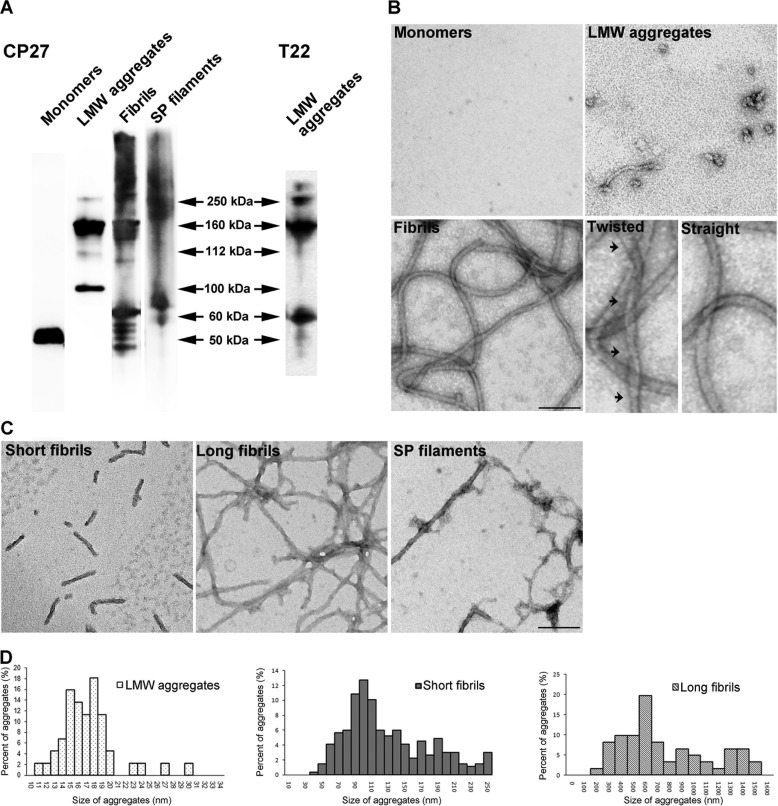
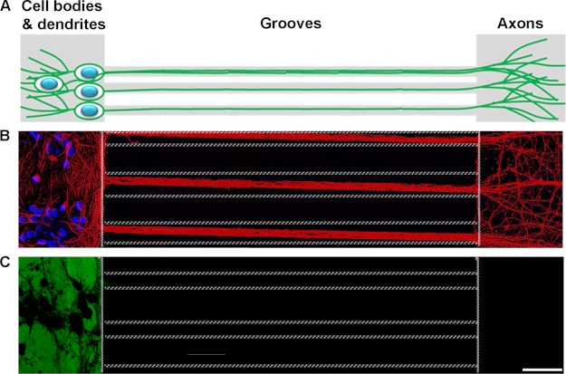
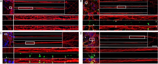
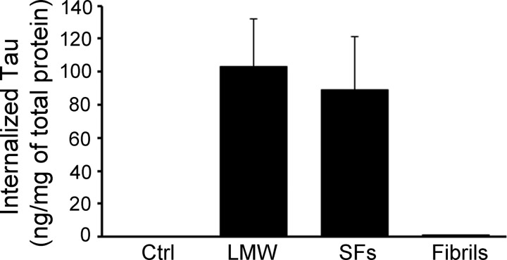
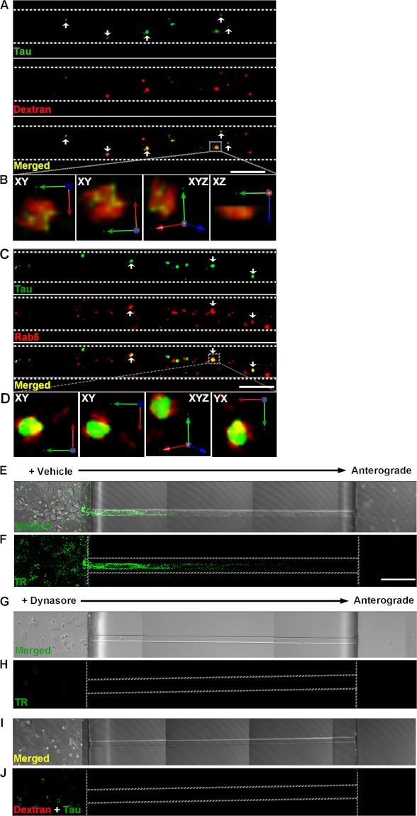
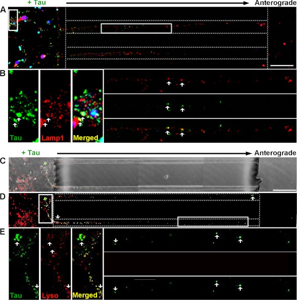
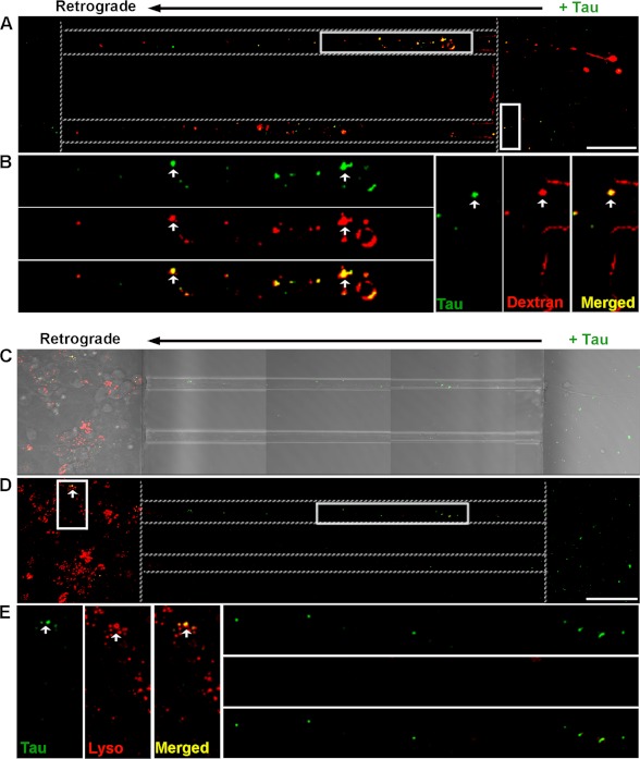
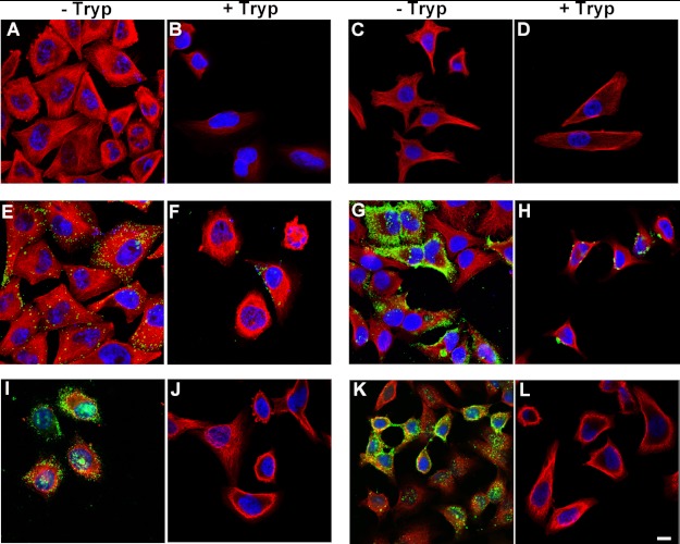
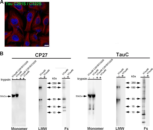
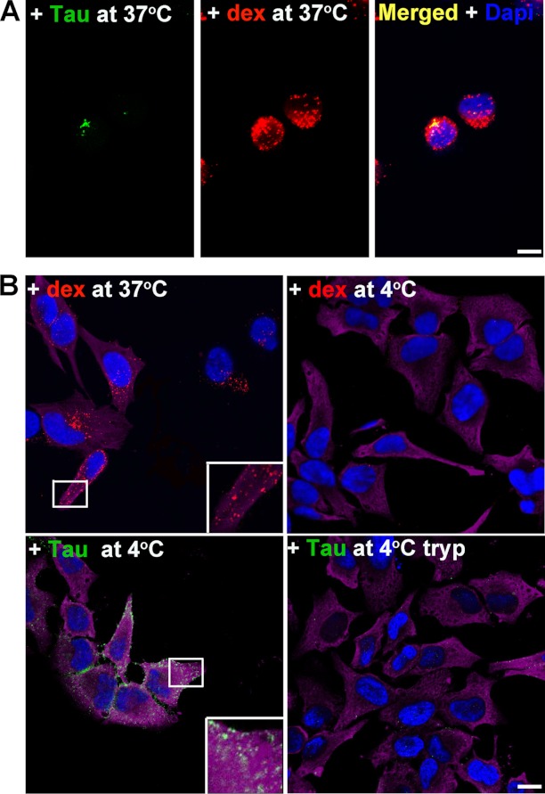
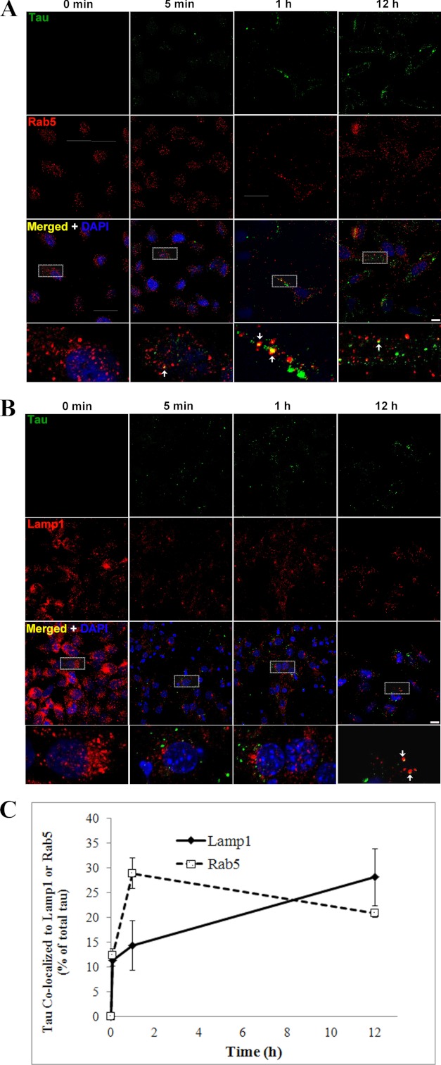
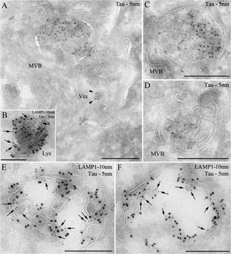
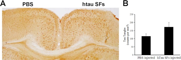
The accumulation of Tau into aggregates is associated with key pathological events in frontotemporal lobe degeneration (FTD-Tau) and Alzheimer disease (AD). Recent data have shown that misfolded Tau can be internalized by cells in vitro (Frost, B., Jacks, R. L., and Diamond, M. I. (2009) J. Biol. Chem. 284, 12845-12852) and propagate pathology in vivo (Clavaguera, F., Bolmont, T., Crowther, R. A., Abramowski, D., Frank, S., Probst, A., Fraser, G., Stalder, A. K., Beibel, M., Staufenbiel, M., Jucker, M., Goedert, M., and Tolnay, M. (2009) Nat. Cell Biol. 11, 909-913; Lasagna-Reeves, C. A., Castillo-Carranza, D. L., Sengupta, U., Guerrero-Munoz, M. J., Kiritoshi, T., Neugebauer, V., Jackson, G. R., and Kayed, R. (2012) Sci. Rep. 2, 700). Here we show that recombinant Tau misfolds into low molecular weight (LMW) aggregates prior to assembly into fibrils, and both extracellular LMW Tau aggregates and short fibrils, but not monomers, long fibrils, nor long filaments purified from brain extract are taken up by neurons. Remarkably, misfolded Tau can be internalized at the somatodendritic compartment, or the axon terminals and it can be transported anterogradely, retrogradely, and can enhance tauopathy in vivo. The internalized Tau aggregates co-localize with dextran, a bulk-endocytosis marker, and with the endolysosomal compartments. Our findings demonstrate that exogenous Tau can be taken up by cells, uptake depends on both the conformation and size of the Tau aggregates and once inside cells, Tau can be transported. These data provide support for observations that tauopathy can spread trans-synaptically in vivo, via cell-to-cell transfer.


Figures

FIGURE 1.
FIGURE 1.
Tau misfolds into biochemically distinct aggregates. A, recombinant hTau40 monomer, low molecular weight species (LMW), fibrils, and Tau filaments purified from rTg4510 mouse brains were prepared and analyzed by Western blot with the CP27 antibody. B, electron microscopy images of negatively stained Tau protein. Fibrils exhibited both twisted helical (arrows) and straight ribbon morphologies. C, fibrils formed at early kinetic points are short, whereas fibrils matured over time are longer in length, similar to Tau filaments purified from the Sarkosyl-insoluble pellet (SP) fraction from rTg4510 mouse brains. Scale bar, 100 nm. D, size distribution of Tau low molecular weight species, short and long fibrils. Data represent measurements from five EM images and >50 fibrils.
FIGURE 2.
FIGURE 2.
Microfluidic chamber system polarizes neurons into somatodendritic and axonal compartments that are fluidically isolated microenvironments. A, schematic diagram of neurons cultured in MF chambers. B, murine hippocampal and cortical neurons grown in chambers extended the axons through microgrooves and into the opposing compartment. β-Tubulin III (neuronal marker), red. DAPI, blue. Dotted lines indicate edges of the microgrooves. C, fluidic isolation of Alexa 488 IgG (green) to the somatodendritic compartment demonstrates that the soma and axonal compartments are isolated and independent microenvironments.
FIGURE 3.
FIGURE 3.
Small, misfolded Tau aggregates are internalized by, and anterogradely transported in neurons. Recombinant hTau40 aggregates were prepared and added to neurons (days in vitro 7). Cells were washed and immunolabeled with anti-β-tubulin III (red) and anti-Tau (CP27, green) antibodies as described under “Experimental Procedures.” Blue, DAPI-stained cell nuclei. Representative confocal images showing Tau LMW (B) aggregates, short (C) and long fibrils (D), but not monomer (A) bound extensively to cells. Furthermore, Tau LMW aggregates and short fibrils were taken up and transported in axons toward the axonal terminals. Multiple insets denote higher magnification of the selected areas in individual and merged channels. Dotted lines indicate the edge of the microgroove. Scale bar, 50 μm.
FIGURE 4.
FIGURE 4.
Tau aggregates are taken up in neurons. Primary neurons from Tau knock-out mice were exposed to Tau LMW aggregates, short fibrils (SFs), or long fibrils for 6 h. Surface-bound Tau aggregates were removed by trypsin treatment and cell lysates were subsequently collected. The amount of internalized Tau was measured by sandwich ELISA using Tau monoclonal antibodies DA31 and DA9. The histogram demonstrates that ∼100 ng of Tau LMW and SFs were internalized per mg of total protein but very little of the long Tau fibrils were internalized.
FIGURE 5.
FIGURE 5.
Exogenously added LMW Tau aggregates are internalized via bulk endocytosis in neurons. Tau LMW aggregates and dextran-Texas Red (marker for bulk endocytosis, red) were added to the somatodendritic compartment of neurons at days in vitro 7, washed, and immunolabeled with anti-Tau antibody (CP27, green) and/or an early endosomal marker antibody (Rab5, red). Tau was transported in axons and co-localized with dextran (A) (arrowheads, yellow) and Rab5 (C) (arrowheads, yellow). B and D, three-dimensional reconstruction of Tau aggregates with dextran or with Rab5 after acquisition at 0.2-μm Z-steps of the selected areas in higher magnification. Distinct co-localization of Tau and dextran is evident from linear and orthogonal perspectives. E and F, transferrin-Alexa 488 uptake was inhibited in cells that were pre-treated with 80 μm dynasore for 15 min but not in vehicle-treated cells (G and H). Plasma membrane-bound transferrin was removed with stringent washes. I and J, bright field and fluorescent images showing inhibition of endocytosis by the small molecule, dynasore effectively inhibited Tau (green) uptake. Dextran was used as a control (red). Dotted lines indicate the edge of the microgroove. Scale bar, 50 μm.
FIGURE 6.
FIGURE 6.
Internalized Tau LMW aggregates are localized to late endosomes and lysosomes. Confocal analysis of aggregates localization to lysosomes in neurons treated with Tau aggregates for 12 h. Tau was immunolabeled with CP27 (green) and lysosomes are marked by Lamp1 (A and B) (late endosomes and lysosomes, red) or with the lysosomal substrate (C–E), DQ-BSA (red). Fluorescent (A and D) and bright field (C) images show distribution of Tau aggregates around the soma, dendrites, and in the axons of neurons. B and E, multiple insets show higher magnifications of the selected area in individual and merged channels. Arrows point to Tau aggregates co-localized to late endosomes/lysosomes (yellow) distributed around the cell body and in the axons. Dotted lines indicate the edge of the microgroove. Scale bar, 50 μm.
FIGURE 7.
FIGURE 7.
Tau LMW aggregates are taken up at axonal terminals and retrogradely transported to lysosomes. Tau aggregates were added to the axonal terminal compartment of neurons for 12 h and immunolabeled with CP27 (green). Endosomes were marked with dextran (red) and lysosomes were labeled by DQ-BSQ (red). Tau aggregates were co-localized with (A and B) dextran (arrowheads, yellow) and retrogradely transported in axons toward the soma. Bright field (C) and fluorescence (D) images showing binding of Tau aggregates to the axonal terminals, in the axons, and around the soma of neurons. E, multiple insets show higher magnification of the selected area in individual and merged channels. Arrowheads point to Tau aggregates that were retrogradely transported in the axons (green) toward the soma and co-localized to late endosomes/lysosomes (yellow) distributed around the cell bodies. Dotted lines indicate the edge of the microgroove. Scale bar, 50 μm.
FIGURE 8.
FIGURE 8.
Exogenous Tau aggregates also bind to non-neuronal cells and are internalized. HeLa cells were incubated with buffer alone (A), Tau monomer (C), LMW aggregates (E), short fibrils (G), long fibrils (I), and filaments (K) purified from rTg4510 mouse brains for 12 h, washed, and immunolabeled with anti-human Tau (green) or anti-α-tubulin (red) antibodies, and analyzed by confocal microscopy. DAPI, blue. As Tau is sensitive to trypsin cleavage, treated cells were also exposed to 0.25% trypsin for 3 min at 37 °C to remove membrane surface-bound Tau in parallel, and then the remaining internalized Tau was immunolabeled as described above (B, D, F, H, J, and L). Only LMW aggregates and short fibrils were taken up by cells (D and F). Scale bar, 50 μm.
FIGURE 9.
FIGURE 9.
Cysteine mutant Tau monomer is not taken up in cells and trypsin effectively digests Tau aggregates. A, cells were exposed to double cysteine mutant Tau monomer for 12 h, immunolabeled with anti-human Tau (green) and anti-α-tubulin (red) antibodies, and analyzed by confocal microscopy. DAPI, blue. Scale bar, 10 μm. Tau monomer was not taken up in cells. B, trypsin effectively digested Tau protein, including monomer formed from cysteine mutant Tau and wild type Tau, LMW aggregates and fibrils formed from wild type Tau protein. Tau was incubated with 0.25% trypsin for 3 min at 37 °C and analyzed by Western blot analysis with CP27 and TauC (total Tau) antibodies.
FIGURE 10.
FIGURE 10.
Uptake of Tau LMW aggregates in non-neuronal cells is regulated by bulk endocytosis and is blocked by temperature shift at 4 °C. A, HeLa cells were exposed to dextran (red) and Tau LMW aggregates (green) for 12 h and immunolabeled as described. Merged image show the punctate vesicular pattern of Tau co-localizing with dextran (yellow). DAPI, blue. B, at 37 °C, dextran was internalized by bulk fluid-phase endocytosis but it was blocked by shifting the cells to 4 °C. At 4 °C, exogenous Tau LMW aggregates bound to the peripheral membrane surface of cells. Trypsinization led to removal of all Tau indicating that none had been internalized when bulk endocytosis had been blocked. α-Tubulin, magenta. Tau, green. DAPI, blue. Scale bar, 10 μm.
FIGURE 11.
FIGURE 11.
Time course of internalization of LMW aggregates into early endosomes and lysosomes. HeLa cells were exposed to exogenous Tau aggregates for 0 min, 5 min, 1 h, and 12 h, washed, and immunolabeled with anti-Tau (CP27, green) and an early endosomal marker (A) (Rab5, red), or a late endosome/lysosomal marker (B), Lamp1. DAPI, blue. Representative images indicate Tau aggregates co-localized to early endosomes (yellow) for different time points. Scale bar, 10 μm. C, histogram showing quantification of Tau aggregates co-localized to endosomes and lysosomes for different time points. Tau was rapidly internalized to early endosomes (5 min, 12.44 ± 1.27%, 1 h, 28.96 ± 3.05%) and later trafficked to lysosomes (28.20 ± 5.74%).
FIGURE 12.
FIGURE 12.
Tau aggregates localize to endosomal compartments. HeLa cells were processed for ultrathin cryosectioning and immunogold labeled for Tau (PAG 5 nm; A, C, and D) or double immunogold labeled for Tau (PAG 5 nm, arrows) and Lamp1 (PAG 10 nm; B, E, and F). Tau aggregates were observed in multivesicular bodies (MVB), small vesicles (A)(Ves, arrowheads), and lysosomes (B)(Lys). Scale bar represents 250 nm.
FIGURE 13.
FIGURE 13.
Injected recombinant hTau SFs exacerbate Tau pathology in rTg4510 mice. A, staining of the cerebral cortex regions of injected rTg4510 mice with Tau antibody, MC1. Injections were performed on 4-week-old rTg4510 mice and tissue was collected 11 weeks later. The MC1 antibody is human specific and it recognizes a misfolded conformational Tau epitope present in NFTs. Tau pathology in the form of cell body accumulation of MC1-positive human Tau was increased in the brain hemisphere that received hTau SFs as compared with the opposing hemisphere that received PBS. B, histogram showing quantification of MCI immunoreactive cells in hTau SFs injected hemispheres compared with sham-injected hemispheres.

Similar articles

Cited by

References

    1. Frost B., Jacks R. L., Diamond M. I. (2009) Propagation of Tau misfolding from the outside to the inside of a cell. J. Biol. Chem. 284, 12845–12852 - PMC - PubMed
    1. Clavaguera F., Bolmont T., Crowther R. A., Abramowski D., Frank S., Probst A., Fraser G., Stalder A. K., Beibel M., Staufenbiel M., Jucker M., Goedert M., Tolnay M. (2009) Transmission and spreading of tauopathy in transgenic mouse brain. Nat. Cell Biol. 11, 909–913 - PMC - PubMed
    1. Lasagna-Reeves C. A., Castillo-Carranza D. L., Sengupta U., Guerrero-Munoz M. J., Kiritoshi T., Neugebauer V., Jackson G. R., Kayed R. (2012) Alzheimer brain-derived tau oligomers propagate pathology from endogenous tau. Sci. Rep. 2, 700. - PMC - PubMed
    1. Ballatore C., Lee V. M., Trojanowski J. Q. (2007) Tau-mediated neurodegeneration in Alzheimer's disease and related disorders. Nat. Rev. Neurosci. 8, 663–672 - PubMed
    1. Hutton M., Lendon C. L., Rizzu P., Baker M., Froelich S., Houlden H., Pickering-Brown S., Chakraverty S., Isaacs A., Grover A., Hackett J., Adamson J., Lincoln S., Dickson D., Davies P., Petersen R. C., Stevens M., de Graaff E., Wauters E., van Baren J., Hillebrand M., Joosse M., Kwon J. M., Nowotny P., Che L. K., Norton J., Morris J. C., Reed L. A., Trojanowski J., Basun H., Lannfelt L., Neystat M., Fahn S., Dark F., Tannenberg T., Dodd P. R., Hayward N., Kwok J. B., Schofield P. R., Andreadis A., Snowden J., Craufurd D., Neary D., Owen F., Oostra B. A., Hardy J., Goate A., van Swieten J., Mann D., Lynch T., Heutink P. (1998) Association of missense and 5′-splice-site mutations in tau with the inherited dementia FTDP-17. Nature 393, 702–705 - PubMed

Publication types

MeSH terms