. 2013 Jan 11;288(2):1135-49.
doi: 10.1074/jbc.M112.417766. Epub 2012 Nov 27.

Rab7 mutants associated with Charcot-Marie-Tooth disease cause delayed growth factor receptor transport and altered endosomal and nuclear signaling

Affiliations

Rab7 mutants associated with Charcot-Marie-Tooth disease cause delayed growth factor receptor transport and altered endosomal and nuclear signaling

Soumik BasuRay et al. J Biol Chem. .

Abstract

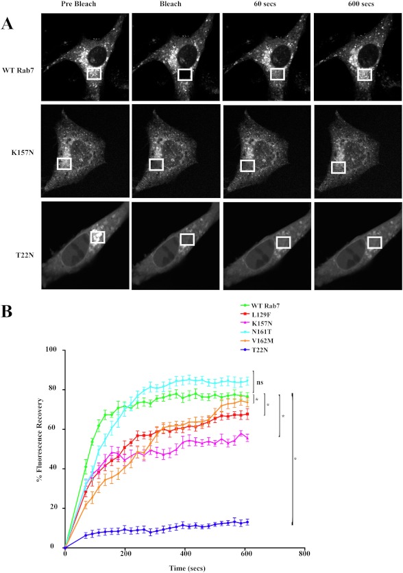
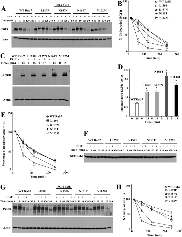
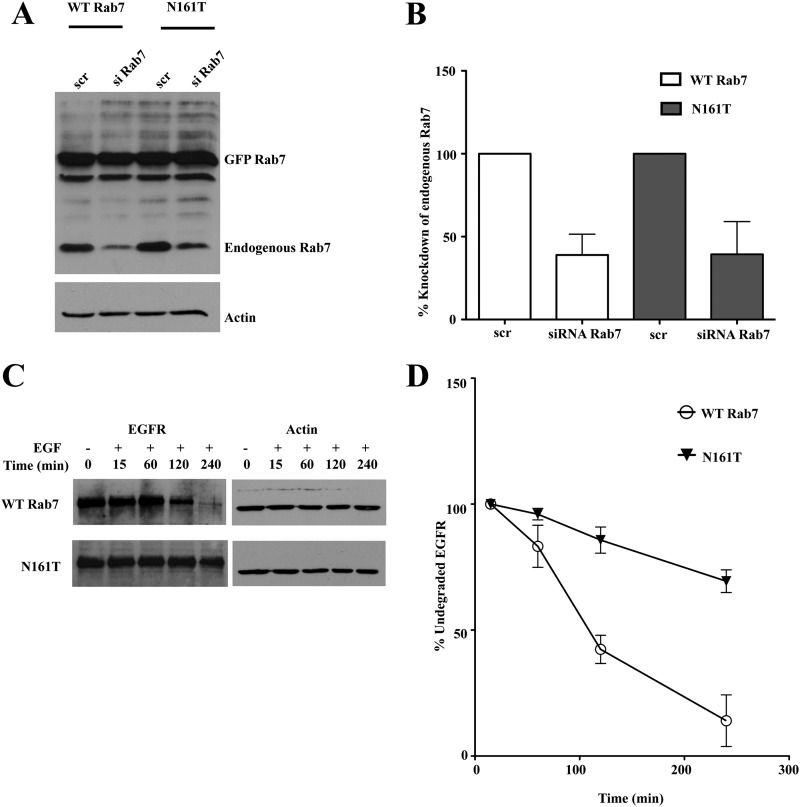
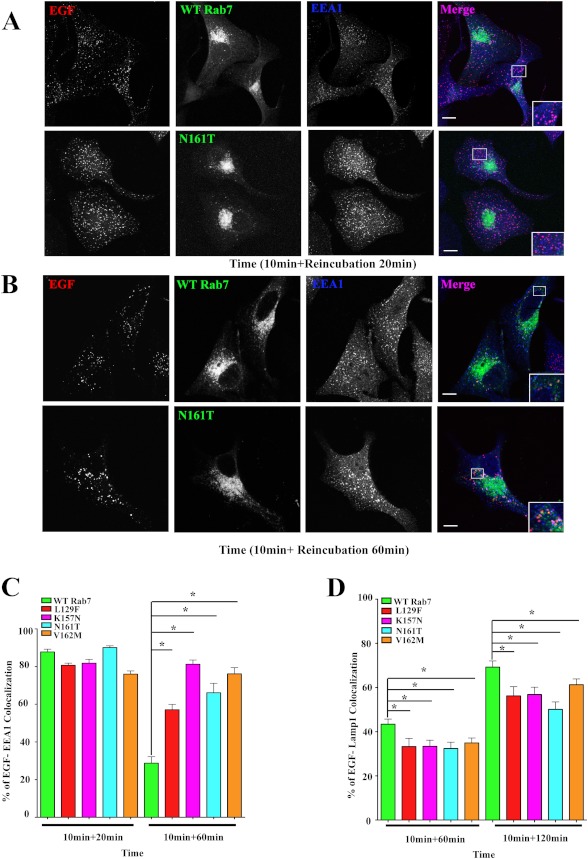
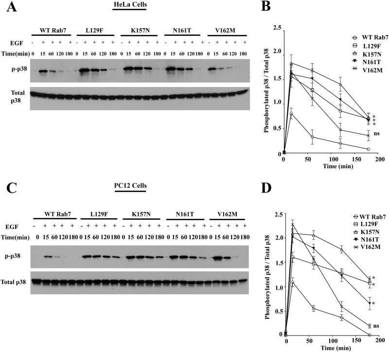
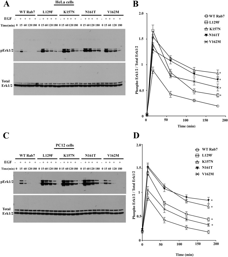
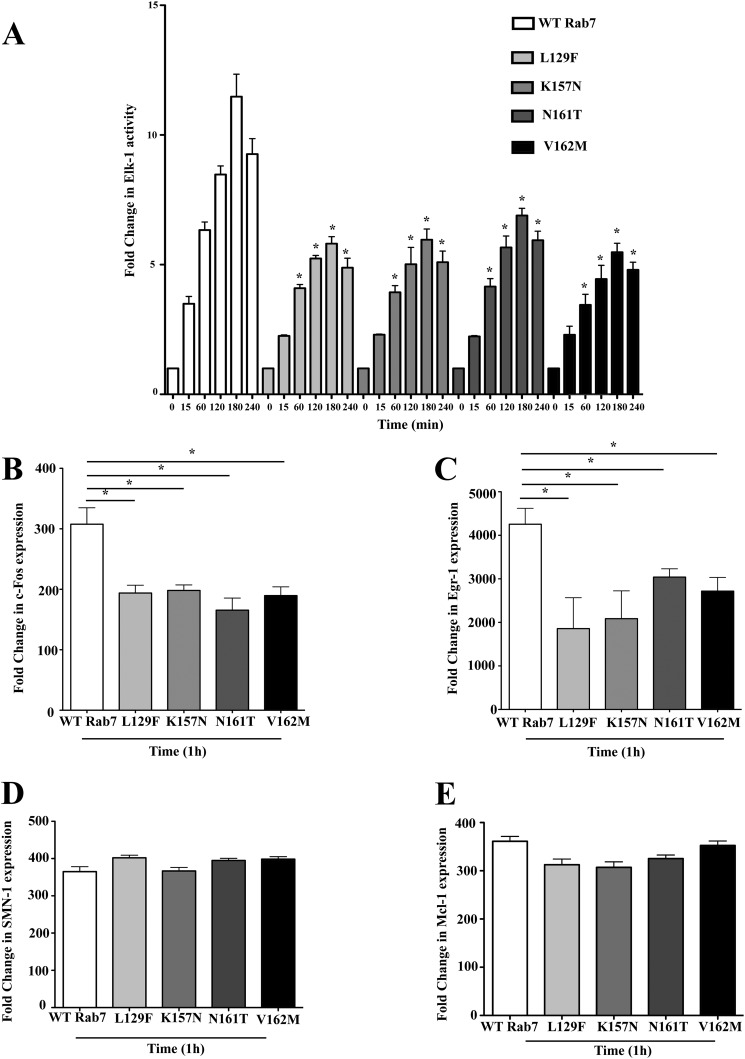
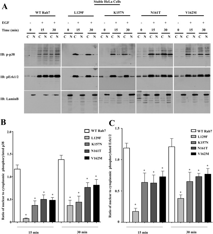
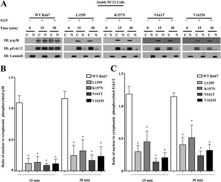
Rab7 belongs to the Ras superfamily of small GTPases and is a master regulator of early to late endocytic membrane transport. Four missense mutations in the late endosomal Rab7 GTPase (L129F, K157N, N161T, and V162M) cause the autosomal dominant peripheral neuropathy Charcot-Marie-Tooth type 2B (CMT2B) disease. As yet, the pathological mechanisms connecting mutant Rab7 protein expression to altered neuronal function are undefined. Here, we analyze the effects of Rab7 CMT2B mutants on epidermal growth factor (EGF)-dependent intracellular signaling and trafficking. Three different cell lines expressing Rab7 CMT2B mutants and stimulated with EGF exhibited delayed trafficking of EGF to LAMP1-positive late endosomes and lysosomes and slowed EGF receptor (EGFR) degradation. Expression of all Rab7 CMT2B mutants altered the Rab7 activation cycle, leading to enhanced and prolonged EGFR signaling as well as variable increases in p38 and ERK1/2 activation. However, due to reduced nuclear translocation of p38 and ERK1/2, the downstream nuclear activation of Elk-1 was decreased along with the expression of immediate early genes like c-fos and Egr-1 by the disease mutants. In conclusion, our results demonstrate that Rab7 CMT2B mutants impair growth factor receptor trafficking and, in turn, alter p38 and ERK1/2 signaling from perinuclear, clustered signaling endosomes. The resulting down-regulation of EGFR-dependent nuclear transcription that is crucial for normal axon outgrowth and peripheral innervation offers a crucial new mechanistic insight into disease pathogenesis that is relevant to other neurodegenerative diseases.


Figures

FIGURE 1.
FIGURE 1.
Rab7 CMT2B mutants show differential membrane cycling. A, BHK-21 cells transfected with GFP-Rab7 WT and CMT2B mutants were used for FRAP studies. Regions of transfected cells were photobleached with a high intensity laser, and recovery of fluorescence was tracked over time. Representative time-lapse images of GFP-Rab7 vesicles before and after photobleaching are shown. At least 30 cells were analyzed for each cell condition in each trial, and results are representative of three independent experiments. B, FRAP analyses show a decreased rate of fluorescence recovery of all of the Rab7 CMT2B mutants compared with the wild type. Rab7T22N mutant showed little or no fluorescence recovery. The differences in fluorescence recovery from three independent trials and 30 cells per trial are plotted ± S.E. (error bars). *, p < 0.05 based on one-way ANOVA with Dunnett's post-test and relative to Rab7 wild-type control.
FIGURE 2.
FIGURE 2.
Rab7 CMT2B mutants delay EGFR degradation. A, HeLa cells stably transfected with GFP-tagged WT Rab7 and CMT2B disease mutants were starved in DMEM for 5 h and pretreated with cycloheximide for 1 h followed by EGF stimulation (100 ng/ml) in a time-dependent manner as indicated. Representative blots from one of three independent experiments are shown. Subsequently, cells were lysed, and equal amounts of protein were immunoblotted and probed for EGFR with actin as the loading control. B, films from three independent experiments were quantified by ImageJ analysis. The line graphs show levels of undegraded EGFR in GFP-WT Rab7- and CMT2B mutant-expressing cells. In each case, the amount of EGFR was normalized to the amount of actin. C, samples from EGFR degradation assays were probed for phospho-EGFR with actin as the loading control. D, films from at least three independent experiments were quantified by ImageJ analysis. The bar graphs show levels of phospho-EGFR in GFP-Rab7 wild type- and CMT2B mutant-expressing cells. In each case, the amount of phospho-EGFR was normalized to the amount of total actin. E, samples from EGFR degradation assays were probed with antibody against tyrosine 1045 of EGFR to analyze the rate of dephosphorylation of the EGFR at the given time points. The line graphs represent the levels of phosphorylated EGFR normalized to actin. F, samples from EGFR degradation assays were probed with antibody directed against GFP to assess the GFP-Rab7 expression levels for all the samples. The levels of GFP Rab7 were found to be similar for all of the samples. G, neuronal PC12 cells stably transfected with GFP-tagged WT Rab7 and CMT2B disease mutants were starved in DMEM for 5 h and pretreated with cycloheximide for 1 h, followed by EGF stimulation (100 ng/ml) in a time-dependent manner as indicated. Representative blots from one of three independent experiments are shown. Subsequently, cells were lysed, and equal amounts of protein were immunoblotted and probed for EGFR with actin as the loading control. H, films from at least three independent experiments were quantified by ImageJ analysis. The line graphs show levels of undegraded EGFR in GFP-WT Rab7- and CMT2B mutant-expressing cells. In each case, the amount of EGFR was normalized to the amount of actin. Values in all cases are from three independent experiments plotted ± S.E. (error bars). *, p < 0.05 based on one-way ANOVA with Dunnett's post-test and relative to Rab7 wild-type control. Non-linear curve fits were used to analyze degradation rates and obtain half-lives in GraphPad Prism version 5.0 (Table 2).
FIGURE 3.
FIGURE 3.
Knockdown of endogenous wild-type Rab7 showed a pronounced delay in EGFR degradation in cells overexpressing Rab7N161T mutant compared with the cells overexpressing Rab7 wild-type. A, HeLa cells stably expressing murine GFP-Rab7 WT and CMT2B mutants were transfected with scrambled siRNA (scr) and siRNA targeted against human Rab7a (siRab7) to knockdown the endogenous Rab7. Cells were kept for 48 h to bring about effective knockdown. A representative Western blot is shown, showing the unaltered expression of murine GFP-Rab7 (∼50 kDa) and knockdown of endogenous Rab7 (∼25 kDa). B, densitometric quantification of the Western blots showed ∼50% knockdown of endogenous Rab7 in both GFP-Rab7 wild type- and N161T-expressing cells. C, HeLa cells stably expressing murine GFP-Rab7 wild type and CMT2B mutants were transfected with siRNA targeted against endogenous Rab7. The cells were incubated for 48 h following transfection to bring about effective knockdown of endogenous Rab7, following which cells were starved for 5 h and pretreated with cycloheximide for 1 h and stimulated with EGF for the indicated times. Rab7N161T showed a pronounced delay in EGFR degradation compared with the wild type. D, densitometric quantitation of films from at least three independent experiments showed a faster kinetics of EGFR degradation in cells expressing wild-type Rab7 compared with those expressing Rab7N161T. Error bars, S.E.
FIGURE 4.
FIGURE 4.
Rab7 CMT2B mutants show a delay in EGF transport from early to late endosomes. HeLa cells transfected with GFP-tagged wild-type (WT) Rab7 and Rab7 CMT2B mutants were starved for 14 h and stimulated with Alexa555-tagged EGF for 10 min followed by reincubation in DMEM for 20 and 60 min (A and B) at 37 °C, permeabilized with Triton X-100, and stained with anti-EEA-1 antibody. C, bar graphs showing the degree of colocalization of EGF and EEA-1 at 20 and 60 min. At least 30 cells were scored for each construct of one experiment for a total of three independent experiments. The degree of colocalization of EGF with EEA-1 was analyzed by Slidebook 4.1 software, and the differences are represented as ±S.E. (error bars). Scale bar, 10 μm. D, bar graphs showing the degree of colocalization of EGF and Lamp1 at 60 and 120 min. At least 30 cells were scored for each construct of one experiment for a total of three independent experiments. The degree of colocalization of EGF with Lamp1 was analyzed by Slidebook 4.1 software, and the differences are represented as ±S.E. Values in all cases are from three independent experiments plotted ± S.E. *, p < 0.05 based on one-way ANOVA with Dunnett's post-test and relative to Rab7 wild-type control.
FIGURE 5.
FIGURE 5.
Rab7 CMT2B mutants show enhanced p38 activation. A, HeLa cells stably expressing GFP-WT Rab7, GFP-Rab7L129F, GFP-Rab7K157N, GFP-Rab7N161T, and GFP-Rab7V162M were starved with DMEM for 5 h and stimulated with EGF (100 ng/ml) for the indicated times. Subsequently, cells were lysed, and equal amounts of protein were immunoblotted and probed for total and phosphorylated p38. B, films from three independent experiments were quantified densitometrically. The line graph shows levels of p-p38 in GFP-Rab7 WT- and CMT2B mutant-expressing cells. In each case, the amount of p-p38 was normalized to the total p38 protein expressed. Differences are represented as ±S.E. (error bars). C, PC12 cells stably expressing GFP-WT Rab7, GFP-Rab7L129F, GFP-Rab7K157N, GFP-Rab7N161T, and GFP-Rab7V162M were starved in DMEM for 5 h and stimulated with EGF (100 ng/ml) for the indicated times. Subsequently, cells were lysed, and equal amounts of protein were immunoblotted and probed for total and phosphorylated p38. D, films from three independent experiments were quantified densitometrically. The line graph shows levels of phosphorylated p38 in GFP-WT Rab7- and CMT2B mutant-expressing cells. In each case, the amount of phosphorylated p38 was normalized to the total p38 protein expressed. Differences are represented as ±S.E. *, p < 0.05 based on one-way ANOVA with Dunnett's post-test and relative to Rab7 wild-type control.
FIGURE 6.
FIGURE 6.
Rab7 CMT2B mutants show enhanced ERK1/2 activation. A, HeLa cells stably expressing GFP-WT Rab7, GFP-Rab7L129F, GFP-Rab7K157N, GFP-Rab7N161T, and GFP-Rab7V162M were starved with DMEM for 5 h and stimulated with EGF (100 ng/ml) for the indicated times. Subsequently, cells were lysed, and equal amounts of protein were immunoblotted and probed for total and phosphorylated ERK1/2. B, films from three independent experiments were quantified densitometrically. The line graph shows levels of pERK1/2 in GFP-Rab7 WT- and CMT2B mutant-expressing cells. In each case, the amount of pERK1/2 was normalized to the total ERK1/2 protein expressed. Differences are represented as ±S.E. (error bars). C, PC12 cells stably expressing GFP-WT Rab7, GFP-Rab7L129F, GFP-Rab7K157N, GFP-Rab7N161T, and GFP-Rab7V162M were starved in DMEM for 5 h and stimulated with EGF (100 ng/ml) for the indicated times. Subsequently, cells were lysed and equal amounts of protein were immunoblotted and probed for total and phosphorylated ERK1/2. D, films from three independent experiments were quantified densitometrically. The line graph shows levels of pERK1/2 in GFP-WT Rab7- and CMT2B mutant-expressing cells. In each case, the amount of pERK1/2 was normalized to the total ERK1/2 protein expressed. Differences are represented as ±S.E. *, p < 0.05 based on one-way ANOVA with Dunnett's post-test and relative to Rab7 wild-type control.
FIGURE 7.
FIGURE 7.
Rab7 CMT2B mutants show lower Elk-1-driven gene activation and c-fos and Egr-1 expression on EGF stimulation. A, HeLa cells stably expressing GFP-tagged WT Rab7 and CMT2B mutants were transfected with plasmids pFR Luc and pFA2 Elk-1 to study Elk-1-driven gene activity. Cells were starved in DMEM for 14 h, followed by EGF stimulation (100 ng/ml) in a time-dependent manner. Following stimulation, cells were kept in serum-free medium for 6 h and lysed, and luciferase activity was determined in a luminometer. Each time point was performed twice and measured in triplicates. Relative light units are normalized to the protein amounts. Disease mutants showed lower Elk-1-driven gene activity. Rab7 CMT2B mutants were compared with the wild-type Rab7 at respective time points. Differences are represented as ±S.E. (error bars). B, HeLa cells stably expressing GFP-Rab7 wild type and CMT2B mutants were serum-starved, EGF-stimulated for 1 h, and processed for RT-PCR as detailed under “Experimental Procedures.” The bar graphs show the lower expression of c-fos upon EGF stimulation in cells expressing GFP-Rab7 CMT2B mutants compared with the wild type. Differences are represented as ±S.E. C, bar graphs showing the lower expression of Egr-1 upon 1-h EGF stimulation in cells expressing GFP-Rab7 CMT2B mutants compared with the wild type. Differences are represented as ±S.E. D and E, bar graphs showing no difference in expression levels of SMN1 and Mcl-1 upon 1-h EGF stimulation in cells expressing GFP-Rab7 CMT2B mutants compared with the wild type. *, p < 0.05 based on one-way ANOVA with Dunnett's post-test and relative to Rab7 wild-type control.
FIGURE 8.
FIGURE 8.
Rab7 CMT2B mutants show delayed nuclear shuttling of activated MAPKs in HeLa cells. A, stable HeLa cells expressing GFP-tagged WT and Rab7 CMT2B mutants were serum-starved for 5 h and stimulated with EGF (100 ng/ml) for 15 and 30 min, respectively. Subsequently, cells were lysed, subjected to subcellular fractionation to separate the cytosolic (C) and nuclear (N) fractions, and immunoblotted with antibodies directed against phosphorylated p38 and phosphorylated ERK1/2. The purity of fractions was confirmed by probing for the nuclear marker lamin B. Rab7 CMT2B mutants showed an impaired nuclear translocation of p-p38. Rab7L129F- and Rab7K157N-expressing cells showed an impaired translocation of pERK1/2, which was also observed in Rab7N161T- and Rab7V162M-expressing cells but to a lesser extent. B and C, bar graphs showing the differences in the nuclear/cytoplasmic ratio of activated p38 and ERK1/2 following 15 and 30 min of EGF stimulation. Differences are represented as ±S.E. (error bars). *, p < 0.05 based on one-way ANOVA with Dunnett's post-test and relative to Rab7 wild-type control. Comparison between CMT2B mutants was based on one-way ANOVA with Tukey's post-test and showed L129F significantly different from N161T and V162M for p38 activation at 15 min of EGF stimulation (p < 0.05).
FIGURE 9.
FIGURE 9.
Rab7 CMT2B mutants show delayed nuclear shuttling of activated MAPKs in neuronal PC12 cells. A, Stable PC12 cells expressing GFP-tagged WT and Rab7 CMT2B mutants were serum-starved for 5 h and stimulated with EGF (100 ng/ml) for 15 and 30 min, respectively. Subsequently, cells were lysed, subjected to subcellular fractionation to separate the cytosolic (C) and nuclear (N) fractions, and immunoblotted with antibodies directed against phosphorylated p38 and phosphorylated ERK1/2. The purity of fractions was confirmed by probing for the nuclear marker lamin B. Rab7 CMT2B mutants showed an impaired nuclear translocation of p-p38 and pERK1/2. B and C, bar graphs showing the differences in the nuclear/cytoplasmic ratio of activated p38 and ERK1/2 following 15 and 30 min of EGF stimulation. Differences are represented as ±S.E. (error bars). *, p < 0.05 based on one-way ANOVA with Dunnett's post-test and relative to Rab7 wild-type control. Comparison between CMT2B mutants based on one-way ANOVA with Tukey's post-test showed no significant differences between the mutants.

References

    1. Barisic N., Claeys K. G., Sirotković-Skerlev M., Löfgren A., Nelis E., De Jonghe P., Timmerman V. (2008) Charcot-Marie-Tooth disease. A clinico-genetic confrontation. Ann. Hum. Genet. 72, 416–441 - PubMed
    1. Shy M. E., Garbern J. Y., Kamholz J. (2002) Hereditary motor and sensory neuropathies. A biological perspective. Lancet Neurol. 1, 110–118 - PubMed
    1. Young P., Suter U. (2003) The causes of Charcot-Marie-Tooth disease. Cell Mol. Life Sci. 60, 2547–2560 - PMC - PubMed
    1. Bienfait H. M., Baas F., Koelman J. H., de Haan R. J., van Engelen B. G., Gabreëls-Festen A. A., Ongerboer de Visser B. W., Meggouh F., Weterman M. A., De Jonghe P., Timmerman V., de Visser M. (2007) Phenotype of Charcot-Marie-Tooth disease Type 2. Neurology. 68, 1658–1667 - PubMed
    1. Snider M. D. (2003) A role for rab7 GTPase in growth factor-regulated cell nutrition and apoptosis. Mol. Cell. 12, 796–797 - PubMed

Publication types

Substances

Supplementary concepts