. 2013 Jan 18;288(3):1500-10.
doi: 10.1074/jbc.M112.439463. Epub 2012 Nov 27.

Direct regulation of nephrin tyrosine phosphorylation by Nck adaptor proteins

Affiliations

Direct regulation of nephrin tyrosine phosphorylation by Nck adaptor proteins

Laura A New et al. J Biol Chem. .

Abstract

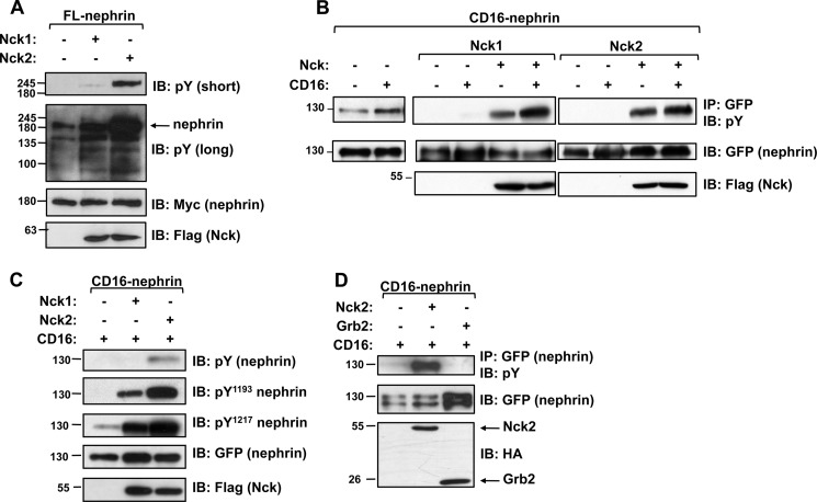
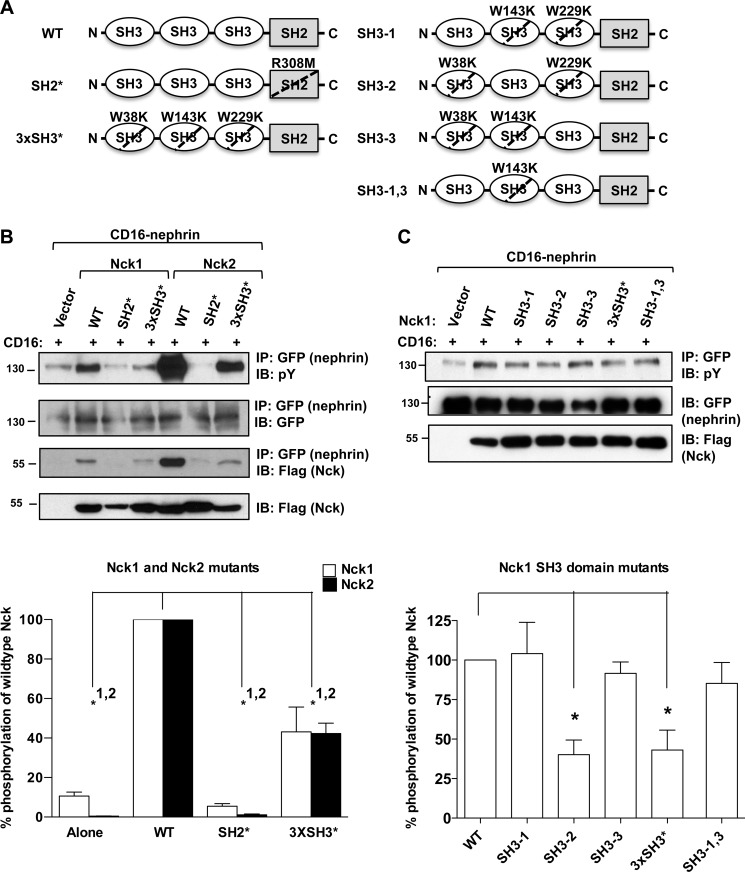
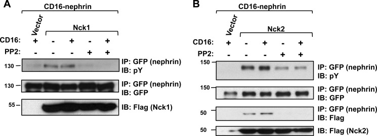
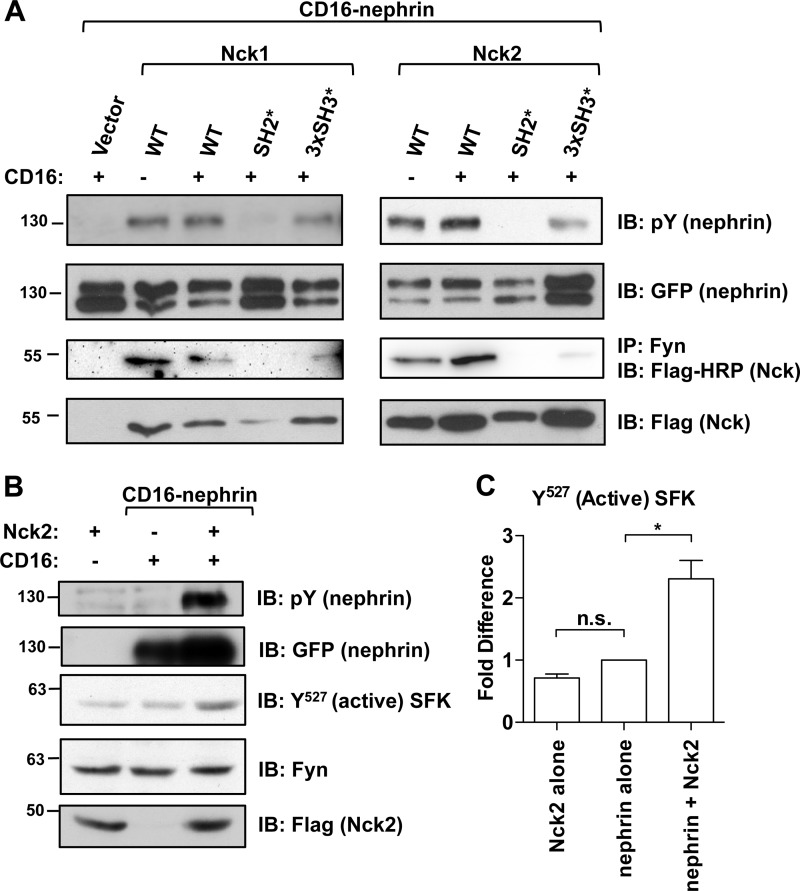
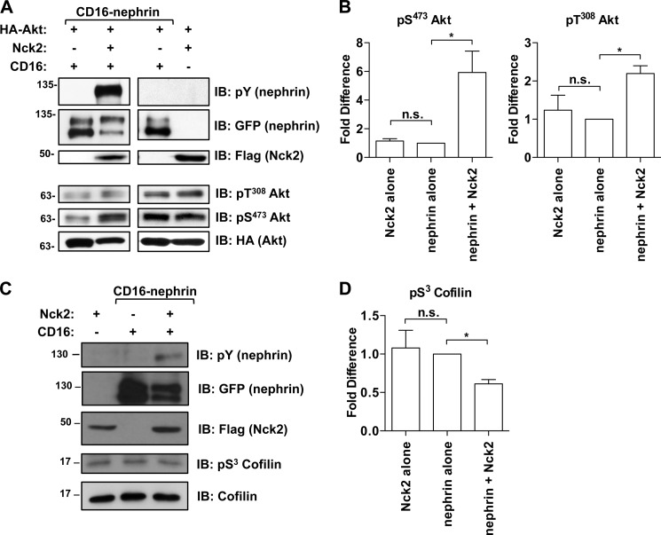
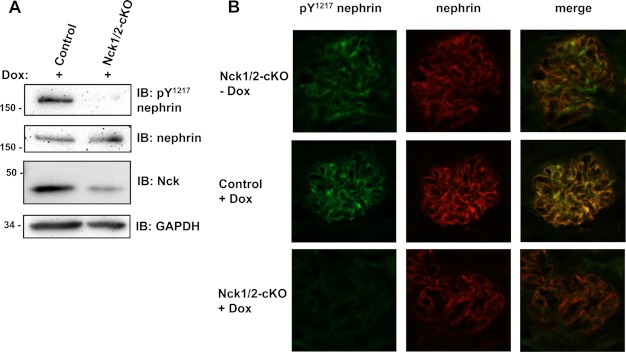
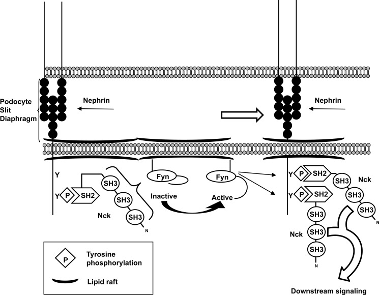
The transmembrane protein nephrin is a key component of the kidney slit diaphragm that contributes to the morphology of podocyte foot processes through signaling to the underlying actin cytoskeleton. We have recently reported that tyrosine phosphorylation of the cytoplasmic tail of nephrin facilitates recruitment of Nck SH2/SH3 adaptor proteins and subsequent actin remodeling and that phosphorylation of the Nck binding sites on nephrin is decreased during podocyte injury. We now demonstrate that Nck directly modulates nephrin phosphorylation through formation of a signaling complex with the Src family kinase Fyn. The ability of Nck to enhance nephrin phosphorylation is compromised in the presence of a Src family kinase inhibitor and when the SH3 domains of Nck are mutated. Furthermore, induced loss of Nck expression in podocytes in vivo is associated with a rapid reduction in nephrin tyrosine phosphorylation. Our results suggest that Nck may facilitate dynamic signaling events at the slit diaphragm by promoting Fyn-dependent phosphorylation of nephrin, which may be important in the regulation of foot process morphology and response to podocyte injury.


Figures

FIGURE 1.
FIGURE 1.
Nck increases nephrin tyrosine phosphorylation. A, HEK293T cells were transfected with full-length (FL) Myc-tagged nephrin and FLAG-tagged Nck1 or Nck2 as indicated, and lysates were immunoblotted (IB) for phosphotyrosine (pY), Myc, or FLAG. Co-expression of Nck1 or Nck2 increases overall nephrin phosphorylation as compared with the base-line phosphorylation seen in the long exposure. B, HEK293T cells were transfected with GFP-tagged CD16-nephrin and Nck1 or Nck2 and then stimulated with CD16 (+) or left untreated (−). CD16 clustering induces an increase in nephrin phosphorylation (left panel), which is enhanced in the presence of Nck1 or Nck2 and further increased after CD16 clustering (middle and right panels). Phosphorylation of CD16-nephrin alone can be seen after long pTyr exposure (left panel), whereas phosphorylation of CD16-nephrin co-expressed with Nck is visible in short pTyr exposures (middle and right panels). C, lysates from CD16-stimulated HEK293T cells expressing CD16-nephrin with or without Nck1 or Nck2 were immunoblotted as indicated to reveal increased levels of nephrin phosphorylation on residues Tyr1193 and Tyr1217 in the presence of Nck1 and Nck2. D, HEK293T cells were transfected with CD16-nephrin and HA-tagged Nck2 or Grb2 as indicated. CD16-induced hyperphosphorylation of nephrin is seen with Nck2 but not with Grb2.
FIGURE 2.
FIGURE 2.
Increased nephrin phosphorylation is dependent on the SH3 domains of Nck. A, schematic illustrating the structure of Nck and the amino acid residues mutated to generate the SH2* and various SH3* mutant constructs in Nck1. The corresponding residues were mutated in Nck2. B, HEK293T cells were transfected with CD16-nephrin and various mutants of Nck1 or Nck2 as indicated. CD16-stimulated (+) lysates were examined for nephrin phosphorylation and Nck binding by Western blotting followed by densitometry to determine the amount of nephrin phosphorylation relative to wild-type Nck. With wild-type (WT) Nck1 (white) or Nck2 (black) normalized to 100%, the SH2 domain mutants (SH2*) do not cause an increase in CD16-nephrin phosphorylation for either Nck1 (5.4 ± 1.4%) or Nck2 (1.1 ± 0.43%) relative to the amount of phosphorylation observed with CD16-nephrin alone (Nck1: 10.6 ± 2.0%; Nck2: 0.47 ± 0.044%). Mutation of all SH3 domains (3×SH3*) leads to a decrease in nephrin phosphorylation to 43.1 ± 12.6% for Nck1 and 42.3 ± 5.3% for Nck2 of that observed with the wild-type Nck. The amount of nephrin phosphorylation correlates with the amount of Nck immunoprecipitated (IP) with nephrin. * indicates p < 0.05 as compared with wild type for Nck1 (indicated by 1) and Nck2 (indicated by 2). IB, immunoblotted; pY, phosphotyrosine. C, transfection of HEK293T cells with additional SH3 domain mutants of Nck1 and CD16-nephrin followed by CD16 stimulation (+) revealed that the first and third SH3 domains either alone (SH3-1, 104.1 ± 19.8%; SH3-3, 91.6 ± 7.2%) or in combination (SH3-1,3, 85.3% ± 13.2%) increase nephrin phosphorylation similarly to wild-type Nck1 (100%), whereas the second SH3 domain alone (SH3-2, 40.2 ± 9.3%) is equivalent to the 3×SH3* mutant (43.1 ± 12.6%). * indicates p < 0.05 as compared with wild type.
FIGURE 3.
FIGURE 3.
Nck-induced hyperphosphorylation of nephrin requires SFK activity. A, HEK293T cells expressing CD16-nephrin and Nck1 were pretreated with 10 μm PP2 as indicated for 3 h prior to stimulation with CD16 (+) to induce CD16-nephrin phosphorylation or left unstimulated (−). PP2 inhibition of SFKs blocks Nck1-mediated nephrin phosphorylation. IP, co-immunoprecipitation; IB, immunoblot; pY, phosphotyrosine. B, identical experiment as in A, except that Nck2 was used instead of Nck1. Treatment with PP2 reduces Nck2-induced hyperphosphorylation of nephrin and the interaction between Nck2 and nephrin as observed by co-immunoprecipitation.
FIGURE 4.
FIGURE 4.
Nck interacts with Fyn, which increases nephrin phosphorylation and Fyn activation. A, HEK293T cells were transfected with CD16-nephrin and Nck1 or Nck2 constructs as indicated and then stimulated with CD16 (+) or left untreated (−). Both Nck1 and Nck2 co-immunoprecipitate (IP) with endogenous Fyn; however, we observed that the interaction with Fyn was much stronger for Nck2 as compared with Nck1. The interaction between Fyn and Nck is very weak when all three SH3 domains are mutated and absent when the SH2 domain is mutated. The strength of these interactions correlates with the amount of nephrin phosphorylation seen in the lysates. IB, immunoblot; pY, phosphotyrosine. B, HEK293T cells were transfected with Nck2 alone as a control, CD16-nephrin alone, or CD16-nephrin and Nck2, and those containing CD16-nephrin were stimulated with CD16 (+). Lysates were immunoblotted for unphosphorylated Tyr527 Src, which recognizes activated SFKs, and then reprobed for Fyn. C, lysates were analyzed by densitometry to determine relative Fyn activity. Expression of Nck2 alone did not alter Fyn activity relative to CD16-nephrin alone. However, when Nck2 was co-expressed with CD16-nephrin, there was a 2.3 ± 0.30-fold increase in the amount of active Fyn relative to CD16-nephrin alone. * indicates p < 0.05 as compared with CD16-nephrin alone; n. s. indicates a nonsignificant difference.
FIGURE 5.
FIGURE 5.
Hyperphosphorylation of nephrin correlates with increased downstream signaling activity. A, HEK293T cells were transfected with HA-tagged Akt, CD16-nephrin and Nck2, as indicated. All cells expressing CD16-nephrin were stimulated with CD16 (+) to induce nephrin phosphorylation. Western blotting (IB) was performed on lysates for phosphorylated and total Akt. Nck2 positively influenced Akt activation on both sites in the presence but not the absence of CD16-nephrin. pY, phosphotyrosine; pS, phosphoserine; pT, phosphothreonine. B, densitometry showing that phosphorylation of Akt on Thr308 increased 2.2 ± 0.21-fold, whereas phosphorylation of Ser473 increased by 5.9 ± 1.5-fold with the addition of Nck2 to CD16-nephrin. Phosphorylation on these sites was not appreciably altered when Nck2 was expressed alone. C, HEK293T cells were transfected with Nck2 alone as a control, CD16-nephrin alone, or CD16-nephrin and Nck2, and those containing CD16-nephrin were stimulated with CD16 (+) to induce nephrin phosphorylation. Western blotting was performed on lysates for phosphorylated and total cofilin. D, densitometry showing that the presence of Nck2 decreased cofilin phosphorylation by 40% (0.61 ± 0.056) as compared with CD16-nephrin alone (1.0) and that expression of Nck2 alone did not alter cofilin activity. * indicates p < 0.05 as compared with CD16-nephrin alone in B and D; n. s. indicates a nonsignificant difference.
FIGURE 6.
FIGURE 6.
Nephrin phosphorylation is reduced in vivo following podocyte-specific depletion of Nck2. A, Western blot analysis of kidney cortex lysates obtained from podocyte-specific Nck1/2 conditional knock-out mice (Nck1/2-cKO) and control littermates exposed to Dox for 1–2 weeks shows a decrease in phosphorylated (Tyr1217) nephrin but not total nephrin in Nck1/2-cKO animals. Reduced Nck expression is also only seen in Nck1/2-cKO animals, demonstrating the specificity of the Dox-induced gene excision. IB, immunoblot; pY, phosphotyrosine. B, dual immunofluorescence analysis of phosphorylated (pTyr1217) and total nephrin expression in glomeruli of Nck1/2-cKO mice and control littermates. Phosphorylated nephrin (green) and total nephrin (red) can be seen in Nck1/2-cKO animals prior to treatment with Dox (top panel). Upon Dox exposure, nephrin phosphorylation can still be readily observed in control animals (middle panel) but not in Nck1/2-cKO animals (bottom panel). Despite the decreased nephrin phosphorylation, total nephrin expression remains unchanged in Dox-treated Nck1/2-cKO animals.
FIGURE 7.
FIGURE 7.
Hypothetical model of Nck-mediated Fyn activation and its role in nephrin signaling at the slit diaphragm. At the podocyte slit diaphragm, nephrin is recruited into lipid rafts where it is initially phosphorylated by Fyn. Nephrin phosphorylation allows for the recruitment of Nck via its SH2 domain, thereby allowing the SH3 domains of Nck to interact with Fyn at the cell membrane and in turn increase Fyn activation. This creates a positive feedback loop, wherein increased Fyn activity leads to the phosphorylation of adjacent nephrin molecules on multiple tyrosine residues. Additional Nck molecules as well as other binding partners can then be recruited to the newly phosphorylated nephrin complex, ultimately leading to the potentiation of downstream signaling pathways required for podocyte function.

Similar articles

Cited by

References

    1. Pavenstädt H., Kriz W., Kretzler M. (2003) Cell biology of the glomerular podocyte. Physiol. Rev. 83, 253–307 - PubMed
    1. Kriz W., Kretzler M., Provoost A. P., Shirato I. (1996) Stability and leakiness: opposing challenges to the glomerulus. Kidney Int. 49, 1570–1574 - PubMed
    1. Welsh G. I., Saleem M. A. (2010) Nephrin—signature molecule of the glomerular podocyte? J. Pathol. 220, 328–337 - PubMed
    1. Verma R., Wharram B., Kovari I., Kunkel R., Nihalani D., Wary K. K., Wiggins R. C., Killen P., Holzman L. B. (2003) Fyn binds to and phosphorylates the kidney slit diaphragm component nephrin. J. Biol. Chem. 278, 20716–20723 - PubMed
    1. Yu C. C., Yen T. S., Lowell C. A., DeFranco A. L. (2001) Lupus-like kidney disease in mice deficient in the Src family tyrosine kinases Lyn and Fyn. Curr. Biol. 11, 34–38 - PubMed

Publication types

MeSH terms

LinkOut - more resources