

Epidermal growth factor promotes protein degradation of epithelial protein lost in neoplasm (EPLIN), a putative metastasis suppressor, during epithelial-mesenchymal transition
- PMID: 23188829
- PMCID: PMC3548460
- DOI: 10.1074/jbc.M112.438341
Epidermal growth factor promotes protein degradation of epithelial protein lost in neoplasm (EPLIN), a putative metastasis suppressor, during epithelial-mesenchymal transition
Abstract
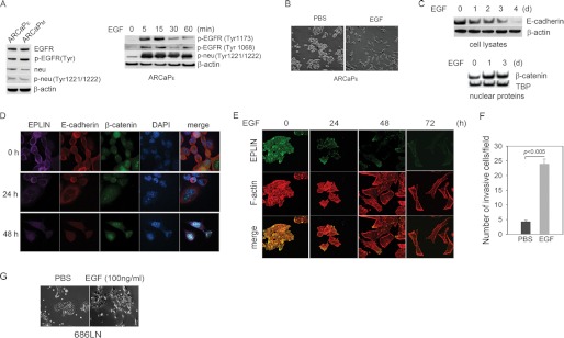
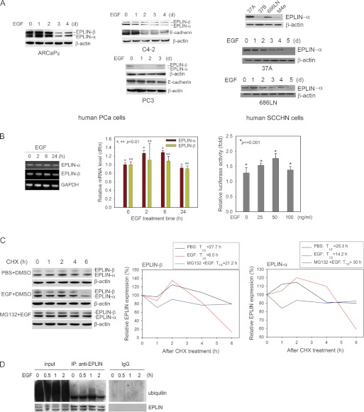
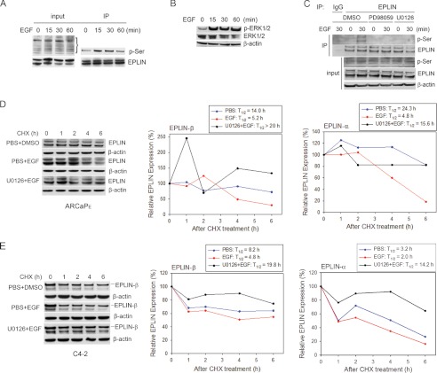
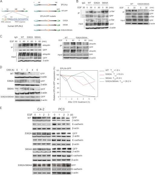
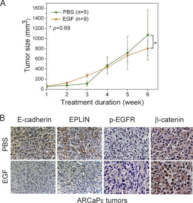
Aberrant expression of EGF receptors has been associated with hormone-refractory and metastatic prostate cancer (PCa). However, the molecular mechanism for EGF signaling in promoting PCa metastasis remains elusive. Using experimental models of PCa metastasis, we demonstrated that EGF could induce robust epithelial-mesenchymal transition (EMT) and increase invasiveness. Interestingly, EGF was found to be capable of promoting protein turnover of epithelial protein lost in neoplasm (EPLIN), a putative suppressor of EMT and tumor metastasis. Mechanistic study revealed that EGF could activate the phosphorylation, ubiquitination, and degradation of EPLIN through an extracellular signal-regulated kinase 1/2 (ERK1/2)-dependent signaling cascade. Pharmacological inhibition of the ERK1/2 pathway effectively antagonized EGF-induced EPLIN degradation. Two serine residues, i.e. serine 362 and serine 604, were identified as putative ERK1/2 phosphorylation sites in human EPLIN, whose point mutation rendered resistance to EGF-induced protein turnover. This study elucidated a novel molecular mechanism for EGF regulation of EMT and invasiveness in PCa cells, indicating that blockade of EGF signaling could be beneficial in preventing and retarding PCa metastasis at early stages.
Figures
Similar articles
-
EPLIN downregulation promotes epithelial-mesenchymal transition in prostate cancer cells and correlates with clinical lymph node metastasis.Oncogene. 2011 Dec 15;30(50):4941-52. doi: 10.1038/onc.2011.199. Epub 2011 May 30. Oncogene. 2011. PMID: 21625216 Free PMC article.
-
A ROS/STAT3/HIF-1α signaling cascade mediates EGF-induced TWIST1 expression and prostate cancer cell invasion.Prostate. 2014 May;74(5):528-36. doi: 10.1002/pros.22776. Epub 2014 Jan 16. Prostate. 2014. PMID: 24435707
-
LIV-1 promotes prostate cancer epithelial-to-mesenchymal transition and metastasis through HB-EGF shedding and EGFR-mediated ERK signaling.PLoS One. 2011;6(11):e27720. doi: 10.1371/journal.pone.0027720. Epub 2011 Nov 16. PLoS One. 2011. PMID: 22110740 Free PMC article.
-
EPLIN: a fundamental actin regulator in cancer metastasis?Cancer Metastasis Rev. 2015 Dec;34(4):753-64. doi: 10.1007/s10555-015-9595-8. Cancer Metastasis Rev. 2015. PMID: 26350886 Free PMC article. Review.
-
[Aberrant Activation Mechanism of TGF-β Signaling in Epithelial-mesenchymal Transition].Yakugaku Zasshi. 2021;141(11):1229-1234. doi: 10.1248/yakushi.21-00143. Yakugaku Zasshi. 2021. PMID: 34719542 Review. Japanese.
Cited by
-
Rab40-Cullin5 complex regulates EPLIN and actin cytoskeleton dynamics during cell migration.J Cell Biol. 2021 Jul 5;220(7):e202008060. doi: 10.1083/jcb.202008060. Epub 2021 May 17. J Cell Biol. 2021. PMID: 33999101 Free PMC article.
-
Human phosphatase CDC14A regulates actin organization through dephosphorylation of epithelial protein lost in neoplasm.Proc Natl Acad Sci U S A. 2017 May 16;114(20):5201-5206. doi: 10.1073/pnas.1619356114. Epub 2017 May 2. Proc Natl Acad Sci U S A. 2017. PMID: 28465438 Free PMC article.
-
Chromatinized protein kinase C-θ directly regulates inducible genes in epithelial to mesenchymal transition and breast cancer stem cells.Mol Cell Biol. 2014 Aug;34(16):2961-80. doi: 10.1128/MCB.01693-13. Epub 2014 Jun 2. Mol Cell Biol. 2014. PMID: 24891615 Free PMC article.
-
Epithelial Protein Lost in Neoplasm, EPLIN, the Cellular and Molecular Prospects in Cancers.Biomolecules. 2021 Jul 16;11(7):1038. doi: 10.3390/biom11071038. Biomolecules. 2021. PMID: 34356662 Free PMC article. Review.
-
Characterization of LIMA1 and its emerging roles and potential therapeutic prospects in cancers.Front Oncol. 2023 May 19;13:1115943. doi: 10.3389/fonc.2023.1115943. eCollection 2023. Front Oncol. 2023. PMID: 37274282 Free PMC article. Review.
References
-
- Shah R. B., Ghosh D., Elder J. T. (2006) Epidermal growth factor receptor (ErbB1) expression in prostate cancer progression: correlation with androgen independence. Prostate 66, 1437–1444 - PubMed
-
- Mimeault M., Pommery N., Hénichart J. P. (2003) New advances on prostate carcinogenesis and therapies: involvement of EGF-EGFR transduction system. Growth Factors 21, 1–14 - PubMed
-
- Di Lorenzo G., Tortora G., D'Armiento F. P., De Rosa G., Staibano S., Autorino R., D'Armiento M., De Laurentiis M., De Placido S., Catalano G., Bianco A. R., Ciardiello F. (2002) Expression of epidermal growth factor receptor correlates with disease relapse and progression to androgen-independence in human prostate cancer. Clin. Cancer Res. 8, 3438–3444 - PubMed
Publication types
MeSH terms
Substances
Grants and funding
LinkOut - more resources
Full Text Sources
Other Literature Sources
Medical
Molecular Biology Databases
Miscellaneous
